Patrícia Correia Oliveira.
Effects of environmental contaminants
on the exotic invasive bivalve
Corbicula fluminea
(Müller, 1774)
Effects o
f e
nvironmental c
ontaminants o
n t
he e
xotic i
nvasive b
ivalve
Corbicula f
luminea
(Müller, 1
774)
Patrícia A
lexandra C
orreia O
liveira
2018 DOUTORAMENTOCIÊNCIAS DO MAR E DO AMBIENTE
Effects of environmental contaminants
on the exotic invasive bivalve
Corbicula fluminea (Müller, 1774)
Patrícia Correia Oliveira
D
UNIVERSIDADES PARTICIPANTES UNIVERSIDADE DO PORTO UNIVERSIDADE DO ALGARVE UNIVERSIDADE DE AVEIROD
.ICBAS
2018
Patrícia Alexandra Correia Oliveira
Effects of environmental contaminants on the exotic invasive
bivalve Corbicula fluminea (Müller, 1774)
Tese de Candidatura ao grau de Doutor em
Ciências do Mar e do Ambiente;
Programa Doutoral da Universidade do Porto
(Instituto de Ciências Biomédicas de Abel Salazar e
Faculdade de Ciências), Universidade de Aveiro e
Universidade do Algarve.
Orientador - Prof. Doutora Lúcia Maria das
Candeias Guilhermino
Categoria - Professora Catedrática
Afiliação - Instituto de Ciências Biomédicas de Abel
Salazar da Universidade do Porto e Centro
Interdisciplinar de Investigação Marinha e Ambiental
da Universidade do Porto
iii The author had a PhD fellowship from the Foundation of Science and Technology (FCT) (SFRH/BD/82402/2011), in the scope of the QREN - POPH - “Tipologia 4.1 - Formação Avançada”, co-founded by the European Social Fund and national funds of the Portuguese Ministry of Education and Science.
The research work included in the present PhD Thesis was developed in the scope of the following projects:
“NISTRACKS - Processes influencing the invasive behaviour of the non-indigenous species Corbicula fluminea (Mollusca: Bivalvia) in estuaries - identification of genetic and
environmental key factors”, funded by the Portuguese “Fundação para a Ciência e a
Tecnologia, I.P.” (FCT) (PTDC/AAC-AMB/102121) and by the COMPETE - Operational Competitiveness Program (“Programa Operational Temático Fatores de Competitividade, FCOMP-01-0124-FEDER-0086556”) co-funded by the European Regional Development Fund (ERDF). GOVERNO DA REPÚBLICA PORTUGUESA FUNDO EUROPEU DE DESENVOLVIMENTO REGIONAL
iv
“INNOVMAR - Innovation and Sustainability in the Management and Exploitation of Marine Resources” (NORTE-01-0145-FEDER-000035), research line 3 “ECOSERVICES - Assessing the environmental quality, vulnerability and risks for the sustainable management of the NW coast natural resources and ecosystem services in a changing world”, funded by
NORTE2020 and ERDF.
“PLASTICGLOBAL - Assessment of plastic-mediated chemicals transfer in food webs of deep, coastal and estuarine ecosystems under global change scenarios”, co-funded by FCT,
Portugal, with national funds (FCT/MCTES, “Orçamento de Estado”, project reference PTDC/MAR-PRO/1851/2014) and the ERDF through the COMPETE 2020 (POCI-01-0145-FEDER-016885) and Lisboa 2020 (LISBOA-01-0145-(POCI-01-0145-FEDER-016885) programmes.
The study was also supported by funds of the Institute of Biomedical Sciences of Abel Salazar of the University of Porto (ICBAS), Portugal, and by the Strategic Funding UID/Multi/04423/2013 through national funds provided by FCT and ERDF in the framework of the programme Portugal 2020 to Interdisciplinary Centre of Marine and Environmental Research - University of Porto (CIIMAR).
v
“The two most powerful warriors are patience and time”
vii
Acknowledgements
Ao longo destes anos tive a felicidade de ter por perto um conjunto de pessoas que me ajudaram e às quais não posso deixar de expressar aqui a minha gratidão.
Agradeço:
- Em primeiro lugar, à Professora Doutora Lúcia Guilhermino pela orientação desta Tese. Foi um enorme privilégio poder contar com o seu apoio e confiança ao longo destes anos de trabalho que marcaram decisivamente o meu crescimento profissional e pessoal.
- Ao Professor Doutor Jorge Machado, Professora Doutora Cristina Canhoto, Professora Doutora Cristina Carvalho, Doutor Vasco Branco e Doutora Neusa Figueiredo pela disponibilidade em colaborar nos trabalhos que integram esta Tese.
- À Ana Lírio, ao Manuel Lopes-Lima, ao Pedro Vilares e ao Gabriel Barboza agradeço a preciosa ajuda em momentos-chave deste trabalho.
- A todos os colegas do CIIMAR/ICBAS que, de uma ou de outra forma, me ajudaram.
- Ao Dr. Paulo Azevedo.
- Ao meu Namorado, à minha Irmã e aos meus Amigos.
- Por fim, aos meus Pais, a quem dedico este trabalho.
Agradeço à Fundação Portuguesa para a Ciência e Tecnologia pelo suporte financeiro através de uma Bolsa de Doutoramento (SFRH/BD/82402/2011). Quero também agradecer às instituições envolvidas na produção desta Tese, nomeadamente o Instituto de Ciências Biomédicas de Abel Salazar (ICBAS) e o Centro Interdisciplinar de Investigação Marinha e Ambiental (CIIMAR) da Universidade do Porto. Deixo também uma nota de agradecimento aos revisores das revistas que elevaram a qualidade deste trabalho com os seus comentários e sugestões.
ix
Author’s declaration
In agreement with the Portuguese law through the article 4th of the “Regulamento Geral dos Terceiros Ciclos de Estudos da Universidade do Porto” of September 12th (GR.08/07/2017), the author states devotion in a major contribution to the conceptual design and technical execution of the work, interpretation of the results and the manuscript preparation of the published, submitted or under preparation publications corresponding to the sections of the present Thesis.
Publications
The following published, accepted for publication or in preparation manuscripts resulted from the experimental research work carried out in the scope of the present Thesis:
Oliveira, P., Lopes-Lima, M., Machado, J., Guilhermino, L. (2015) Comparative sensitivity of European native (Anodonta anatina) and exotic (Corbicula fluminea) bivalves to mercury.
Estuarine, Coastal and Shelf Science 167, Part A: 191-198.
https://doi.org/10.1016/j.ecss.2015.06.014 (Corresponds to Chapter III, with the permission of Elsevier included in the Annex I).
Oliveira, P., Lírio, A.V., Canhoto, C., Guilhermino, L. (2018) Toxicity of mercury and post-exposure recovery in Corbicula fluminea: neurotoxicity, oxidative stress and oxygen consumption. Ecological Indicators 91: 503-510.
https://doi.org/10.1016/j.ecolind.2018.04.028 (In press, corresponds to Chapter IV, with the permission of Elsevier included in the Annex I).
Oliveira, P., Barboza, L.G.A., Branco, V., Figueiredo, N., Carvalho, C., Guilhermino, L. Effects of microplastics and mercury in the freshwater bivalve Corbicula fluminea (Müller, 1774): filtration rate, biochemical biomarkers and mercury bioconcentration. (Accepted in Ecotoxicology and Environmental Safety, corresponds to Chapter V, with permission of Elsevier included in the Annex I).
Oliveira, P., Guilhermino, L. Acclimation conditions for the use of the exotic invasive species
xi
Contents Index
Abstract ... xv
Resumo ... xxi
Figures index ... xxvii
Tables index ... xxxi
List of abbreviations ... xxxv
CHAPTER I ... 1
General Introduction 1.1. Bioinvasions ... 3
1.2. Corbicula fluminea (Müller, 1774) ... 6
1.2.1. Biology and ecology of C. fluminea ... 6
1.2.2. C. fluminea bioinvasions ... 8
1.2.3. Factors influencing the invasive behaviour of C. fluminea ... 9
1.2.4. Impacts of C. fluminea ... 10
1.2.5. Use of C. fluminea in environmental studies ... 13
1.3. Objectives and Outline of the Thesis ... 22
CHAPTER II ... 27
Acclimation conditions for the use of the exotic invasive species Corbicula fluminea in toxicity bioassays Abstract ... 29
2.1. Introduction ... 30
2.2. Material and methods ... 31
2.2.1. Chemicals ... 31
2.2.2. Collection of animals and transport to the laboratory ... 31
2.2.3. Experimental conditions and sample collection ... 31
2.2.4. Analyses of biomarkers ... 32
2.2.5. Statistical analysis ... 34
2.3. Results and discussion ... 34
xii
CHAPTER III ... 39
Comparative sensitivity of European native (Anodonta anatina) and exotic (Corbicula fluminea) bivalves to mercury Abstract ... 41
3.1. Introduction ... 42
3.2. Material and methods ... 44
3.2.1. Chemicals ... 44
3.2.2. Collection and laboratory maintenance of organisms ... 44
3.2.3. Mercury bioassay ... 45
3.2.3.1. Experimental design and exposure conditions...45
3.2.3.2. Biomarkers determination... 46
3.2.4. Statistical analysis ... 47
3.3. Results and discussion ... 48
3.3.1. Comparative sensitivity to mercury ... 48
3.3.2. Effects of mercury on C. fluminea biomarkers ... 50
3.4. Conclusions ... 54
Acknowledgements ... 55
CHAPTER IV ... 57
Toxicity of mercury and post-exposure recovery in Corbicula fluminea: neurotoxicity, oxidative stress and oxygen consumption Abstract ... 59
4.1. Introduction ... 60
4.2. Material and methods ... 61
4.2.1. Chemicals ... 61
4.2.2. Collection and maintenance of C. fluminea in the laboratory ... 61
4.2.3. Bioassays ... 62
4.2.4. Oxygen consumption rate ... 64
4.2.5. Biochemical biomarkers ... 64
4.2.6. Statistical analysis ... 65
4.3. Results ... 66
xiii
4.3.2. Effects of mercury and recovery in C. fluminea from the L- est ... 72
4.4. Discussion ... 73
4.5. Conclusions ... 75
Acknowledgements ... 76
CHAPTER V ... 77
Effects of microplastics and mercury in the freshwater bivalve Corbicula fluminea (Müller, 1774): filtration rate, biochemical biomarkers and mercury bioaccumulation Abstract ... 79
5.1. Introduction ... 80
5.2. Material and methods ... 81
5.2.1. Chemicals ... 81
5.2.2. Sampling of C. fluminea and acclimation to laboratory conditions ... 81
5.2.3. Experimental design and exposure conditions of the bioassay ... 82
5.2.4. Endpoints ... 83
5.2.5. Microplastics and mercury in test media and mercury in C. fluminea ... 86
5.2.6. Statistical analysis ... 87
5.3. Results and discussion ... 87
5.3.1. Microplastics and mercury in test media ... 87
5.3.2. Microplastics and mercury in the body of C. fluminea ... 92
5.3.3. Effects of microplastics, mercury and mixture in biomarkers and post-exposure recovery ... 95
5.4. Conclusions ... 101
Acknowledgements ... 101
Supplementary material ... 102
CHAPTER VI ... 103
General discussion and concluding remarks CHAPTER VII ... 111
References ANNEX I ... 155
xv
Abstract
The protection of aquatic ecosystems and their resources is a priority of the European Union Water Framework Directive (WFD) (EC, 2000). To achieve a good ecological status, the reduction of the anthropogenic pressures exerted on water bodies, the prevention and mitigation of adverse effects due to global changes, including in relation to bioinvasions are of most importance.
Bioinvasions are considered a global problem because invasive species can change the structure and functioning of ecosystems and reduce biodiversity. The influence of anthropogenic pressures on the success of invasive species has been recognized, thus more investigation on the effects of environmental contaminants on exotic invasive species is essential for the establishment of plans for the prevention, management and control of bioinvasions.
Corbicula fluminea, commonly known as the Asiatic clam, is an exotic invasive freshwater
bivalve species in Europe, United States of America and other regions. C. fluminea presents a strong invasive potential that allows the establishment of large populations in the invaded ecosystems, causing important ecological impacts and considerable economic losses. The species is used for human consumption in some areas in its native range.
The main objective of this Thesis was to investigate the effects of environmental contaminants on C. fluminea. Mercury was selected as a model contaminant because it is a priority hazardous substance under the WFD (EU, 2013), has a global distribution, long environmental persistence and is very toxic, posing a threat to animal, ecosystem and human health.
The specimens of C. fluminea used in this work were adult individuals collected in the Minho River upper estuary (Northwest Iberian Peninsula). This estuary was selected because it is included in the NATURA 2000 network, is considered a low impacted estuary and its C.
fluminea population has been studied for several years. In the third study were also used
adult specimens from the Lima River estuary.
A first study was carried out to determine the time period of acclimation to laboratorial conditions that should be used before using C. fluminea from wild populations in toxicity bioassays based on a set of sub-individual biomarkers. To achieve this objective, the activities of the enzymes cholinesterases (ChE), NADP-dependent isocitrate dehydrogenase (IDH), octopine dehydrogenase (ODH), catalase (CAT), glutathione reductase (GR), glutathione peroxidase (GPx) and glutathione S-transferases (GST), and the lipid
xvi peroxidation levels (LPO) were determined immediately after arrival to laboratory, and after 7 and 14 days in controlled acclimation conditions. Bivalves were maintained in a room with a temperature of 16 ± 1 ºC and a photoperiod of 16 hours light/ 8 hours dark in tanks filled with dechlorinated tap water (hereafter indicated as clean medium). Changes of clean medium were carried out every 48 hours and bivalves were fed with a mixture of Chlorella vulgaris and Chlamidomonas reinhardtii (50%: 50% cells/cells) in a final concentration of 8
×
105cells/mL/bivalve. After 7 days in such conditions, all biomarkers except ODH were significantly altered in relation to the corresponding levels determined immediately after arrival to the laboratory: LPO levels and the activity of the enzymes ChE, IDH and CAT were significantly increased, whereas GR, GPx and GST activities were significantly decreased. Such alterations indicate that after 7 days in the laboratory, bivalves were under stress. After 14 days of acclimation, all biomarkers returned to baseline levels determined immediately after arrival to the laboratory. Therefore, 14 days was found to be an adequate acclimation period before using C. fluminea from wild populations in toxicity bioassays using the tested biomarkers as effect criteria, and was selected as the acclimation period in the following experiments.
In a second study, the sensitivities of C. fluminea and of Anodonta anatina (native bivalve in Europe) to mercury were compared. After 14 days of acclimation in the conditions previously indicated (first study) individuals of the two species were independently exposed for 96 hours to mercury (31‒500 µg/L) in laboratory semi-static conditions. No food was provided. The effect criteria were mortality and the biomarkers used in the first study. In the range of concentrations tested, 96 hours of exposure to mercury induced high mortality on A. anatina (up to 100% at 125 µg/L), whereas no mortality on C. fluminea was recorded. These results indicate that the native species was more sensitive to mercury than the invasive one, suggesting that the higher tolerance metal may beneficiate C. fluminea in scenarios of competition with A. anatina in mercury contaminated ecosystems. The biomarkers determined in C. fluminea indicated induction of defence mechanisms (up to 63 µg/L), and a significant (p ≤ 0.05) and almost complete inhibition of IDH activity (96% at 500 µg/L) that could possibly be related with low oxygen levels resulting from long periods of valve closure, observed during the bioassay. Thus, valve closure and the effective activation of antioxidant defence mechanisms may have contributed to the relatively high tolerance of C. fluminea to mercury.
In the third study, the toxicity induced by 14 days of exposure to mercury on C. fluminea and the post-exposure recovery were investigated in relation to the potential influence of
xvii environmental conditions of wild populations natural habitats. The approach consisted in comparing the responses of bivalves collected in the estuaries of Minho and Lima rivers. These ecosystems have several environmental differences, including in abiotic conditions and levels of some nutrients and contaminants, with the Lima River estuary being in general more contaminated than the Minho River estuary. Two independent semi-static bioassays were carried out simultaneously: one with bivalves from the Minho River estuary and the other with bivalves from the Lima River estuary. During the exposure period, bivalves were fed with a mixture of Chlorella vulgaris and Chlamidomonas reinhardtii (50%: 50% cells/cells) with a final concentration of 8 × 10⁵ cells/mL/bivalve. The effect criteria were the following
biomarkers: the oxygen consumption rate, the activities of ChE, IDH, ODH, CAT, GR, GPx and GST enzymes and the LPO levels. The biomarkers were determined in groups of animals at the end of the acclimation period and after the exposure to the following treatments: clean medium for 8 days; 31 µg/L of mercury for 8 days; 31 µg/L of mercury for 8 days followed by 6 days in clean medium (post-exposure recovery); clean medium for 14 days; and 31 µg/L of mercury for 14 days. For bivalves of both estuaries, no significant differences in any biomarker among the control groups were found. The integrated analysis of data (Three-way Analysis of Variance, fixed factors: estuary, time and mercury) indicated for several biomarkers: significant differences (p ≤ 0.05) between bivalves from distinct estuaries; significant differences (p ≤ 0.05) among animals exposed to distinct periods of time; significant differences (p ≤ 0.05) between animals exposed to mercury and those not exposed to the metal; significant (p ≤ 0.05) interaction between estuary and time; significant (p ≤ 0.05) interaction between estuary and mercury; significant (p ≤ 0.05) interaction between time and time mercury; and significant (p ≤ 0.05) interaction among estuary, time and mercury. The further analyses of data indicated that after 8 days of exposure to mercury, bivalves from the Minho River estuary had significantly (p ≤ 0.05) decreased GR activity while animals from the Lima River estuary had no alterations in any biomarker. The post-exposure recovery group of the Minho River estuary had significantly (p ≤ 0.05) decreased oxygen consumption rate, inhibited IDH and GR activities and significantly increased LPO levels. No significant differences were found in animals from the Lima River estuary. Therefore, mercury induced delayed toxicity in bivalves from the Minho River estuary but not in those from the Lima River estuary. After 14 days of exposure to mercury, animals from both populations had significantly (p ≤ 0.05) depressed oxygen consumption rate and IDH activity, suggesting changes in the cellular energy production pathways and reduced individual fitness. Moreover, at this period, decreased GR activity, increased GST activity and increased LPO levels were
xviii observed in bivalves from Minho River estuary but not in those from Lima River estuary. Overall, the findings of this study indicated that: i) the exposure to 31 µg/L of mercury for 8 days and 14 days induced toxic effects on C. fluminea, ii) 6 days in clean medium was not sufficient to recover from 8 days of mercury exposure, a finding that has implications for human food safety, and iii) bivalves from the Minho River estuary were more sensitive to mercury exposure than those of the Lima River estuary.
Finally, a bioassay was carried out to investigate the combined effects of mercury and microplastics (another global pollutant of environmental, animal and human health concern) on C. fluminea. Bivalves were collected in the estuary of the Minho River estuary. The mercury body burden (whole soft body, hereafter indicated as body) was determined in a group of animals. The other bivalves were acclimated to laboratory conditions for 14 days (as previously described). At the end of that period, the body concentrations of mercury and the following biomarkers were determined in a group of animals: the activities of ChE, IDH, ODH, CAT, GR, GPx and GST and the LPO levels. The other bivalves were exposed to the following treatments: clean medium for 8 days; 0.13 mg/L of microplastics for 8 days; 0.03 mg/L of mercury for 8 days; mixture of microplastics (0.13 mg/L) and mercury (0.03 mg/L), hereafter indicated as mixture, for 8 days; clean medium for 14 days; 0.13 mg/L of microplastics for 8 days + clean medium for 6 days (post-exposure recovery); 0.03 mg/L of mercury for 8 days + clean medium for 6 days; and mixture for 8 days + clean medium for 6 days. Test medium was renewed every 24 hours, and animals were fed with a mixture (50%: 50% cells/cells) of Chlorella vulgaris and Chlamidomonas reinhardtii, in a final concentration of 8 × 10⁵ cells/mL/bivalve. The concentrations of microplastics and mercury in test medium
were determined at beginning, at the end and along the bioassay. After the exposure period, the concentrations of mercury in the body of animals and the biomarkers were determined. After 8 days, bivalves exposed to the metal alone and to the mixture had significantly (p ≤ 0.05) increased body mercury concentrations. However the mercury bioconcentration was significantly lower in animals exposed to the mixture. After 8 days of exposure, mercury alone caused a significant (p ≤ 0.05) decrease in the filtration rate (FR), in IDH, GR and GPx activities, as well as a significant increase in CAT and GST activities and in LPO levels. After 8 days of exposure to microplastics alone, particles were found in the digestive tract and in the gills. Moreover, animals exposed to microplastics alone had significant (p ≤ 0.05) decreased FR, inhibited ChE, and increased LPO levels. After 8 days of exposure to the mixture, bivalves had significantly (p ≤ 0.05) decreased FR, inhibited GR and GPx activities and increased CAT activity and LPO levels. Six days of post-exposure recovery in clean
xix medium was not sufficient for a complete recover of bivalves completely exposed to microplastics, mercury and mixture, since recovery was observed only in some biomarkers. Together, the results of this study indicate that microplastics influence the bioaccumulation and toxicity of mercury to C. fluminea and suggest antagonism between the two pollutants in this species.
Overall, the findings of the present Thesis provided a more in-depth view on the effects induced by mercury exposure in C. fluminea, on the mechanisms involved in the tolerance to mercury-induced stress and the post-exposure recovery capacity of this species. The knowledge of these aspects is intended to be a relevant contribution to a more effective management of C. fluminea bioinvasions and also to provide important data regarding public health by helping to establish or improve safety criteria for C. fluminea consumption.
xxi
Efeitos de contaminantes ambientais no bivalve exótico invasor
Corbicula fluminea (Müller, 1774)
Resumo
A proteção dos ecossistemas aquáticos e dos seus recursos é uma prioridade da Diretiva-Quadro da Água da União Europeia (DQA) (EC, 2000). Para atingir um bom estado ecológico é fundamental a redução das pressões antropogénicas exercidas sobre as massas de água, bem como a prevenção e mitigação de efeitos adversos decorrentes das alterações globais, incluindo das bioinvasões.
As bioinvasões são consideradas um problema global, uma vez que as espécies invasoras podem alterar a estrutura e o funcionamento dos ecossistemas e reduzir a biodiversidade. A influência das pressões antropogénicas no sucesso das espécies invasoras tem vindo a ser reconhecida, pelo que o estudo dos efeitos de contaminantes ambientais nestas espécies é essencial para o estabelecimento de planos para a prevenção, gestão e controlo das bioinvasões.
Corbicula fluminea, também conhecida por amêijoa-asiática, é uma espécie de bivalve de
água doce, exótica e invasora na Europa, Estados Unidos da América, entre outras regiões.
C. fluminea apresenta um forte potencial invasor que permite o estabelecimento de grandes
populações nos ecossistemas invadidos, provocando impactos ecológicos importantes e prejuízos económicos consideráveis. C. fluminea é utilizada para consumo humano em algumas regiões onde a espécie é nativa.
A presente Tese teve como objetivo principal investigar os efeitos de contaminantes ambientais em C. fluminea. O mercúrio foi selecionado como contaminante modelo porque é uma substância perigosa prioritária no âmbito da DQA (EU, 2013), tem uma distribuição global, elevada persistência ambiental e apresenta uma elevada toxicidade, constituindo, assim, uma ameaça à saúde ambiental, animal e humana.
Os espécimes de C. fluminea utilizados nos trabalhos a seguir apresentados foram recolhidos no seu estado adulto na parte superior do estuário do Rio Minho (Noroeste da Península Ibérica). Este estuário foi selecionado porque está incluído na Rede NATURA 2000, é considerado um estuário com baixo nível de pressão antropogénica, e porque a população de C. fluminea tem vindo a ser investigada há vários anos. Num dos estudos
xxii foram também utilizados espécimes adultos de C.fluminea da população do estuário do Rio Lima.
O primeiro estudo teve como objetivo a determinação do período de aclimatação laboratorial adequado para a utilização de C. fluminea proveniente de populações selvagens em bioensaios de toxicidade baseados num conjunto de biomarcadores sub-individuais. Imediatamente após a chegada ao laboratório, e após 7 e 14 dias em condições laboratoriais controladas, foram determinadas as atividades das enzimas colinesterases (ChE), isocitrato desidrogenase dependente de NADP (IDH), octopina desidrogenase (ODH), catalase (CAT), glutationa redutase (GR), glutationa peroxidase (GPx) e glutationa S-transferases (GST) e os níveis de peroxidação lipídica (LPO). Os bivalves foram mantidos numa sala com temperatura de 16 ± 1 ºC e fotoperíodo de 16 horas de luz/8 horas de escuridão em tanques com água da torneira desclorada (doravante designada por meio limpo). As mudanças de meio limpo foram realizadas a cada 48 horas e os bivalves foram alimentados com uma mistura de Chlorella vulgaris e Chlamidomonas reinhardtii (50%: 50% células/células) numa concentração final de 8
× 10
5 células/mL/bivalve. Após 7 dias nestas condições, todos osbiomarcadores, exceto a ODH, encontravam-se significativamente alterados em relação aos níveis correspondentes determinados imediatamente após a chegada ao laboratório: os níveis de LPO e a atividade das enzimas ChE, IDH e CAT encontravam-se significativamente aumentados, enquanto as actividades da GR, GPx e GST encontravam-se significativamente diminuídas. Estas alterações sugerem que após 7 dias no laboratório os bivalves encontravam-se sob stress. Após 14 dias de aclimatação às condições laboratoriais definidas, todos os biomarcadores regressaram aos níveis basais determinados imediatamente após a chegada ao laboratório. Concuiu-se, assim, que 14 dias é o período de aclimatação adequado para a utilização de C. fluminea proveniente de populações selvagens em bioensaios de toxicidade que utilizem como critérios de efeito os biomarcadores testados neste trabalho. Por esse motivo, foi também definido como o período de aclimatação dos bioensaios a seguir apresentados.
No segundo estudo foi comparada a sensibilidade de C. fluminea e de Anodonta anatina (bivalve nativo na Europa) ao mercúrio. Após 14 dias de aclimatação às condições previamente indicadas (primeiro estudo), os indivíduos das duas espécies foram expostos independentemente durante 96 horas a mercúrio (31‒500 µg/L) em condições laboratoriais semi-estáticas. Não foi fornecido qualquer alimento no decorrer do ensaio. A taxa de mortalidade e os biomarcadores utilizados no primeiro estudo foram utilizados como critérios de efeito. No intervalo de concentrações testadas, a exposição ao mercúrio durante 96 horas
xxiii induziu uma elevada mortalidade em A. anatina (100% nos bivalves expostos a 125 µg/L), enquanto não foi registada qualquer mortalidade em C. fluminea. Estes resultados indicam uma maior sensibilidade ao mercúrio da espécie nativa comparativamente à espécie invasora, sugerindo que a tolerância mais elevada de C. fluminea poderá, eventualmente, beneficiá-la em cenários de competição com A. anatina em ecossistemas contaminados por mercúrio. Os biomarcadores determinados em C. fluminea indicaram a indução de mecanismos de defesa (até 63 µg/L) e a diminuição significativa (p ≤ 0.05) da atividade da IDH (96% nos bivalves expostos a 500 µg/L). Esta inibição poderá estar relacionada com baixos níveis de oxigénio resultantes de longos períodos de fechamento das valvas observados no decorrer do bioensaio. Assim, o fechamento das valvas e a ativação efetiva de mecanismos de defesa antioxidante parecem estar na base da tolerância relativamente elevada de C. fluminea ao mercúrio.
No terceiro estudo, foi investigada a toxicidade induzida pela exposição ao mercúrio durante 14 dias e a recuperação pós-exposição de C. fluminea em relação à potencial influência das condições ambientais dos habitats naturais de duas populações selvagens. A abordagem consistiu na comparação das respostas de bivalves provenientes das populações dos estuários dos rios Minho e Lima. Estes ecossistemas apresentam várias diferenças ambientais, incluindo nas condições abióticas e nos níveis de alguns nutrientes e contaminantes, sendo o estuário do Rio Lima, em geral, mais contaminado do que o estuário do Rio Minho. Foram realizados em simultâneo dois bioensaios independentes, em condições semi-estáticas: um com bivalves do estuário do Rio Minho e outro com bivalves do estuário do Rio Lima. Durante o período de exposição, os animais foram alimentados com uma mistura de Chlorella vulgaris e Chlamidomonas reinhardtii (50%: 50% células/células) numa concentração final de 8 × 10⁵ células/mL/bivalve. Os critérios de efeito foram os
seguintes biomarcadores: a taxa de consumo de oxigénio; as actividades das enzimas ChE, IDH, ODH, CAT, GR, GPx e GST; e os níveis de LPO. Os biomarcadores foram determinados em grupos de animais após o período de aclimatação e após a exposição aos seguintes tratamentos: meio limpo durante 8 dias; 31 µg/L de mercúrio durante 8 dias; 31 µg/L de mercúrio durante 8 dias, seguidos de 6 dias em meio limpo (recuperação pós-exposição); meio limpo durante 14 dias; e 31 µg/L de mercúrio durante 14 dias. Não foram encontradas diferenças significativas em qualquer biomarcador entre os grupos controlo dos bivalves dos estuários dos rios Minho e Lima. A análise integrada dos dados (Análise de Variância de três fatores; fatores fixos: estuário, tempo e mercúrio) indicou para vários biomarcadores: diferenças significativas (p ≤ 0.05) entre bivalves dos dois estuários;
xxiv diferenças significativas (p ≤ 0.05) entre animais expostos durante distintos períodos de tempo; diferenças significativas (p ≤ 0.05) entre animais expostos ao mercúrio e aqueles não expostos ao metal; interação significativa (p ≤ 0.05) entre estuário e tempo; interação significativa (p ≤ 0,05) entre estuário e mercúrio; interação significativa (p ≤ 0,05) entre tempo e mercúrio; e interação significativa (p ≤ 0.05) entre estuário, tempo e mercúrio. As análises posteriores mostraram uma diminuição significativa (p ≤ 0.05) da atividade da GR nos bivalves do estuário do Rio Minho após 8 dias de exposição ao mercúrio, enquanto os animais do estuário do Rio Lima não apresentaram quaisquer alterações. O grupo de recuperação pós-exposição do estuário do estuário do Rio Minho apresentou uma diminuição significativa (p ≤ 0.05) da taxa de consumo de oxigénio, das actividades da IDH e da GR e um aumento significativo dos níveis de LPO. Nos bivalves do estuário do rio Lima não foi encontrada qualquer diferença significativa, concluindo-se, assim, que o mercúrio induziu toxicidade retardada nos bivalves do estuário do Rio Minho, mas não nos animais do estuário do Rio Lima. Após 14 dias de exposição ao mercúrio, os animais de ambas as populações apresentaram uma diminuição significativa (p ≤ 0.05) da taxa de consumo de oxigénio e inibição da atividade da IDH, resultado que sugere alterações nas vias celulares de produção de energia e uma redução do estado geral de saúde individual. Além disso, neste período os bivalves do estuário do Rio Minho apresentaram a atividade da GR significativamente inibida e a atividade da GST e os níveis de LPO significativamente aumentados, o que não se verificou nos bivalves do estuário do Rio Lima. Em conclusão, os resultados deste estudo indicaram que: i) a exposição a 31 µg/L de mercúrio durante 8 e 14 dias induziu efeitos tóxicos em C. fluminea, ii) um período de 6 dias em meio limpo não foi suficiente para recuperar da exposição ao mercúrio durante 8 dias (um dado que tem implicações para a segurança alimentar humana) e iii) os bivalves do estuário do Rio Minho são mais sensíveis à exposição ao mercúrio do que os do estuário do Rio Lima.
Por último, foi realizado um bioensaio com o objetivo de investigar os efeitos combinados de mercúrio e microplásticos (outro poluente global preocupante a nível da saúde ambiental, animal e humana) em C. fluminea. Os bivalves foram recolhidos na parte superior do estuário do Rio Minho. A concentração de mercúrio no corpo total de C. fluminea (corpo mole inteiro, doravante designado por corpo) foi determinada num grupo de animais. Os restantes bivalves foram aclimatados durante 14 dias às condições laboratoriais descritas anteriormente. No fim desse período, as concentrações corporais de mercúrio e os seguintes biomarcadores foram determinados num grupo de animais: atividades das enzimas ChE, IDH, ODH, CAT, GR, GPx e GST e os níveis de LPO. Os restantes bivalves foram expostos
xxv aos seguintes tratamentos: meio limpo durante 8 dias; 0.13 mg/L de microplásticos durante 8 dias; 0.03 mg/L de mercúrio durante 8 dias; mistura de microplásticos (0.13 mg/L) e mercúrio (0.03 mg/L) durante 8 dias, a seguir indicada como mistura; meio limpo durante 14 dias; 0.13 mg/L de microplásticos durante 8 dias + meio limpo durante 6 dias (recuperação pós-exposição); 0.03 mg/L de mercúrio durante 8 dias + meio limpo durante 6 dias e mistura durante 8 dias + meio limpo durante 6 dias. Os meios de teste foram renovados a cada 24 horas e os animais foram alimentados com uma mistura (50%: 50% células/células) de
Chlorella vulgaris e Chlamidomonas reinhardtii, numa concentração final de 8 × 10⁵
células/ml/bivalve. As concentrações de mercúrio e microplásticos nos meios de teste foram determinadas no início, no fim e ao longo do bioensaio. Após o período de exposição foram determinadas as concentrações de mercúrio no corpo dos animais e os biomarcadores. Os bivalves expostos apenas ao metal e à mistura apresentaram concentrações de mercúrio significativamente (p ≤ 0.05) aumentadas. Contudo, a bioconcentração de mercúrio foi significativamente inferior nos animais expostos à mistura. Após 8 dias de exposição ao mercúrio verificou-se uma redução significativa (p ≤ 0.05) na taxa de filtração, nas atividades da IDH, GR e GPx, bem como um aumento significativo das atividades da CAT e da GST e dos níveis de LPO. Após 8 dias de exposição a microplásticos, foi detetada a presença de partículas no trato digestivo e nas brânquias. Além disso, os animais expostos a este tratamento apresentaram uma diminuição significativa (p ≤ 0.05) da taxa de filtração e da atividade da ChE e um aumento significativo dos níveis de LPO. Após 8 dias de exposição à mistura, foi observada uma diminuição significativa (p ≤ 0.05) da taxa de filtração, das atividades da GR e da GPx e um aumento significativo da atividade da CAT e dos níveis de LPO. O período de 6 dias em meio limpo revelou-se insuficiente para a recuperação completa dos bivalves às exposições a microplásticos, mercúrio e mistura, uma vez que se observou recuperação apenas em alguns biomarcadores. Em conjunto, os resultados deste estudo indicam que os microplásticos influenciaram a bioacumulação e a toxicidade do mercúrio em C. fluminea e sugerem antagonismo entre os dois poluentes nesta espécie. No geral, os resultados da presente Tese apresentam uma visão mais aprofundada sobre os efeitos induzidos pela exposição ao mercúrio em C. fluminea, sobre os mecanismos envolvidos na tolerância ao stress induzido pelo metal e sobre a capacidade de recuperação da espécie. Pretende-se que o conhecimento destes aspetos seja um contributo relevante para uma gestão mais eficiente das bioinvasões de C. fluminea, e que possa também fornecer dados importantes para a saúde pública, tendo em vista o melhoramento ou o estabelecimento de critérios de segurança para o consumo desta espécie.
xxvii
Figures index
Fig. 1. Corbicula fluminea with visible inhalant and exhalant siphons ... 7
Fig. 2. A - Activity of cholinesterase enzymes (ChE) and B - Activity of NADP-dependent
isocitrate dehydrogenase (IDH) of Corbicula fluminea determined after arrival to laboratory (day 0) and after acclimation to laboratory conditions for 7 and 14 days. The values are the mean ± standard error of the mean of 9 organisms. Significant differences between treatments are identified by different letters above the bars (one-way ANOVA and the Tukey’s test, p ≤ 0.05) ... 35 Fig. 3. A - Activity of catalase (CAT), B - Activity of glutathione reductase (GR), C - Activity of glutathione peroxidase (GPx), D - Activity of glutathione S-transferases (GST) and E - Lipid peroxidation (LPO) levels of Corbicula fluminea determined after arrival to laboratory (day 0) and after acclimation to laboratory conditions for 7 and 14 days. The values are the mean ± standard error of the mean of 9 organisms. Significant differences between treatments are identified by different letters above the bars (one-way ANOVA and the Tukey’s test, p ≤ 0.05) ... 36 Fig. 4. Effects of mercury on biomarkers of neurotoxicity and energetic metabolism of
Corbicula fluminea. The values are the mean of 9 clams with the corresponding S.E.M. bars.
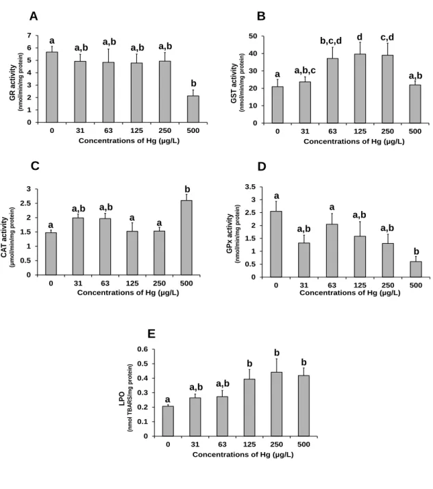
A - Activity of cholinesterase enzymes (ChE) determined in the adductor muscle. B - Activity of NADP-dependent isocitrate dehydrogenase (IDH) determined in the foot. C - Activity of octopine dehydrogenase (ODH) determined in the foot. Significant differences between treatments are identified by different letters above the bars (one-way ANOVA and the Tukey's test, p ≤ 0.05) ... 51 Fig. 5. Effects of mercury on biomarkers of oxidative stress and damage of Corbicula
fluminea. The values are the mean of 9 bivalves with the corresponding S.E.M. bars. A -
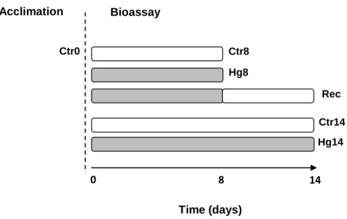
Activity of glutathione reductase (GR), B - activity of glutathione S-transferases (GST), C - activity of catalase (CAT), D - Activity of glutathione peroxidase (GPx) and E - Levels of lipid peroxidation (LPO). Significant differences between treatments are identified by different letters above the bars (one-way ANOVA and the Tukey's test, p ≤ 0.05) ... 52 Fig. 6. Experimental design adopted to study the effects of mercury exposure and recovery (rec) in Corbicula fluminea from Minho and Lima estuaries. In both bioassays, organisms
xxviii were analysed after the acclimation period (Ctr0) and after 8 and 14 days of experiment. Bivalves were exposed to the following treatments: 8 days to dechlorinated tap water for human consumption (clean medium) (Ctr8), 14 days to clean medium (Ctr14), 8 days to 31 µg/L of Hg (Hg8), 14 days to 31 µg/L of Hg (Hg14) and 31 µg/L of Hg for 8 days + 6 days to clean medium (Rec) ... 63 Fig. 7. A - Oxygen consumption rate (OCR), B - Activity of cholinesterase enzymes (ChE) and C - Activity of NADP-dependent isocitrate dehydrogenase (IDH) determined in Corbicula
fluminea from Minho River estuary at day 0 (Ctr0), after 8 and 14 days of exposure to
mercury (Hg) and after a period of recovery. Values are the mean ± standard error of 9 organisms. Significant differences between treatments are identified by different letters above the bars (one-way ANOVA and the Tukey's test, p ≤ 0.05) ... 70 Fig. 8. A - Activity of glutathione reductase (GR), B - Activity of glutathione S-transferases (GST) and C - Lipid peroxidation (LPO) levels determined in Corbicula fluminea from Minho River estuary at day 0 (Ctr0), after 8 and 14 days of exposure to mercury (Hg) and after a period of recovery. Values are the mean ± standard error of 9 organisms. Significant differences between treatments are identified by different letters above the bars (one-way ANOVA and the Tukey's test, p ≤ 0.05) ... 71 Fig. 9. A - Oxygen consumption rate (OCR), B - Activity of NADP-dependent isocitrate dehydrogenase (IDH), C - Activity of glutathione S-transferases (GST) and D - Lipid peroxidation (LPO) levels determined in Corbicula fluminea from Lima River estuary at day 0 (Ctr0), after 8 and 14 days of exposure to mercury (Hg) and after a period of recovery. Values are the mean ± standard error of 9 organisms. Significant differences between treatments are identified by different letters above the bars (one-way ANOVA and the Tukey's test, p ≤ 0.05) ... 72 Fig. 10. Microplastic particles detected in the body of Corbicula fluminea exposed to microplastics alone for 8 days. A - Digestive tract (outlined in a box; scale bar = 10 mm) and B - Gill tissue (indicated by arrows; scale bar = 500 µm) ... 93 Fig. 11. Biomarkers determined in Corbicula fluminea after 8 days of exposure to microplastics (MP), mercury (Hg) and mixture (Mix) (grey bars) and after the post-exposure recovery period (striped bars). A - Filtration rate (FR), B - Cholinesterase enzymes (ChE) activity, C - NADP-dependent isocitrate dehydrogenase (IDH) activity, D - Catalase (CAT)
xxix activity, E - Glutathione reductase (GR) activity, F - Glutathione peroxidase (GPx) activity, G - Glutathione S-transferases (GST) activity and D - Lipid peroxidation (LPO) levels. Significant differences between treatments are identified by different letters above the bars (one-way ANOVA and the Tukey's test, p ≤ 0.05) ... 98
Fig. 12. Calibration curve of fluorescence versus concentration of microplastics (MP, mg/L) in clean medium, and the linear regression model: MP concentration = - 0.02 + 0.01 x fluorescence. RFU – Relative fluorescence units. ... 102
Fig. 13. Answers to the specific questions (SQ) formulated in the present Thesis (Chapter I) ... 108
xxxi
Tables index
Table 1. Parameters evaluated in Corbicula fluminea exposed to heavy metals in field (F) and laboratory (L) studies. Catalase (CAT); Cyclooxygenase 1 (cox1); Glutathione, reduced form (GSH); glutathione peroxidase (GPx); Glutathione S-transferases (GST); Heat shock protein (Hsp); Lipid peroxidation (LPO); Messenger ribonucleic acid (mRNA); Metallothioneins (MT); Multixenibiotic resistance (MXR); Peroxidase (POD); Retinoblastoma gene (RB); selenium-dependent glutathione peroxidase (Se-GPx); 12S ribosomal RNA (12S); Ribosomal S9 protein gene (rps9); Sodium potassium adenosine triphosphatase (Na+/K+-ATPase);
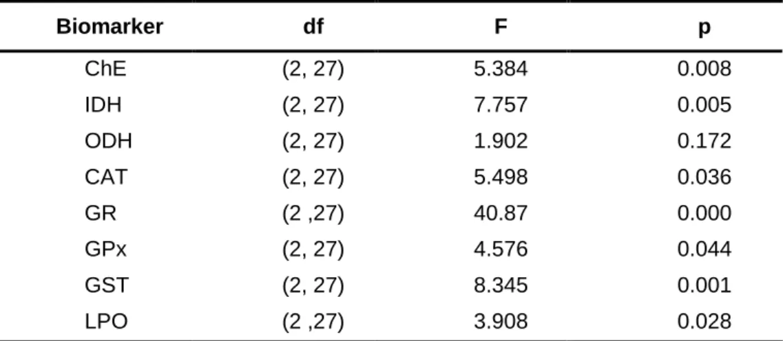
Superoxide dismutase (SOD) ... 16 Table 1. (Continued) ... 17 Table 1. (Continued) ... 18 Table 1. (Continued) ... 19 Table 1. (Continued) ... 20 Table 2. Results of the one-way ANOVA carried out with each biomarker data set of
Corbicula fluminea to investigate the effect of the acclimation period. Cholinesterase
enzymes (ChE) activity; NADP-dependent isocitrate dehydrogenase (IDH) activity; Octopine dehydrogenase (ODH) activity; Catalase (CAT) activity; Glutathione reductase (GR) activity; Glutathione peroxidase (GPx) activity; Glutathione S-transferases (GST) activity; Lipid peroxidation (LPO) levels; df - Degrees of freedom... 34 Table 3. Percentages of mortality induced by different concentrations of mercury on
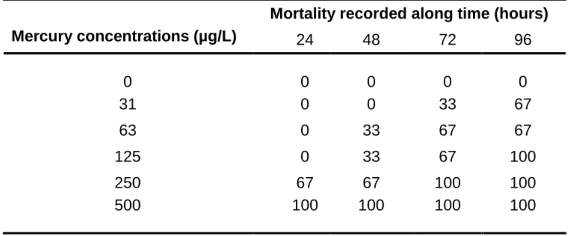
Anodonta anatina over 96 hours of exposure through test medium. For ethical reasons, only 3
specimens were used per treatment ... 48 Table 4. Results of the one-way ANOVA carried out with the data of each biomarker to compare different treatments. ChE - Cholinesterase enzymes activity; IDH - NADP-dependent isocitrate dehydrogenase activity; ODH - Octopine dehydrogenase activity; GR - Glutathione reductase activity; GST - Glutathione S-transferases activity; CAT - Catalase activity; GPx - Glutathione peroxidase activity; LPO - Lipid peroxidation levels; df - Degrees of freedom ... 50
xxxii
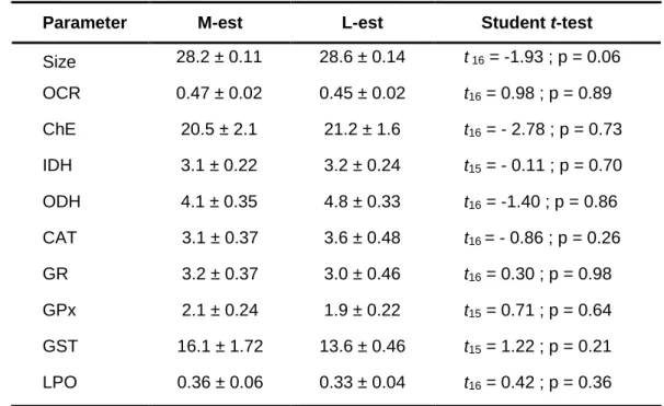
Table 5. Results of the Student’s t-test performed to compare the size and biomarkers of
Corbicula fluminea from the Minho (M-est) and Lima (L-est) River estuaries at the beginning
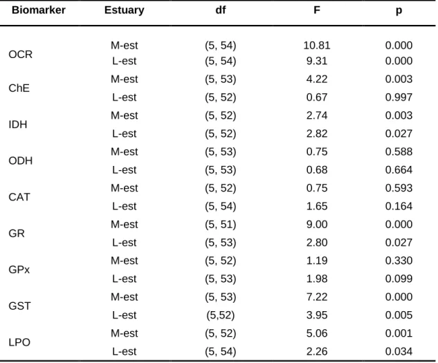
of the bioassays (Ctr0). Values are the mean ± standard error of anterior-posterior shell length (size), oxygen consumption rate (OCR), cholinesterase enzymes (ChE) activity, NADP-dependent isocitrate dehydrogenase (IDH) activity, octopine dehydrogenase (ODH) activity, catalase (CAT) activity, glutathione reductase (GR) activity, glutathione peroxidase (GPx) activity, glutathione S-transferases (GST) activity, and lipid peroxidation (LPO) levels .. ... 66 Table 6. Results of the three-way ANOVA performed to investigate the effects of estuary (Est), time of exposure (Time) and type of exposure (Exp) on the biomarkers of Corbicula
fluminea. Oxygen consumption rate (OCR), activities of cholinesterase enzymes (ChE),
NADP-dependent isocitrate dehydrogenase (IDH), octopine dehydrogenase (ODH), catalase (CAT), glutathione reductase (GR), glutathione peroxidase (GPx), glutathione S-transferases (GST), and lipid peroxidation (LPO) levels. Df - Degrees of freedom ... 68 Table 7. Results of the one-way ANOVA carried out with the data of each biomarker to compare different experimental treatments. M-est - Minho River estuary; L-est - Lima River estuary; OCR - Oxygen consumption rate; ChE - Cholinesterase enzymes activity; IDH - NADP-dependent isocitrate dehydrogenase activity; ODH - Octopine dehydrogenase activity; CAT - Catalase activity; GR - Glutathione reductase activity; GPx - Glutathione peroxidase activity; GST - Glutathione S-transferases activity; LPO - Lipid peroxidation levels; df - Degrees of freedom ... 69 Table 8. Actual concentrations of microplastics (MP, mg/L) obtained from fluorescence (relative fluorescence units - RFU) determined in fresh (0 h) and old (24 h) media, in the absence or presence of mercury (Hg) and in the absence or presence of Corbicula fluminea. The values are the mean ± standard deviation. A two-way ANOVA was performed to investigate the effect of Hg and animals in MP concentrations. The MP estimated exposure concentrations in test media with or without Hg were compared by the Student’s t-test. The significant level was 0.05 ... 89 Table 9. Actual concentrations of mercury (Hg, mg/L) in fresh (0 h) and old media (24 h) in the absence or presence of microplastics (MP) and in the absence or presence of Corbicula
fluminea. Values are the mean ± standard deviation. Hg concentrations in fresh media with
and without MP were compared by Student’s t-test. A two-way ANOVA was performed to
xxxiii investigate the effect of MP and animals in Hg concentrations in old media. The Hg estimated exposure concentrations in test media with or without MP were compared by the Student’s t-test. The significant level was 0.05 ... 91 Table 10. Results of the three-way ANOVA performed to investigate the effects of mercury (Hg), microplastics (MP) and Recovery on Hg body concentrations of Corbicula fluminea. The Hg concentrations and the bioconcentration factors were determined in bivalves collected from the field, after 14 days of acclimation and after exposure to different treatments of the bioassay ... 94 Table 11. Results of the three-way ANOVA of the biochemical biomarkers of Corbicula
fluminea performed to investigate the effects of microplastics (MP), mercury (Hg) and
recovery on: filtration rate (FR), cholinesterase enzymes (ChE) activity, NADP-dependent isocitrate dehydrogenase (IDH) activity, octopine dehydrogenase (ODH) activity, catalase (CAT) activity, glutathione reductase (GR) activity, glutathione peroxidase (GPx) activity, glutathione S-transferases (GST) activity and lipid peroxidation (LPO) levels. The significant level was 0.05 ... 96 Table 12. Results of the one-way ANOVA carried out with the data of each biomarker to compare different experimental treatments. FR - Filtration rate; ChE - Cholinesterase enzymes activity; IDH - NADP-dependent isocitrate dehydrogenase activity ; ODH - Octopine dehydrogenase activity ; CAT - Catalase activity; GR - Glutathione reductase activity; GPx - Glutathione peroxidase activity; GST - Glutathione S-transferases activity; LPO - Lipid peroxidation levels. Df - Degrees of freedom ... 97 Table 13. Obtained and certified concentrations of mercury (Hg, µg/g, dry weight) in certified reference material (CRM) BCR 463 (mercury and methylmercury in tuna fish) and the respective recovery percentage ... 102
xxxv
List of abbreviations
ANOVA - Analysis of VarianceAPA - Agência Portuguesa do Ambiente BCF - Bioconcentration factor
CAT - Catalase enzyme
CDNB - 1-Chloro-2,4-dinitrobenzene ChE - Cholinesterase enzymes DO - Dissolved oxygen
DTT - DL-1,4-Dithiothreitol Dw - Dry weight
EC - European Commission
EDTA - Ethylenediaminetetraacetic acid EU - European Union
GSH - Glutathione (reduced form) GSSG - Glutathione (oxidized form) GPx - Glutathione peroxidase enzyme GR - Glutathione reductase enzyme
GST - Glutathione S-transferases enzymes IDH - isocitrate dehydrogenase enzyme
LC50 - Median lethal concentration: the concentration of the tested substance estimated to
cause 50% of mortality in the tested population in the specific conditions of the toxicity bioassay.
LC20 - 20% lethal concentration: the concentration of the tested substance estimated to cause
20 % of mortality in the tested population in the specific conditions of the toxicity bioassay. LC10 - 10% lethal concentration: the concentration of the tested substance estimated to cause
20% of mortality in the tested population in the specific conditions of the toxicity bioassay. LT50 - Median lethal time: the estimated time (hours) necessary to induce 50% of mortality in
the tested population under exposure to a certain concentration of the tested substance in the specific conditions of the toxicity bioassay.
L-est - Lima River estuary LPO - Lipid peroxidation M-est - Minho River estuary MP - Microplastics
xxxvi MT - Metallothionein
MXR - Multixenobiotic resistance
NAD+ - Nicotinamide adenine dinucleotide (oxidized form)
NADH Nicotinamide adenine dinucleotide (reduced form)
NADP+ - Nicotinamide adenine dinucleotide phosphate (oxidized form)
NADPH - Nicotinamide adenine dinucleotide phosphate (reduced form) NIS - Non-indigenous species
NW - Northwest OD - Optical density
ODH - Octopine dehydrogenase enzyme
OECD - Organisation for Economic Co-operation and Development
OPSPAR Convention - Oslo and Paris Convention for the Protection of the Marine Environment of the North-East Atlantic
Ppm - Parts per million Psu - Practical salinity units ROS - Reactive oxygen species RFU - Relative fluorescence units SD - Standard deviation
S.E.M - Standard error of the mean SOD - Superoxide dismutase enzyme
TBARS - Thiobarbituric acid reactive substances UNEP - United Nations Environment Programme WFD - Water Framework Directive
CHAPTER I
3
1.1. Bioinvasions
“The real thing is that we are living in a period of the world's history when the mingling of thousands of kinds of organisms from different parts of the world is setting up terrific dislocations in nature” (Elton, 1958)
Freshwater and estuarine ecosystems have been continuously subjected to critical threats including habitat destruction, climate changes, pollution and bioinvasions (Meybeck, 2003). Bioinvasions are one of the most significant problems to ecosystem integrity and biodiversity (Sousa et al., 2011; Gangloff et al., 2016; O’Brien et al., 2016). Although bioinvasions are part of Earth's evolutionary processes, they are now a global paradigm with implications to environmental, animal and human health, mainly because they are occurring at unprecedented rates (Ricciardi, 2007; Simberloff et al., 2013; Ochocki and Miller, 2017). Since the publication of the monograph “The Ecology of Invasions by Animals and Plants” by Charles Elton in 1958 (Elton, 1958) and the emergence of invasion ecology as a discipline, the anthropogenic dimension of bioinvasions has been unquestionably recognized (Pyšek and Richardson, 2010). The development of a wide range of diverse human activities has led to the emergence of new routes allowing the introduction of several species into territories where these species did not exist before. The globalization of trade has been pointed as the principal driver of bioinvasions in aquatic systems (Levine and D’Antonio, 2003; Karatayev et
al., 2007; Hulme, 2009). Moreover, climate changes have the potential to increase the
likelihood of expansion of some invasive species beyond their native distribution (Hulme, 2017).
One of the proposed frameworks for bioinvasions divides the process into four main stages: transport, introduction, establishment and spread (Blackburn et al., 2011). The first step is the overcoming of an obstacle that prevents the movement of the species. This can occur without assistance, when the species has natural dispersal ability, but sometimes it can happen through human-mediated transport, whether in accidental or deliberate ways. Vectors and pathways through which exotic species can be introduced in a new area include ballast water, aquaculture, fish baits, aquarium and ornamental trades, tourism and recreational activities (Padilla and Williams, 2004; Gollasch, 2006; Williams et al., 2013; Patoka et al., 2017; Rhyne
et al., 2017). The introduction per se happens with the arrival of the species at the new
location. After a successful establishment, the dispersion may occur to a greater or lesser extent, depending on a combination of the characteristics of the recipient ecosystem and the
4 species itself (Chapple et al., 2012). Measures to combat invasive species can be applied at any stage of bioinvasions but their effectiveness will be higher if implemented at early stages of introduction and establishment phases, whereas their efficacy tend to decrease and costs tend to increase at later stages of establishment and spread (Epanchin-Niell, 2017).
A species introduced into a new environment is referred as non-native, non-indigenous, exotic or alien, and when it establishes and spreads very rapidly is also called invasive (Colautti and MacIsaac, 2004). The introduction and spread of non-indigenous species (NIS) in terrestrial and aquatic habitats is well documented and became a topic of special concern in the field of Ecology. The impacts of NIS assume variable forms and can affect the recipient biota at different organizational levels (Strayer, 2010; Ricciardi et al., 2013). The ecological impacts of NIS in the abundance and diversity of existing communities can be direct (e.g. predator-prey interactions, parasitism, hybridization or diseases) or indirect, when the NIS alters the habitat structure or interferes with trophic webs and energy fluxes (Crooks, 2002; Schmidlin and Baur, 2007; Gallardo et al., 2016). Moreover, NIS can threat human health by acting as potential vectors of diseases (Conn, 2014) and also cause considerable economic losses. The annual economic costs associated with damage and control of NIS are estimated to be ~120 billion $ in the U.S.A and 12.5 billion € in Europe (Pimentel et al., 2005; Kettunen
et al. 2008). According to the project “Delivering Alien Invasive Species Inventory for Europe”
(DAISIE, 2018), the number of NIS successfully established in Europe has been increasing exponentially, currently reaching more than 12000. From these, around 15% are adversely affecting the biodiversity and causing losses of billions of Euros every year (Hulme et al., 2009; Latombe et al., 2016). In these regards, the monitoring of NIS is one of the descriptors of the European Union (EU) Marine Strategy Framework Directive (EC, 2008a) that aims to achieve or maintain a “Good Environmental Status” of the EU marine waters by 2020. Moreover, the European Parliament adopted the EU Biodiversity Strategy to 2020 (EC, 2011) whose Target 5 is to combat NIS by minimizing their negative impacts on biodiversity through measures that include an early detection and eradication of recently arrived NIS, and the effective management of those already established. As part of the Convention on Biological Diversity, the EU provided the legal framework to combat NIS, through the Regulation on the “Prevention and Management of the Introduction and Spread of Invasive Alien Species” (EU, 2014). This regulation aims the protection of the biodiversity, ecosystem services and human health and establishes that the detection, early eradication and management must be carried out by Member States. More recently, the European Commission (EC) also adopted the first
5 list of “Invasive Alien Species of Union concern” (EU 2016; EU, 2017). This list includes all the species subjected to restrictions of keeping, importing, selling, breeding and growing. Aquatic bioinvasions are of special concern because the current and future extinction rates are estimated to be five times higher than those occurring in terrestrial environments (Ricciardi et al., 1998). Moreover, due to intense anthropogenic pressures and a high number of dispersal vectors, freshwaters and transitional waters are considered particularly susceptible to bioinvasions (Ricciardi and Kipp, 2008). Among aquatic faunal groups, bivalve molluscs stand out for their ability to disrupt trophic chains, alter nutrient fluxes and control the structure and functioning of the invaded ecosystems (Vaughn and Hakenkamp, 2001). Furthermore, there is evidence of a relationship between the introduction of bivalve NIS and declines of the native ones (Ricciardi et al., 1998; Ricciardi and Whoriskey, 2004). In view of these considerations, one of the main goals of invasion biology is to identify the factors that influence the likelihood of bioinvasions and the success of NIS in the recipient ecosystems (Kolar and Lodge, 2001; Walther et al., 2009; Mächler and Altermatt, 2012), which is crucial to define and implement strategies to prevent or manage the impacts of these species (Epanchin-Niell, 2017).
In general, bionvasions are known to occur more frequently in human-altered habitats (Dafforn et al., 2009; Sullivan et al., 2015), and a positive correlation between the invasiveness and the degree of disturbance has been established (Preisler et al. 2009; Tamburello et al., 2014; Bulleri et al., 2016). Anthropogenic pressures can lead to a reduction of habitat quality, affecting the lifecycle and health status of resident species (Bogan, 1993). Several human-generated disturbances such as regularization of rivers by man-made structures, draining activities and pollution facilitate the invasion and establishment of NIS (Lozon and MacIsaac, 1997; Salomidi et al., 2013). Chemical contamination from anthropogenic sources is a particularly important form of disturbance of aquatic environments that may favor the success of NIS in different ways and at different stages of bioinvasions (McKenzie et al., 2012). Environmental contaminants may favour the introduction of NIS because they can cause significant degradation of habitats, negatively affecting native species (Crooks et al., 2011). Additionally, NIS often present characteristics that represent advantages over their native competitors (Piola and Johnston 2008, 2009). In fact, tolerance to environmental contamination is pointed out as one of the factors contributing to the success of NIS over native species in aquatic ecosystems (Bielen et al., 2016). However, the results of several studies comparing tolerances of invasive species and native taxonomically related ones are contradictory (Prenter et al., 2004; Faria et al., 2010; Lenz et al., 2011; Velez
6
et al., 2016), showing that invasive species are not always the most tolerant. Thus, this topic
requires further investigation.
1.2. Corbicula fluminea (Müller, 1774)
Corbicula fluminea (Müller, 1774) (Bivalvia: Corbiculidae), also known as the Asiatic clam, is
a native species to Asia, Africa and Australia that has been spreading to multiple ecosystems all over the world (Mouthon 1981; Counts 1986; Araujo et al., 1993; Lucy et al., 2012; Crespo
et al., 2015). It has a marked invasive behaviour and is included in the list of the 100 worst
NIS in Europe (DAISIE, 2018). The first record of C. fluiminea outside its native range was in 1924 in Vancouver Island, British Columbia, Canada (Mcmahon, 1983). The first introduction of C. fluminea in South America occurred in Argentina around 1960s - 1970s (Ituarte, 1981) and since then its presence has been reported in Uruguay, Paraguay and Southern Brazil (Cataldo and Boltovskoy, 1998). In late 1970s - early 1980s, C. fluminea was introduced in Europe, being reported for the first time in France and Portugal by Mouthon (1981).
1.2.1. Biology and ecology of C. fluminea
C. fluminea is generally described as a hermaphroditic species, which reproduction occurs
mainly through cross-fertilization (Rajagopal et al., 2000; Park and Chung, 2004) but self-fertilization can also be observed (Kraemer et al., 1986). The reproduction is initiated by favourable environmental conditions, especially increased water temperatures and high food availability (Doherty et al., 1987; Mouthon, 2001; Beekey and Karlson, 2003). Although depending on the ecosystem, C. fluminea usually presents a bivoltine reproductive cycle, with two spawning periods: one between the the late spring and the early summer and the other between the late summer and the autumn. (Rajagopal et al. 2000; Mouthon and Parghentanian 2004; Sousa et al., 2008a). Nevertheless, some studies report an almost continuous reproduction with no clear patterns of gamete release and spawning (Oliveira, 2015; Cao et al., 2017). The fertilization occurs inside the paleal cavity of adult individuals, and the fertilized eggs and pediveliger larvae are kept in the inner demibranch (Kraemer and Galloway, 1986). The incubation period depends on environmental conditions, varying between 6 to 60 days, usually taking two weeks (King et al. 1986, McMahon 1999). After veliger and pediveliger stages, the larvae of C. fluminea have a “D” shape configuration with straight hinged shells measuring about 250 µm (anterior-posterior length) (King et al., 1986;
7 McMahon, 1999). At this stage, larvae are released from the gills’ chambers via the exhalant siphon into the surrounding water, and after four days they settle in the sediment (Araujo et
al. 1993; McMahon, 1999). Under favourable hydrological conditions, they can be released
from sediments back to the water column (McMahon, 1999). The sexual maturation is reached between 3 to 6 months of age (McMahon, 1999). The lifespan of C. fluminea is variable but usually ranges from 1 to 5 years (Sousa et al. 2008a). In the adult stage, the anterior-posterior length of the shell is, in average, 30 mm (McMahon, 2002) (Fig. 1).
Fig. 1. Corbicula fluminea with visible inhalant and exhalant siphons.
The high metabolic rates and rapid growth of C. fluminea are due, in part, to its feeding strategy (Hakenkamp and Palmer 1999). It feeds mainly on phytoplankton and bacteria present in the water column through water filtration (Beaver et al., 1991; Boltovskoy et al., 1995) but when planktonic food is not abundant it can also assimilate organic matter from the sediment using the foot, a mechanism designated as pedal feeding (Hakenkamp and Palmer, 1999). Pedal feeding is the primary feeding mechanism in larvae until the development of filtration structures is complete (McMahon, 1991; Reid et al., 1992; Hakenkamp et al., 2001).
C. fluminea is a freshwater bivalve species (McMahon, 1999) that tolerates salinity levels up
to 17 psu (Britton and Morton, 1982; Franco et al., 2012; Verbrugge et al., 2012; Modesto et
al., 2013; Crespo et al., 2017). This indicates a good adaptation to brackish conditions, which
is possibly related to efficient osmoregulation mechanisms (Morton and Tong, 1985, McMahon, 1991). This tolerance allows the species to colonize downstream estuarine areas (Sousa et al., 2008b; Franco et al., 2012; Ilarri et al., 2014).