Effects of Chronic Exposure to Mercury on Angiotensin-Converting
Enzyme Activity and Oxidative Stress in Normotensive and
Hypertensive Rats
Dalton Valentim Vassallo,
1,2Maylla Ronacher Simões,
1Karina Giuberti,
1Bruna Fernandes Azevedo,
1Rogerio Faustino Ribeiro Junior,
1Mercedes Salaices,
3,4Ivanita Stefanon
1Departamento de Ciências Fisiológicas - Universidade Federal do Espírito Santo,1 Vitória, ES – Brazil
Centro de Ciências da Saúde de Vitória - Escola Superior de Ciências da Santa Casa de Misericórdia de Vitória (EMESCAM),2 Vitória, ES – Brazil Departamento de Farmacologia - Universidade Autônoma de Madri – Espanha Instituto de Investigación Sanitária Hospital La Paz,3 Madri – Spain CIBER de Enfermidades Cardiovasculares,4 Madri – Spain
Mailing Address: Maylla Ronacher Simões •
Departamento de Ciências Fisiológicas - Universidade Federal do Espírito Santo - Av. Marechal Campos, 1468. Postal Code 29043-900, Maruípe, Vitória, ES – Brazil
E-mail: [email protected], [email protected]
Manuscript received April 27, 2018, revised manuscript July 04, 2018, accepted August 02, 2018
DOI: 10.5935/abc.20180271
Abstract
Background: Mercury’s deleterious effects are associated with increased cardiovascular risk.
Objective: To determine whether chronic exposure to inorganic mercury increases the activity of angiotensin-converting enzyme and its relationship with oxidative stress in several organs and tissues.
Methods: We studied male Wistar and spontaneously hypertensive rats (SHR) (3-month-old) exposed or not to HgCl2
for 30 days. At the end of treatment, we investigated the following: changes in body weight, hemodynamic parameters, angiotensin-converting enzyme (ACE) activity and oxidative stress in the heart, aorta, lung, brain and kidney in hypertensive compared to normotensive animals. A value of p < 0.05 was considered significant.
Results: Chronic exposure to HgCl2 did not affect weight gain in either group. Systolic blood pressure, measured weekly,
did not increase in Wistar rats but showed a small increase in SHR rats. We also observed increases in left ventricular end-diastolic pressure and ACE activity in the plasma and hearts of normotensive rats. In the SHR+Hg group, ACE activity increased in plasma but decreased in kidney, lung, heart, brain and aorta. Oxidative stress was assessed indirectly by malondialdehyde (MDA) production, which increased in Hg-treated rats in both plasma and heart. In the SHR+Hg group, MDA increased in heart and aorta and decreased in lungs and brain.
Conclusion: These results suggest that chronic exposure to inorganic mercury aggravates hypertension and produces more expressive changes in ACE activity and oxidative stress in SHRs. Such exposure affects the cardiovascular system, representing a risk factor for the development of cardiovascular disorders in normotensive rats and worsening of pre-existing risks for hypertension. (Arq Bras Cardiol. 2018; [online].ahead print, PP.0-0)
Keywords: Mercury Poisoning; Oxidative Stress/radiation effects; Peptidyl-Dipeptidase A; Hypertension; Rats.
Introduction
Mercury is a toxic metal that causes harmful effects on the cardiovascular system. Blood concentrations levels of 8 ng/ mL are found in exposed individuals,1,2 which might have a
relationship with hypertension development.3
Several reports showed that mercury induces oxidative stress and might damage several organs and systems.4-9
In addition, increased mercury exposure has been associated with cardiovascular diseases, such as hypertension, carotid
atherosclerosis, myocardial infarction and coronary heart disease.10,11 Moreover, oxidative stress is reported to be an
efficient mechanism for generation of oxidized low-density lipoprotein and subsequently atherosclerosis;12,13 then,
generation of advanced glycation end-products and participation of inflammatory cells take place, sustaining vascular injury.14
One of the main harmful actions of mercury is the
generation of oxygen free radicals. NADPH oxidase activation and cyclooxygenase (COX) stimulation induced by mercury may trigger the production of reactive oxygen species (ROS).11,15,16 Moreover, in animal models chronic mercury
exposure for 30 days promoted contractility dysfunction in isolated hearts as a result of decreased Na+-K+-ATPase (NKA)
activity, reduction in sodium/calcium exchanger (NCX) and sarco/endoplasmic reticulum calcium ATPase (SERCA) activity and increased phospholamban (PLB) expression.17 Although no
Additionally, at the vascular level, the vasoconstrictor response to phenylephrine was increased in caudal, mesenteric, coronary arteries and in the rat aorta, effects commonly related to reduced bioavailability of nitric oxide (NO) and increased oxidative stress.4,18,19 Interacting with NO,
superoxide anion (O2.-) forms peroxynitrite, decreasing NO
availability for smooth muscle relaxation.20-22
We reported that mercury administration increases local angiotensin converting enzyme (ACE) activity,18 releasing more
angiotensin II that enhances the production of free radicals.23
These results show that mercury pressor effects might depend on angiotensin II generation and are involved in oxidative stress generation. Previous studies showed that mercury could increase local ACE activity and oxidative stress with subsequent oxidative damage in several organs and systems,5,11,24-27 but the
in vivo effects of mercury chronic exposure on cardiovascular activity are not yet completely understood.
Moreover, investigations on mercury effects have been mainly focused on the cardiovascular systems of normotensive animals. However, little information exists about the chronic effects of low doses of inorganic mercury regarding ACE activity in organs and tissues of normotensive and hypertensive animals. To investigate such effects, increased mercury levels were induced to produce blood level concentrations similar to those of exposed individuals. Therefore, we aimed to determine whether chronic exposure to inorganic mercury increases the activity of ACE and the relationship of such exposure with oxidative stress on heart, aorta, lung, brain and kidney in hypertensive compared to normotensive animals.
Methods
Animals
Three-month-old male normotensive Wistar rats and SHRs (spontaneously hypertensive rats) were obtained from the Federal University of Espirito Santo breeding laboratories. During treatment, rats were housed at a constant room temperature, humidity, and 12:12-h light-dark cycle. Rats had free access to tap water and were fed with standard chow ad libitum.All experiments were conducted in compliance with the guidelines for biomedical research as stated by Conselho Nacional de Controle de Experimentação Animal-CONCEA, and in accordance with the Guide for the Care and Use of Laboratory Animals of the National Institute of Health. The protocols were approved by the Ethics Committee of Escola Superior de Ciências da Santa Casa de Misericórdia de Vitória, Brazil (CEUA-EMESCAM 003/2007). Wistar rats and SHRs were divided into four groups: control Wistar rats (n = 6) and SHRs (n = 9) treated with vehicle (saline solution, im), and Wistar rats (n = 8) and SHRs (n = 9) treated with mercury chloride (HgCl2)
for 30 days (1st dose 4.6 µg/kg, subsequent dose 0.07 µg/kg/day,
im to cover daily loss). We used the model described by Wiggers et al.4 to reach blood level concentrations (7,97 ng/ml)
similar to those of exposed individuals.
Blood pressure measurements
Indirect systolic blood pressure was measured at both the beginning and the end of the treatment using tail-cuff plethysmography (IITC Life Science Inc.). For this measurement,
conscious rats were restrained for 5–10 min in a warm and quiet room and were conditioned to numerous cuff inflation-deflation cycles by a trained operator. Subsequently, systolic blood pressure was measured, and the mean of three measurements was recorded.
Hemodynamic parameter measurements
At the end of treatment, control and HgCl2-treated rats (n = 26) were anaesthetized with urethane (1.2 g/kg, Sigma, St Louis, MO, USA), and the carotid artery and jugular vein were cannulated. A polyethylene catheter (PE50/Clay-Adams) filled with heparinized saline (50 U/mL) was introduced into the carotid artery to measure systolic blood pressure (SBP) and diastolic blood pressure (DBP). The carotid artery catheter was introduced into the left ventricle, and the jugular vein cannula was advanced into the right ventricular chamber to measure the left and right ventricular systolic pressures (LVSP and RVSP) and their positive and negative time derivatives (+dP/dt and - dP/dt, respectively) along with the left and right ventricular end-diastolic pressures (LVEDP and RVEDP). Recordings were performed over 30 min with a pressure transducer (TSD 104A-Biopac) and with an interface and software for computer data collection (MP100A, Biopac System, Inc., Santa Barbara, CA, USA). Heart rate (HR) was determined in the interbeat intervals.
Measurement of malondialdehyde (MDA) production. Levels of MDA in plasma, heart, aorta, brain, kidney and lung were measured using a modified thiobarbituric acid (TBA) assay.28 Plasma and tissue samples were mixed with 20%
trichloroacetic acid in 0.6 M HCl (1:1, v/v), and tubes were kept on ice for 20 min to precipitate plasma components to avoid possible interferences. Samples were centrifuged at 1500 x g for 15 minutes before adding TBA (120 mM in Tris 260 mM, pH 7) to the supernatant in a proportion of 1:5 (v/v); then, the mixture was boiled at 97°C for 30 min. Spectrophotometric measurements at 535 nm were taken at 20° C.
ACE activity assay
ACE activity was measured in plasma, heart, aorta, brain, kidney and lung using a fluorometric method adapted from Friedland and Silverstein.29 Briefly, triplicate tissue and
plasma samples (3 µL) were incubated for 15-90 minutes at 37°C with 40 µL of assay buffer containing the ACE substrate 5 mM Hip-His-Leu (Sigma). The reaction was stopped by the addition of 190 µL of 0.35 M HCl. The generated product, His-Leu, was measured fluorometrically following 10 min of incubation with 100 µL of 2% o-Phthalaldehyde in methanol. Fluorescence measurements were taken at 37°C in a FLUOstar Optima plate reader (BMG Labtech, Offenburg, Germany) with 350 nm excitation and 520 nm emission filters. The fluorescence plate reader was controlled using the FLUOstar Optima Software. Black 96-Well polystyrene microplates (Biogen Cientifica, Madrid, Spain) were used. A calibration curve with ACE from the rabbit lung (Sigma) was included in each plate.
Data analysis and statistics
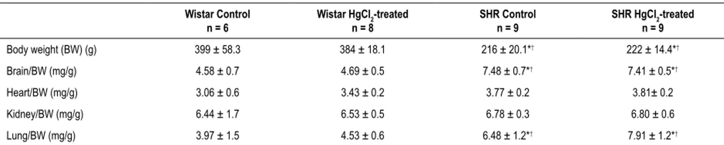
Kolmogorov-Table 1 – Body Weight (BW), Brain/BW, Heart/BW, Kidney/BW, Lung/BW, Adrenals/BW, Spleen/PC and Liver/PC from HgCl2-treated and non-treated Wistar rats and spontaneously hypertensive rats (SHRs)
Wistar Control n = 6
Wistar HgCl2-treated
n = 8
SHR Control n = 9
SHR HgCl2-treated
n = 9
Body weight (BW) (g) 399 ± 58.3 384 ± 18.1 216 ± 20.1*† 222 ± 14.4*†
Brain/BW (mg/g) 4.58 ± 0.7 4.69 ± 0.5 7.48 ± 0.7*† 7.41 ± 0.5*†
Heart/BW (mg/g) 3.06 ± 0.6 3.43 ± 0.2 3.77 ± 0.2 3.81± 0.2
Kidney/BW (mg/g) 6.44 ± 1.7 6.53 ± 0.5 6.78 ± 0.3 6.80 ± 0.6
Lung/BW (mg/g) 3.97 ± 1.5 4.53 ± 0.6 6.48 ± 1.2*† 7.91 ± 1.2*†
Results represent mean ± SD; n: number of animals used. One-way ANOVA, post hoc Tukey’s.*p < 0.05 compared with the Wistar control and † p < 0.05 compared
with HgCl2: treated Wistar rats.
Smirnov test. Differences were analysed using one-way ANOVA, followed by a post hoc Tukey test (GraphPad Prism Software, San Diego, CA). A p value < 0.05 was considered significant.
Results
At 30 days of mercury treatment, Wistar controls, Wistar treated rats, and treated and untreated SHRs had similar body weights, although the SHRs had lower body weights when compared with Wistar rats (Table 1).
Table 1 also shows that several organs, including the brain, heart, kidney and lungs, presented similar weights, normalized by body weight, which did not change after mercury treatment.
Indirect SBP measured at day zero in awake rats showed that SHRs had a higher mean arterial pressure compared with Wistar rats (Table 2). However, at the end of the treatment, mercury produced a significant increment of blood pressure only in HgCl2-treated SHR rats (Table 2).
Arterial blood pressures, ventricular pressures and their respective derivatives, and HR measurements in anaesthetized rats were not different between groups (Table 3), but the LVEDP increased after Hg treatment in the Wistar group, as previously reported.17
It has been reported in animal and human studies that mercury increases free radical production leading to an oxidative stress.4,24,30,31 We then evaluated the oxidant state in
the blood and in several other tissues by measuring MDA levels (Table 4). MDA plasma levels were greater in mercury-treated than in untreated Wistar rats but did not change in SHRs. Mercury treatment increased MDA levels in the heart in both Wistar and SHRs. In the aorta, different from plasma, MDA levels were increased in mercury-treated SHRs but not in Wistar rats. For brain and lungs, no changes were observed for MDA levels in mercury-treated Wistar rats, but a reduction occurred in SHRs. For kidneys, mercury treatment reduced MDA levels in both Wistar and SHR mercury-treated groups.
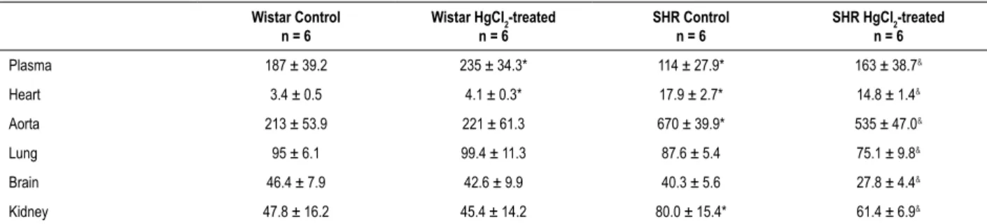
Since angiotensin II is reported to increase ROS and mercury increases ACE,32,33 we investigated whether ACE
activity was altered after 30 days of mercury treatment in Wistar and SHR groups. Table 5 shows that plasma ACE levels increased in both groups after mercury treatment. In the hearts of Wistar rats, mercury induced a slight ACE activity increment, but no changes were observed in the
aorta, lungs, brain or kidneys. However, in mercury-treated SHRs, ACE activity was reduced in the heart, aorta, lungs, brain and kidneys. Interestingly, ACE activity was higher in the heart, aorta and kidneys and lower in plasma of SHR controls compared with Wistar controls.
Discussion
The results presented here suggest that Wistar rats and SHRs, submitted to chronic exposure to inorganic mercury for 30 days, haveblood concentrations similar to exposed individuals.1,2
In addition, HgCl2-treated SHR, but not Wistar rats have increased blood pressure at the end of treatment. The intervention also influenced ACE activity and oxidative stress, by increasing or decreasing them, mainly in SHRs.
Previous reports showed that changes resulting from chronic exposure to mercury have been focused on its toxic effects on the cardiovascular system and the associations with hypertension, carotid atherosclerosis, myocardial infarction and coronary heart disease.9,10,34 Mercury exposure, both
acute and chronic, affects the heart and endothelial function, reducing NO bioavailability and increasing ACE and NADPH activities.15,18,19 Moreover, studies in rats showed that body
weight gain and arterial pressure were not affected when chronic exposure was performed,4,17 suggesting that this
treatment was not sufficient, in either amount or time, to produce changes. Our results reproduced those findings, showing no changes in body weight gain; additionally, similar behaviour was observed for the heart, brain, kidneys and lung, reinforcing the suggestion that this treatment is not sufficient to produce these changes, although cardiovascular function began to be affected.
Regarding the hemodynamic evaluation, no changes were observed in the left or right ventricle in Wistar rats or SHRs. Only an increment of LVEDP was observed in normotensive rats treated with mercury, indicating some deleterious effects of mercury on ventricular function.35 Right ventricular pressures
were investigated because of our previous report showing that under acute mercury exposure (0.5 mg/kg), there was an increase in right ventricular systolic pressure because of pulmonary hypertension,3,36-39 which was not observed with
Table 2 – Values of systolic blood pressure (SBP in mmHg) measured by tail plethysmography in Wistar rats and spontaneously hypertensive rats (SHRs) before and after treatment for 30 days with HgCl2.
Wistar CT n = 5
Wistar Hg n = 5
SHR CT n = 5
SHR Hg n = 5
SBP – Day 0 (mmHg) 123 ± 13 131 ± 15 205 ± 15 198 ± 22
SBP – Day 7 (mmHg) 119 ± 4 132 ± 9 221 ± 18 197 ± 18
SBP – Day 14 (mmHg) 115 ± 10 135 ± 9 219 ± 9 199 ± 29
SBP – Day 21 (mmHg) 132 ± 17 142 ± 14 200 ± 13 199 ± 9
SBP – Day 30 (mmHg) 117 ± 6 143 ± 11 220 ± 21 232 ± 19#
Results represent the mean ± SD; n: number of animals used. One-way ANOVA, post hoc Tukey’s for all groups. #p < 0.05 vs. SHR treated with mercury at day 0
Table 3 – Hemodynamic parameters from untreated and mercury (HgCl2)-treated Wistar rats and spontaneously hypertensive rats (SHRs)
Wistar Control n = 6
Wistar HgCl2-treated
n = 7
SHR Control n = 6
SHR HgCl2-treated
n = 7
SBP (mmHg) 105 ± 10 97 ± 11 105 ± 7 113 ± 8
DBP (mmHg) 71 ± 10 67 ± 11 58 ± 5 68 ± 11
HR (bpm) 324 ± 88 325 ± 58 343 ± 32 341 ± 34
LVSP (mmHg) 114 ± 20 107 ± 16 117 ± 22 112 ± 8
LVEDP (mmHg) 0.256 ± 1 3.31 ± 1* 1.11 ± 0.2 0.493 ± 0.5
+dP/dt LV (mmHg/s) 8627 ± 3378 8500 ± 2419 7360 ± 1854 7001 ± 1921
-dP/dt LV -6270 ± 1232 -6249 ± 1234 -7169 ± 1173 -6524 ± 1131
RVSP (mmHg) 32 ± 10 29 ± 5 29 ± 5 33 ± 5
RVEDP (mmHg) -1.080 ± 1 1.10 ± 2 -0.472 ± 1 0.459 ± 0.3
+dP/dt RV (mmHg/s) 3339 ± 2202 1758 ± 435 2776 ± 1056 2171 ± 405
– dP/dt RV (mmHg/s) -2560 ± 1553 -1387 ± 469 -1833 ± 478 -1695 ± 368
Changes in systolic (SBP) and diastolic (DBP) pressure, heart rate (HR), left and right ventricle systolic pressure (LVSP, RVSP), left and right ventricle end diastolic pressure (LVEDP, RVEDP) and positive (+dP/dt) and negative first-time derivatives (-dP/dt) from the left and right ventricles of Control and HgCl2-treated rats. The results represent the mean ± SD. n-Number of animals used. One-way ANOVA, post hoc Tukey’s. *p < 0.05 vs Wistar Control.
The reduction of NO bioavailability is a hallmark resulting from the increase in ROS generation contributing to the development of cardiovascular diseases such as atherosclerosis and hypertension.10,11,34 The interaction of superoxide anion with
NO generates peroxynitrite that decreases NO bioavailability increasing vascular reactivity.20-22 In fact, our previous studies
have associated mercury exposure with increased oxidative stress and the reduction of NO bioavailability.15,19In addition,
it has been shown that an increase of the local ACE activity could increase NADPH oxidase activity16 40 and ROS in the
aortas of normotensive and SHRs. Therefore, we investigated whether mercury effects alter the renin-angiotensin system and oxidative stress in the organs and tissues of hypertensive and normotensive rats. The increase in ACE activity induced by mercury could lead to increased activity of NADPH oxidase, which could, in turn, increase the release of ROS, generating an oxidative stress, as observed in this study.
Considering that both Hg and increased ACE activity can induce oxidative stress, we should observe a correlation between the amount of oxidative stress and ACE activity measured by MDA. An interesting aspect is that ACE activity levels and MDA concentrations showed similar behavior in plasma and organs investigated. Also, it is of note that both ACE activity and MDA concentrations showed more expressive changes in HgCl2
-treated SHRs. Similarly, inorganic mercury treatment aggravated hypertension in SHRs, suggesting that a pre-existing hypertensive condition enhances inorganic mercury action.
ROS are damped in the plasma of all locations where they are produced, and consequently, it is expected an increase in MDA. We have shown that plasma ACE activity increases after acute exposure to low mercury concentrations and reduces after exposure to high concentrations.18,39 However, we might
speculate that in the SHR group, when exposed to mercury, tissues that produce more ROS, such as the aorta, lung and kidney, ACE activity is reduced. Similarly, in the brain tissue, which concentrates mercury, ACE activity also decreased. LVEDP increments in Wistar rats could be explained by the local increase in ACE activity and oxidative stress in the heart. These two factors might explain the small, but significant increase in LVEDP, probably induced by a calcium overload.
Although we cannot give a proper explanation for all the events, it can be suggested that mercury, even at concentrations that do not affect arterial pressure and weight gainin normotensive rats, affects ACE activity and oxidative stress. However, in hypertensive animals, inorganic mercury actions were more expressive.
Table 4 – Malondialdehyde (MDA) (mM/mg of protein) concentrations in plasma, heart, aorta, lung, brain and kidney of untreated and Mercury (HgCl2)-treated Wistar rats and spontaneously hypertensive rats (SHRs)
Wistar Control n = 6
Wistar HgCl2-treated
n = 6
SHR Control n = 6
SHR HgCl2-treated
n = 7
Plasma 0.93 ± 0.15 1.28 ± 0.44* 0.89 ± 0.22 0.92 ± 0.05
Heart 0.22 ± 0.03 0.28 ± 0.03* 0.45 ± 0.05 0.55 ± 0.05&
Aorta 0.13 ± 0.03 0.12 ± 0.05 0.96 ± 0.27 1.51 ± 0.37&
Lung 0.18 ± 0.05 0.14 ± 0.03 0.21 ± 0.03 0.12 ± 0.03&
Brain 0.13 ± 0.03 0.09 ± 0.03 0.54 ± 0.07 0.34 ± 0.03&
Kidney 0.38 ± 0.07 0.14 ± 0.03* 0.96 ± 0.07 0.51 ± 0.03&
Values are expressed in mM/mg of protein (MDA). The results represent the mean ± SD. N-Number of animals used. One-way ANOVA, post hoc Tukey’s. *p < 0.05 vs Wistar Control and &p < 0.05 vs SHR Control.
Table 5 – Angiotensin converting enzyme (ACE) activity levels in plasma, heart, aorta, lung, brain and kidney of untreated and Mercury (HgCl2)-treated Wistar rats and spontaneously hypertensive rats (SHRs)
Wistar Control n = 6
Wistar HgCl2-treated
n = 6
SHR Control n = 6
SHR HgCl2-treated
n = 6
Plasma 187 ± 39.2 235 ± 34.3* 114 ± 27.9* 163 ± 38.7&
Heart 3.4 ± 0.5 4.1 ± 0.3* 17.9 ± 2.7* 14.8 ± 1.4&
Aorta 213 ± 53.9 221 ± 61.3 670 ± 39.9* 535 ± 47.0&
Lung 95 ± 6.1 99.4 ± 11.3 87.6 ± 5.4 75.1 ± 9.8&
Brain 46.4 ± 7.9 42.6 ± 9.9 40.3 ± 5.6 27.8 ± 4.4&
Kidney 47.8 ± 16.2 45.4 ± 14.2 80.0 ± 15.4* 61.4 ± 6.9&
Values are expressed in nmol/mL/min/mg of protein in tissues and in nmol/mL of plasma/min in plasma (ACE). Results represent the mean ± SD. N-Number of animals used. One-way ANOVA, post hoc Tukey’s. *p < 0.05 vs Wistar Control and &p < 0.05 vs SHR Control.
depend on mercury concentration in each of them? Would a pre-existing cardiovascular disorder be aggravated by exposure to inorganic mercury? These questions can be considered limitations of our study, and issues for further studies.
Conclusions
Results described here allow us to affirm that chronic exposure to inorganic mercury, similarly to thatwe previously reported, produces blood concentrations compatible with those found in exposed humans, and do represent a cardiovascular risk factor. Such exposure influenced ACE activity, increased oxidative stress and promoted hypertension in SHRs (which had a higher blood pressure increment compared with untreated SHRs), as well as increased the LVEDP in Wistar rats. This controlled exposure affected the cardiovascular system, produced more expressive changes of ACE activity and oxidative stress in SHRs representing a risk factor for the development of cardiovascular disorders in normotensive rats and a contributing factor to pre-existing risks in high blood pressure condition.
Author contributions
Conception and design of the research: Vassallo DV, Simões MR, Giuberti K, Stefanon I; acquisition of data: Giuberti K, Azevedo BF, Ribeiro Junior RF; analysis and interpretation of the data and statistical analysis: Vassallo
DV, Simões MR, Giuberti K, Azevedo BF, Ribeiro Junior RF, Salaices M, Stefanon I; obtaining funding: Vassallo DV, Salaices M; writing of the manuscript: Vassallo DV, Simões MR; critical revision of the manuscript for intellectual content: Vassallo DV, Simões MR, Salaices M, Stefanon I.
Potential Conflict of Interest
No potential conflict of interest relevant to this article was reported.
Sources of Funding
This study was funded by FAPES, CAPES, CNPq, Ministério da Economia e Competitividade (SAF 2016-80305-P).
Study Association
This article is part of the thesis of Doctoral submitted by Maylla Ronacher Simões, from Universidade Federal do Espírito Santo.
Ethics approval and consent to participate
1. Gupta M, Bansal JK, Khanna CM. Blood mercury in workers exposed to the preparation of mercury cadmium telluride layers on cadmium telluride base. Ind Health. 1996;34(4):421-5.
2. Asgary S, Movahedian A, Keshvari M, Taleghani M, Sahebkar A, Sarrafzadegan N. Serum levels of lead, mercury and cadmium in relation to coronary artery disease in the elderly: a cross-sectional study. Chemosphere. 2017;180:540-4.
3. Torres AD, Rai AN, Hardiek ML. Mercury intoxication and arterial hypertension: report of two patients and review of the literature. Pediatrics. 2000;105(3):E34.
4. Wiggers GA, Peçanha FM, Briones AM, Pérez-Girón JV, Miguel M, Vassallo DV, et al. Low mercury concentrations cause oxidative stress and endothelial dysfunction in conductance and resistance arteries. Am J Physiol Heart Circ Physiol. 2008;295(3):H1033-43.
5. Mahboob M, Shireen KF, Atkinson A, Khan AT. Lipid peroxidation and antioxidant enzyme activity in different organs of mice exposed to low level of mercury. J Environ Sci Health B. 2001;36(5):687-97.
6. Azevedo BF, Simões MR, Fiorim J, Botelho T, Angeli JK, Vieira J, et al. Chronic mercury exposure at different concentrations produces opposed vascular responses in rat aorta. Clin Exp Pharmacol Physiol. 2016;43(7):712-9.
7. Rizzetti DA, Altermann CD, Martinez CS, Peçanha FM, Vassallo DV, Uranga-Ocio JA, et al. Ameliorative effects of egg white hydrolysate on recognition memory impairments associated with chronic exposure to low mercury concentration. Neurochem Int. 2016;101:30-7.
8. Rizzetti DA, Martinez CS, Escobar AG, da Silva TM, Uranga-Ocio JA, Peçanha FM, et al. Egg white-derived peptides prevent male reproductive dysfunction induced by mercury in rats. Food Chem Toxicol. 2017;100:253-64.
9. Rizzetti DA, Martín Á, Corrales P, Fernandez F, Simões MR, Peçanha FM, et al. Egg white-derived peptides prevent cardiovascular disorders induced by mercury in rats: role of angiotensin-converting enzyme (ACE) and NADPH oxidase. Toxicol Lett. 2017;281:158-74.
10. Salonen JT, Seppanen K, Lakka TA, Salonen R, Kaplan GA. Mercury accumulation and accelerated progression of carotid atherosclerosis: a population-based prospective 4-year follow-up study in men in eastern Finland. Atherosclerosis. 2000;148(2):265-73.
11. Houston MC. The role of mercury and cadmium heavy metals in vascular disease, hypertension, coronary heart disease, and myocardial infarction. Altern Ther Health Med. 2007;13(2):S128-33.
12. Mitra S, Deshmukh A, Sachdeva R, Lu J, Mehta JL. Oxidized low-density lipoprotein and atherosclerosis implications in antioxidant therapy. Am J Med Sci. 2011;342(2):135-42.
13. Münzel T, Camici GG, Maack C, Bonetti NR, Fuster V, Kovacic JC. Impact of oxidative stress on the heart and vasculature: part 2 of a 3-part series. J Am Coll Cardiol. 2017;70(2):212-29.
14. Harja E, Bu DX, Hudson BI, Chang JS, Shen X, Hallam K, et al. Vascular and inflammatory stresses mediate atherosclerosis via RAGE and its ligands in apoE-/- mice. J Clin Invest. 2008;118(1):183-94.
15. Pecanha FM, Wiggers GA, Briones AM, Perez-Giron JV, Miguel M, Garcia-Redondo AB, et al. The role of cyclooxygenase (COX)-2 derived prostanoids on vasoconstrictor responses to phenylephrine is increased by exposure to low mercury concentration. J Physiol Pharmacol. 2010;61(1):29-36.
16. Aguado A, Galán M, Zhenyukh O, Wiggers GA, Roque FR, Redondo S, et al. Mercury induces proliferation and reduces cell size in vascular
smooth muscle cells through MAPK, oxidative stress and cyclooxygenase-2 pathways. Toxicol Appl Pharmacol. 2013;268(2):188-200.
17. Furieri LB, Fioresi M, Junior RF, Bartolomé MV, Fernandes AA, Cachofeiro V, et al. Exposure to low mercury concentration in vivo impairs myocardial contractile function. Toxicol Appl Pharmacol. 2011;255(2):193-9.
18. Wiggers GA, Stefanon I, Padilha AS, Peçanha FM, Vassallo DV, Oliveira EM. Low nanomolar concentration of mercury chloride increases vascular reactivity to phenylephrine and local angiotensin production in rats. Comp Biochem Physiol C Toxicol Pharmacol. 2008;147(2):252-60.
19. Furieri LB, Galán M, Avendaño MS, García-Redondo AB, Aguado A, Martínez S, et al. Endothelial dysfunction of rat coronary arteries after exposure to low concentrations of mercury is dependent on reactive oxygen species. Br J Pharmacol. 2011;162(8):1819-31.
20. Frisbee JC, Maier KG, Stepp DW. Oxidant stress-induced increase in myogenic activation of skeletal muscle resistance arteries in obese Zucker rats. Am J Physiol Heart Circ Physiol. 2002;283(6):H2160-8.
21. Zou MH. Peroxynitrite and protein tyrosine nitration of prostacyclin synthase. Prostaglandins Other Lipid Mediat. 2007;82(1-4):119-27.
22. Förstermann U, Sessa WC. Nitric oxide synthases: regulation and function. Eur Heart J. 2012;33(7):829-37.
23. Touyz RM. Reactive oxygen species and angiotensin II signaling in vascular cells-- implications in cardiovascular disease. Braz J Med Biol Res. 2004;37(8):1263-73.
24. Huang YL, Cheng SL, Lin TH. Lipid peroxidation in rats administrated with mercuric chloride. Biol Trace Elem Res. 1996;52(2):193-206.
25. Miller DM, Woods JS. Urinary porphyrins as biological indicators of oxidative stress in the kidney. Interaction of mercury and cephaloridine. Biochem Pharmacol. 1993;46(12):2235-41.
26. Reus IS, Bando I, Andrés D, Cascales M. Relationship between expression of HSP70 and metallothionein and oxidative stress during mercury chloride induced acute liver injury in rats. J Biochem Mol Toxicol. 2003;17(3):161-8.
27. Kim SH, Sharma RP. Mercury-induced apoptosis and necrosis in murine macrophages: role of calcium-induced reactive oxygen species and p38 mitogen-activated protein kinase signaling. Toxicol Appl Pharmacol. 2004;196(1):47-57.
28. Rodriguez-Martinez MA, Ruiz-Torres A. Homeostasis between lipid peroxidation and antioxidant enzyme activities in healthy human aging. Mech Ageing Dev. 1992;66(2):213-22.
29. Friedland J, Silverstein E. A sensitive fluorimetric assay for serum angiotensin-converting enzyme. Am J Clin Pathol. 1976;66(2):416-24.
30. Kobal AB, Horvat M, Prezelj M, Briski AS, Krsnik M, Dizdarevic T, et al. The impact of long-term past exposure to elemental mercury on antioxidative capacity and lipid peroxidation in mercury miners. J Trace Elem Med Biol. 2004;17(4):261-74.
31. Wolf MB, Baynes JW. Cadmium and mercury cause an oxidative stress-induced endothelial dysfunction. Biometals. 2007;20(1):73-81.
32. García-Redondo AB, Briones AM, Avendaño MS, Hernanz R, Alonso MJ, Salaices M. Losartan and tempol treatments normalize the increased response to hydrogen peroxide in resistance arteries from hypertensive rats. J Hypertens.2009;27(9):1814-22.
33. Vassallo DV, Simões MR, Furieri LB, Fioresi M, Fiorim J, Almeida EA, et al. Toxic effects of mercury, lead and gadolinium on vascular reactivity. Braz J Med Biol Res. 2011;44(9):939-46.
This is an open-access article distributed under the terms of the Creative Commons Attribution License 34. Virtanen JK, Voutilainen S, Rissanen TH, Mursu J, Tuomainen TP,
Korhonen MJ, et al. Mercury, fish oils, and risk of acute coronary events and cardiovascular disease, coronary heart disease, and all-cause mortality in men in eastern Finland. Arterioscler Thromb Vasc Biol. 2005;25(1):228-33.
35. Boffetta P, Sällsten G, Garcia-Gómez M, Pompe-Kirn V, Zaridze D, Bulbulyan M, et al. Mortality from cardiovascular diseases and exposure to inorganic mercury. Occup Environ Med. 2001;58(7):461-6.
36. García Gómez M, Boffetta P, Caballero Klink JD, Español S, Gómez Quintana J. Cardiovascular mortality in mercury miners. Med Clin (Barc). 2007;128(20):766-71.
37. Wakita Y. Hypertension induced by methyl mercury in rats. Toxicol Appl Pharmacol. 1987;89(1):144-7.
38. Carmignani M, Boscolo P, Artese L, Del Rosso G, Porcelli G, Felaco M, et al. Renal mechanisms in the cardiovascular effects of chronic exposure to inorganic mercury in rats. Br J Ind Med. 1992;49(4):226-32.
39. Rossoni LV, Amaral SM, Vassallo PF, França A, Oliveira EM, Varner KJ, et al. Effects of mercury on the arterial blood pressure of anesthetized rats. Braz J Med Biol Res. 1999;32(8):989-97.