Video-assisted thoracoscopy as an option in the surgical
treatment of chylothorax after cardiac surgery in children*
,**
Videotoracoscopia como uma opção no tratamento cirúrgico do quilotórax após cirurgia cardíaca pediátrica
Paulo Manuel Pego-Fernandes, Mauro Boldrini Nascimbem, Otávio T. Ranzani, Mônica Satsuki Shimoda, Rosângela Monteiro, Fábio Biscegli Jatene
Abstract
Objective: To evaluate the use of video-assisted thoracoscopy in the surgical treatment of chylothorax developed after the surgical correction of congenital heart disease in children. Methods: We reviewed the medical charts of 3,092 children who underwent surgery for congenital heart disease between February of 2002 and February of 2007 at the Heart Institute of the University of São Paulo School of Medicine Hospital das Clínicas, in São Paulo, Brazil. Results: Of the 3,092 children, 64 (2.2%) presented with chylothorax as a postoperative complication. In 50 (78.1%) of those patients, the clinical management was successful, whereas it failed in 14 (21.9%), all of whom were then submitted to thoracic duct ligation by video-assisted thoracoscopy. The thoracic duct ligation was successful in 12 patients (86%) but failed in 2. In the postoperative period, additional clinical measures, such as a low-fat diet and parenteral nutrition, were required in order to resolve those 2 cases. There was no surgical morbidity or mortality. Of the 14 patients who underwent thoracic duct ligation, 5 (35%) died due to cardiac or infectious complications. Conclusions: Video-assisted thoracic duct ligation can be safely performed in patients with severe heart disease, and the outcomes are favorable.
Keywords: Chylothorax; Heart defects, congenital; Thoracic duct.
Resumo
Objetivo: Avaliar o uso de videotoracoscopia no tratamento cirúrgico do quilotórax após cirurgia para correção de cardiopatias congênitas em crianças. Métodos: Revisamos os prontuários médicos de 3.092 crianças operadas para a correção de cardiopatias congênitas no Instituto do Coração/Hospital das Clínicas da Faculdade de Medicina da Universidade de São Paulo, São Paulo (SP) entre fevereiro de 2002 e fevereiro de 2007. Resultados: Das 3.092 crianças, 64 (2,2%) apresentaram quilotórax como complicação pós-operatória. Em 50 (78,1%) dessas, o tratamento clínico foi bem-sucedido, enquanto esse falhou em 14 (21,9%), as quais foram submetidas à ligação do ducto torácico por videotoracoscopia. A ligação do ducto torácico obteve sucesso em 12 pacientes (86%) e falhou em 2 casos, os quais foram resolvidos com medidas clínicas adicionais, como dieta pobre em gorduras e nutrição parenteral. Não houve morbidade ou mortalidade relacionada à operação. Dos 14 pacientes, 5 (35%) faleceram em decorrência de complicações cardíacas ou infecciosas. Conclusões: A ligadura videoassistida do ducto torácico pode ser realizada com segurança em pacientes gravemente enfermos e com doença cardíaca grave, com resultados favoráveis.
Descritores: Quilotórax; Cardiopatias congênitas; Ducto torácico.
* Study carried out at the Heart Institute of the University of São Paulo School of Medicine Hospital das Clínicas, São Paulo, Brazil. Correspondence to: Paulo Manuel Pêgo Fernandes. Instituto do Coração, Serviço de Cirurgia Torácica, Avenida Dr. Enéas de Carvalho Aguiar, 44, Bloco II, 2º andar, Sala 9, CEP 05403-000, São Paulo, SP, Brazil.
Tel 55 11 3069-5248. E-mail: [email protected] Financial support: None.
Submitted: 23 February 2010. Accepted, after review: 21 September 2010.
in the treatment.(4,10-12) Surgical treatment is
reserved for the few cases in which the initial conservative management fails. The timing of surgery is controversial. However, in most studies, surgical interventions are recommended when there is no resolution after 1-3 weeks of clinical treatment, or if the daily drainage exceeds 100 mL/kg.(4,8,12-15) Thoracic duct
ligation, performed for the first time by Lampson in 1948,(16) is an efficient treatment option for
patients with low morbidity. The use of video-assisted techniques has recently been described. When such techniques are applied, thoracic duct ligation can be accomplished with less surgical trauma and fewer postoperative complications.
(3,8,17-20)
Methods
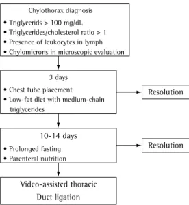
This was a retrospective, descriptive, observational study in which data were collected from clinical charts. Our study population included patients treated at our facility between February of 2002 and February of 2007. All had been submitted to surgical correction of congenital heart disease and had subsequently developed chylothorax. The diagnosis of chylothorax was based on the detection of a high triglyceride concentration (> 110 mg/dL) or a triglyceride/cholesterol ratio > 1 in the pleural fluid. The presence of chylomicrons and a leukocyte count under microscopic examination of the pleural fluid were also indicative of chylothorax. The algorithm for the management of these patients is shown in Figure 1. The institutional review board approved the study and, considering its observational nature, waived requirements for informed consent.
All of the patients diagnosed with chylothorax were submitted to the clinical treatment. If the clinical treatment failed, video-assisted thoracic duct ligation was performed. The surgical procedures were all performed by the same surgeon, in a standard fashion. After orotracheal intubation, the children received olive oil thro-ugh a nasogastric tube (10 mL/kg) to enhance the visualization of the thoracic duct and the fistula in the operative field. The procedure was performed on the side of the effusion or in the right hemithorax when it was bilateral. A 5-cm posterolateral minithoracotomy was performed at the 5th intercostal space for dissection and lung retraction. For visualization,
Introduction
Chylothorax is defined as the presence of lymph in the pleural space secondary to leakage from the thoracic duct or from one of its main tributaries. Chylothorax has various causes, including malignancy, trauma (including surgery), and miscellaneous disorders (such as deep vein thrombosis, sarcoidosis, and congestive heart failure), and can also be idiopathic.(1,2)
Postoperative chylothorax in childhood is a potentially serious complication of any thoracic surgical procedure. The reported incidence of postoperative chylothorax following congenital cardiac surgery ranges from 1.5% to 4.7%.(3-5)
It is associated with extrapericardial procedures, such as patent ductus arteriosus closure, the Blalock-Taussig procedure, and correction of aortic coarctation. In such cases, chylothorax is due to direct trauma to the thoracic duct or its tributaries. Chylothorax also occurs after intrapericardial procedures, such as Glenn and Fontan procedures, without obvious trauma to lymphatic vessels.(6,7) These operations generate
high central venous pressure, leading to high lymphatic pressure and lymphorrhea. The loss of lymph after drainage, sometimes in great quantities, can lead to nutritional depletion, electrolyte disturbances, hypolipemia, and lymphocytopenia with immunodeficiency. Such complications are poorly tolerated in small infants with severe heart disease and are associated with high mortality when left untreated.(3-8)
Patients usually remain asymptomatic, presenting with pronounced chylous effusion only after being started on a regular diet. The initial management consists of pleural drainage, allowing lung expansion, and dietary strategies to decrease lymphorrhea. A low-fat diet with medium-chain triglycerides is the initial approach, followed by prolonged fasting and parenteral nutrition, if the former is unsuccessful in decreasing the chylous fistula. This approach is successful in most cases (66%-100%).(4,6-9)
to surgery were reviewed. The data collected included demographics; heart disease; cardiac surgery performed; duration of chylothorax; clinical treatment; and recovery after thoracic duct ligation.
Results
Between February of 2002 and February of 2007, 3,092 patients underwent surgery for congenital heart disease at our facility. Of those, 64 (2.2%) presented with chylothorax as a complication. In 50 (78.1%) of those patients, the clinical management was successful, whereas it failed in 14 (22.1%), all of whom were therefore submitted to video-assisted thoracic duct ligation. In all such patients, the chest tube was maintained from the day of diagnosis of chylothorax until the day of surgery.
Tables 1 and 2 show data related to the 14 patients who underwent thoracic duct ligation. There were 8 girls (57%) and 6 boys (43%), and, among those patients, the mean age at diagnosis of chylothorax was 2.3 years (range, 1 month-10 years).. The mean elapsed time from the time of cardiac surgery to the diagnosis of chylothorax was 13.5 ± 11.9 days (range, 1-42 days). Central venous hypertension was observed in 12 patients (85.7%; not present in patients 9 and 11). The mean duration of conservative management prior to thoracic duct ligation was 39.5 ± 19.6 days (range, 15-79 days). In most cases, the surgery was postponed due to clinical instability (cardiac or septic problems). Prior to the operation, patients 4, 7, and 13 received octreotide, without any decrease in lymphorrhea in the three. Patients with chylopericardium were approached with pericardiocentesis or video-assisted pericardioscopy and drainage when there were signs of cardiac tamponade, and the evolution was favorable in all such patients. In patients 1 and 4, pleurodesis was used in combination with pleural abrasion. In patients 12 and 14, decortication was performed due to trapped lung.
There were no intraoperative complications. After the surgery, the chest tube was removed at the mean time of 9.5 days (range, 5-16 days), except in 2 patients (14.2%), both of whom had recurrence of the chylothorax. The recurrence was diagnosed on postoperative day 3 in both of those patients, and they were treated with a 30° thoracoscope was introduced at the 8th
intercostal space in the middle of the axillary line. The thoracic duct was dissected between the esophagus and the thoracic spine and ligated with a double suture line buttressed with Teflon pledgets, and a chest tube was maintained in the thoracoscopic access (Figure 2). Considering that, in addition to the duct ligation, there can be significant anatomical variations to the normal pattern, including accessory ducts, the fat between the esophagus and the spine was ligated. On postoperative day 1, a regular diet was started and was maintained thereafter as possible. The chest tube was removed when the daily output dropped to < 10 mL/kg, assuming that there was no sign of chylothorax recurrence. The medical data of all these patients submitted
Figure 1 - Chylothorax management algorithm.
to bidirectional cavopulmonary anastomosis (Glenn operation) accompanied by very high venous pressure. The other 2 presented with chylothorax due to direct trauma to the thoracic duct following patent ductus arteriosus ligation and Blalock-Taussig shunt, respectively. They all presented dyspnea and unilateral/bilateral effusion, sometimes with chylopericardium.
In patients with chylothorax, thoracocentesis shows a milky fluid, and laboratory test results are frequently diagnostic. Chylothorax is occasionally complicated by pleural adhesion or pleural thickening, and the consequent respiratory embarrassment can by resolved by inserting a chest tube, resulting in lung expansion.(3,6-8) Two patients developed pleural
thickening and trapped lung, primarily because of prolonged drainage (for 43 and 79 days, respectively). It should be borne in mind that chylous fluid is bacteriostatic, rich in lymphocytes and immunoglobulins, and secondary empyema is therefore unusual in such cases.(22) Despite the
additional parenteral nutrition and fasting for 25 days (patient 3) and 30 days (patient 6), resulting in resolution in both cases. Of the 14 patients undergoing thoracic duct ligation, 5 (35.0%) died during the hospital stay. No deaths were attributed to the surgery. In those 5 patients, there was no sign of chylothorax recurrence. The most common cause of death was heart failure with cardiogenic shock (patients 1, 7, and 13). Patient 4 died of pneumonia complicated by septic shock, and patient 9 had ventricular tachyarrhythmia evolving to ventricular fibrillation.
Discussion
Chylothorax usually develops in 1-4 weeks after cardiac surgery, when the chest tube has already been removed, and the patient is on a regular diet.(6,8,21) In our patients, only 4 developed
chylothorax within the first postoperative week. One of those 4 patients was submitted to a Fontan procedure, and 1 was submitted
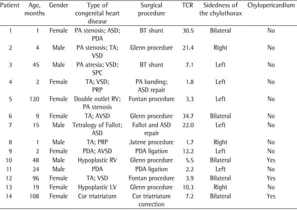
Table 1 - Clinical data of the 14 pediatric patients developing chylothorax after cardiac surgery and submitted to further surgical procedures after not responding to clinical treatment of the chylothorax.
Patient Age, months
Gender Type of congenital heart
disease
Surgical procedure
TCR Sidedness of the chylothorax
Chylopericardium
1 1 Female PA stenosis; ASD; PDA
BT shunt 30.5 Bilateral No
2 4 Male PA stenosis; TA; VSD
Glenn procedure 21.4 Right No
3 45 Male PA atresia; VSD; SPC
BT shunt 7.1 Left No
4 2 Female TA; VSD; PRP
PA banding; ASD repair
1.8 Left No
5 120 Female Double outlet RV; PA stenosis
Fontan procedure 3.3 Left No
6 9 Female TA; AVSD Glenn procedure 34.7 Bilateral No 7 15 Male Tetralogy of Fallot;
ASD
Fallot and ASD repair
22.0 Left No
8 1 Male TA; PRP Jatene procedure 1.7 Right No
9 2 Female PDA; AVSD PDA ligation 12.2 Left No
10 48 Male Hypoplastic RV Glenn procedure 5.5 Bilateral Yes
11 24 Male PDA PDA ligation 2.2 Left No
12 96 Female TA; VSD Fontan procedure 3.9 Bilateral Yes 13 19 Female Hypoplastic LV Glenn procedure 10.3 Right No 14 108 Female Cor triatriatum Cor triatriatum
correction
7.2 Bilateral Yes
Prolonged periods of parenteral nutrition have numerous complications, such as cholestasis, central line infection, and vein thrombosis.
(24-26) Later complications might, in turn, further
enhance chylothorax. There have been reports of chylothorax caused by venous thrombosis secondary to central line insertion, when no other procedure had been performed.(27,28)
Pediatric chylothorax patients can show persistent high output fistulas, despite parenteral nutrition and fasting, some patients even presenting with daily losses > 200 mL/kg. The management of fluid and electrolyte disturbances in these small infants with such losses is quite difficult.(3-8,23) Somatostatin and its analogue,
long periods of clinical treatment in our study, none of the patients developed pleural infection.
In chylothorax, the most difficult task is the closure of the thoracic duct lymph fistula. A low-fat diet is used in order to decrease lymph flow, thereby decreasing the output of the fistula.(3-8) It is also common to prescribe oral
administration of medium-chain triglycerides, because they enter directly the venous system in the small bowel, and not into the splanchnic lymph flow.(3-8) Total parenteral nutrition
is frequently used when a low-fat diet fails to reduce the leakage. How long parenteral nutrition should be used before a more aggre ssive approach is taken is a matter of debate.(3,4,8,9,23)
Table 2 - Time to diagnosis, as well as treatments and outcomes, of the 14 pediatric patients developing chylothorax after cardiac surgery and submitted to further surgical procedures after not responding to clinical treatment of the chylothorax.
Patient Time to diagnosis of chylothorax,
days
Duration of conservative clinical treatment,
days
Use of octreotide
Time from thoracic duct
ligation to resolution of chylothorax, days
Additional management
Time from thoracic duct
ligation to oral intake,
days
Outcome
1 13 78 No 6 Pleurodesis with
pleural abrasion
No oral intake Death on POD 7 due to
cardiogenic shock
2 30 38 No 10 No 7 Discharge
3 1 22 No Recurrence POD 3 Parenteral nutrition and
fasting
25 Discharge
4 42 15 Yes 5 Pleurodesis with
pleural abrasion
18 Death on the POD 60 due to
septic shock
5 24 43 No 6 No 3 Discharge
6 8 23 No Recurrence POD 3 Parenteral nutrition and
fasting
30 Discharge
7 9 35 Yes 10 No No oral intake Death on the POD 12 due to cardiogenic
shock
8 15 38 No 10 No 5 Discharge
9 13 41 No 7 No 6 Death on the
POD 8 due to cardiac arrhythmia
10 5 49 No 16 No 20 Discharge
11 4 15 No 8 No 5 Discharge
12 4 79 No 11 Decortication 18 Discharge
13 2 35 Yes 5 No 8 Death on the
POD 30 due to cardiogenic
shock
14 20 43 No 8 Decortication 4 Discharge
patients, it is even necessary to perform pleural decortication, as was the case for our 2 patients with trapped lung. At our facility, if the effusion is bilateral, we operate through the right side because it provides a better visualization of the thoracic duct. On the left side, the presence of the descending aorta makes the dissection of the thoracic duct more difficult.
In the past, thoracic duct ligation was achieved through classic posterolateral thoracotomy. Because of the increasing use of minimally invasive techniques, the video-assisted approach has become the method of choice. Various reports have shown the effectiveness of video-assisted thoracoscopy for thoracic duct ligation.(3,8,17-19,23)
In the present study, we used a minithoracotomy (4-5 cm), because single-lung ventilation is not possible in infants with cyanotic diseases and low body weights (≤ 4 kg in some of the infants evaluated here). This approach, with minimal trauma, allows a better postoperative course, with less pain and better pulmonary function. Two children were submitted to pleurodesis as an adjuvant treatment to thoracic duct ligation. Pleurodesis is one way to obtain pleural adhesion, thus obliterating chylous leakage. However, we realized that this procedure could be eliminated because it added no advantage over single duct ligation. Due to the fragility of the thoracic duct wall, as well as to prevent chylous fistula, a double suture line buttressed with Teflon pledgets was preferred.
The percutaneous embolization of the thoracic duct represents an additional therapeutic modality and a viable alternative to open surgery, showing effective results in adults. However, in view of the success of thoracic duct ligation in children, it is unclear what advantages percutaneous embolization of the thoracic duct might offer in pediatric populations.
At our facility, we use a treatment algorithm, as shown in Figure 1. In most of the patients (78.1%), chylothorax was resolved with chest tube insertion and dietary changes, showing the effectiveness of this approach. However, the other 21.9% did not improve with dietary changes, and video-assisted thoracic duct ligation was therefore performed. Our recommended strategy was to submit the patients to clinical treatment for 2-3 weeks prior to opting for surgical treatment. However, due to the clinical profile of the patients, the surgery octreotide, have been used in these cases,
and some reports in the literature have shown favorable results, with chylothorax resolution.
(4,10-12) In the present study, octreotide was used
together with parenteral nutrition in 3 patients, without success. It is noteworthy that all 3 of those patients died during their hospital stay. We consider octreotide as an adjuvant clinical treatment but not the definitive answer to the treatment of chylothorax.
High central venous pressure and venous thrombosis are recognized factors associated with refractory chylothorax.(6,7) In
an experimental dog model, chylothorax was induced by superior vena cava ligation, although chylothorax was avoided if the thoracic duct was previously ligated.(29) Patients submitted to
cavopulmonary anastomosis are at an increased risk of chylothorax, and some present with high output and are unresponsive to clinical treatment.(4,6,7) In the present study, 85.7% of the
patients had high venous pressure, and 42.8% were submitted to cavopulmonary anastomosis. We believe that these patients should have been managed in a more aggressive way in order to avoid long periods of unsuccessful parenteral nutrition and pleural drainage.
Surgical treatment is indicated when the clinical management fails, which is often not detected until 2-3 weeks later.(4,6-9) Various
surgical techniques have been employed, such as pleurodesis with pleural abrasion (or with pleurectomy), pleuroperitoneal shunt, shunt between the pleural cavity and the subclavian vein, anastomosis between the thoracic duct and the azygos vein, and thoracic duct ligation, the last being the most commonly performed.
(4,8,12-15) The thoracic duct ligation technique has
intervention could avoid prolonged periods of starvation in this group, thereby improving outcomes and increasing survival.
References
1. Torrejais JC, Rau CB, de Barros JA, Torrejais MM. Spontaneous chylothorax associated with light physical activity. J Bras Pneumol. 2006;32(6):599-602. 2. Vaz MA, Pego-Fernandes P. Quilotórax. J Bras Pneumol.
2006;32(Suppl 4):S197-S203.
3. Nguyen DM, Shum-Tim D, Dobell AR, Tchervenkov CI. The management of chylothorax/chylopericardium following pediatric cardiac surgery: a 10-year experience. J Card Surg. 1995;10(4 Pt 1):302-8.
4. Chan EH, Russell JL, Williams WG, Van Arsdell GS, Coles JG, McCrindle BW. Postoperative chylothorax after cardiothoracic surgery in children. Ann Thorac Surg. 2005;80(5):1864-70.
5. Cormack BE, Wilson NJ, Finucane K, West TM. Use of Monogen for pediatric postoperative chylothorax. Ann Thorac Surg. 2004;77(1):301-5.
6. Bond SJ, Guzzetta PC, Snyder ML, Randolph JG. Management of pediatric postoperative chylothorax. Ann Thorac Surg. 1993;56(3):469-72; discussion 472-3. 7. Beghetti M, La Scala G, Belli D, Bugmann P, Kalangos
A, Le Coultre C. Etiology and management of pediatric chylothorax. J Pediatr. 2000;136(5):653-8.
8. Fahimi H, Casselman FP, Mariani MA, van Boven WJ, Knaepen PJ, van Swieten HA. Current management of postoperative chylothorax. Ann Thorac Surg. 2001;71(2):448-50; discussion 450-1.
9. Cerfolio RJ, Allen MS, Deschamps C, Trastek VF, Pairolero PC. Postoperative chylothorax. J Thorac Cardiovasc Surg. 1996;112(5):1361-5; discussion 1365-6.
10. Landvoigt MT, Mullett CJ. Octreotide efficacy in the treatment of chylothoraces following cardiac surgery in infants and children. Pediatr Crit Care Med. 2006;7(3):245-8.
11. Barili F, Polvani G, Topkara VK, Dainese L, Roberto M, Aljaber E,^Set^Sal. Administration of octreotide for management of postoperative high-flow chylothorax. Ann Vasc Surg. 2007;21(1):90-2.
12. Chan SY, Lau W, Wong WH, Cheng LC, Chau AK, Cheung YF. Chylothorax in children after congenital heart surgery. Ann Thorac Surg. 2006;82(5):1650-6. 13. Liu CS, Tsai HL, Chin TW, Wei CF. Surgical treatment
of chylothorax caused by cardiothoracic surgery in children. J Chin Med Assoc. 2005;68(5):234-6.
14. Pêgo-Fernandes PM, Jatene FB, Tokunaga CC, Simão DT, Beirutty R, Iwahashi ER,^Set^Sal. Ligation of the thoracic duct for the treatment of chylothorax in heart diseases. Arq Bras Cardiol. 2003;81(3):309-17.
15. Selle JG, Snyder WH 3rd, Schreiber JT. Chylothorax: indications for surgery. Ann Surg. 1973;177(2):245-9. 16. Lampson RS. Traumatic chylothorax; a review of the
literature and report of a case treated by mediastinal ligation of the thoracic duct. J Thorac Surg. 1948;17(6):778-91.
17. Christodoulou M, Ris HB, Pezzetta E. Video-assisted right supradiaphragmatic thoracic duct ligation for non-traumatic recurrent chylothorax. Eur J Cardiothorac Surg. 2006;29(5):810-4.
18. Aerts NR, Erling N Jr, Fontes PR. Thoracoscopic thoracic duct ligation for chylothorax after traumatic
was frequently postponed so that their general status could be improved first. In 12 patients (85.7%), chylothorax was resolved, and there were only two failures. Our patients represented a group of severe patients, and all other forms of chylothorax treatment were used. Although there were patients with high venous pressure and others with cavopulmonary anastomosis, we achieved positive outcomes in those situations. The two failures observed at our facility were resolved with additional parenteral nutrition, and the patients were discharged after resolution of the chylothorax. Variations in lymphatic pathways and the presence of accessory or minor lymphatic channels in the mediastinum, as well as the fact that it is not possible to determine the cause of chylothorax on the basis of clinical data,(30) might explain the failure of the thoracic
duct ligation procedure.
In the present study, most of the deaths occurring after thoracic duct ligation were related to cardiac complications, the overall postoperative mortality rate being 35.7%. We suppose that chylothorax and its severity are linked to the general prognosis. Severe congenital heart defects and complex heart operations predispose to refractory chylothorax. In our study, the mortality rate might have merely reflected the prognosis of these heart diseases. A better strategy in the management of these patients is warranted. A multidisciplinary approach, involving the thoracic surgeon, the pediatric cardiologist, and the nutritionist, is important to define a better treatment plan and to determine the appropriate timing of the surgical intervention.
25. Marra AR, Opilla M, Edmond MB, Kirby DF. Epidemiology of bloodstream infections in patients receiving long-term total parenteral nutrition. J Clin Gastroenterol. 2007;41(1):19-28.
26. Johnson T, Sexton E. Managing children and adolescents on parenteral nutrition: Challenges for the nutritional support team. Proc Nutr Soc. 2006;65(3):217-21. 27. Thomas R, Christopher DJ, Roy A, Rose A, Chandy
ST, Cherian RA,^Set^Sal. Chylothorax following innominate vein thrombosis--a rare complication of transvenous pacemaker implantation. Respiration. 2007;74(3):338-40.
28. Manghat N, Hancock J, Walsh M, Puckett M, Noble R, Travis S. Thrombolysis for central venous occlusion causing bilateral chylothorax in a patient with down syndrome. J Vasc Interv Radiol. 2004;15(5):511-5. 29. Blalock A, Cunningham RS, Robinson CS. Experimental
production of chylothorax by occlusion of the superior vena cava. Ann Surg. 1936;104(3):359-64.
30. Milonakis M, Chatzis AC, Giannopoulos NM, Contrafouris C, Bobos D, Kirvassilis GV,^Set^Sal. Etiology and management of chylothorax following pediatric heart surgery. J Card Surg. 2009;24(4):369-73. subclavian artery injury. J Thorac Cardiovasc Surg.
2006;131(3):752-3.
19. Buchan KG, Hosseinpour AR, Ritchie AJ. Thoracoscopic thoracic duct ligation for traumatic chylothorax. Ann Thorac Surg. 2001;72(4):1366-7.
20. Wurnig PN, Hollaus PH, Ohtsuka T, Flege JB, Wolf RK. Thoracoscopic direct clipping of the thoracic duct for chylopericardium and chylothorax. Ann Thorac Surg. 2000;70(5):1662-5.
21. Hughes RL, Mintzer RA, Hidvegi DF, Freinkel RK, Cugell DW. The management of chylothorax. Clinical conference in pulmonary disease from Northwestern University Medical School, Chicago. Chest. 1979;76(2):212-8. 22. Natrajan S, Hadeli O, Quan SF. Infected spontaneous
chylothorax. Diagn Microbiol Infect Dis. 1998;30(1):31-2. 23. Büttiker V, Fanconi S, Burger R. Chylothorax in children: guidelines for diagnosis and management. Chest. 1999;116(3):682-7.
24. Carter BA, Shulman RJ. Mechanisms of disease: update on the molecular etiology and fundamentals of parenteral nutrition associated cholestasis. Nat Clin Pract Gastroenterol Hepatol. 2007;4(5):277-87.
About the authors
Paulo Manuel Pego-Fernandes
Associate Professor. Department of Cardiorespiratory Diseases, University of São Paulo School of Medicine, São Paulo, Brazil.
Mauro Boldrini Nascimbem
Resident in Thoracic Surgery. University of São Paulo School of Medicine, São Paulo, Brazil.
Otávio T. Ranzani
Resident. University of São Paulo School of Medicine, São Paulo, Brazil.
Mônica Satsuki Shimoda
Attending Physician. Department of Cardiology, Heart Institute of the University of São Paulo School of Medicine Hospital das Clínicas, São Paulo, Brazil.
Rosângela Monteiro
Head Biologist. Department of Thoracic Surgery, University of São Paulo School of Medicine Hospital das Clínicas, São Paulo, Brazil.
Fábio Biscegli Jatene