FINDINGS OF MULTIPROFESSIONAL EVALUATION
OF MOUTH BREATHING CHILDREN
Achados da avaliação multiprofissional de crianças respiradoras orais
Mariana da Costa (1), Amanda Freitas Valentim (2), Helena Maria Gonçalves Becker (3), Andréa Rodrigues Motta (4)
(1) Universidade Federal de Minas Gerais. UFMG. Belo
Hori-zonte, Minas Gerais, Brasil.
(2) Prefeitura Municipal de Nova Lima. Nova Lima, Minas
Gerais, Brasil.
(3) Departamento de Otorrinolaringologia da Faculdade de
Medicina da Universidade Federal de Minas Gerais. UFMG. Belo Horizonte, Minas Gerais, Brasil.
(4) Departamento de Fonoaudiologia da Universidade
Fede-ral de Minas Gerais. UFMG. Belo Horizonte, Minas Gerais, Brasil.
Research done with the support of Fundep/Santander, by
provi-ding scholarship to the irst author. Conlict of interest: non-existent
studies of this disorder are scant in the literature, rates of 55%3 and 56.8% have been reported4 in schoolchildren aged 6–9 years. Mouth breathing may be related to genetic factors, inadequate oral habits, and nasal obstruction of varying severity and duration3. Among the obstructive presentations are adeno-tonsillar hyperplasia, allergic and nonallergic rhinites, and hypertrophic inferior nasal conchas. Allergic rhinitis is one of the most common causes of an abnormal breathing mode5.
Mouth breathing propitiates alterations in several organs and systems, which compromises quality of life6. When mouth breathing replaces or comple-ments nasal breathing, it can lead to severe morpho-logical, functional, and behavioral disturbances depending on the duration, intensity, and time of onset of this change in breathing mode6. Some
examples of alterations are frequent tiredness, INTRODUCTION
Mouth breathing is a change in the breathing mode that takes place when breathing occurs continuously through the mouth1,2. Mouth breathing is a frequent dysfunction in childhood. Although prevalence
ABSTRACT
Purpose: to describe orofacial myofunctional indings, as well as the main otolaryngological,
allergological and orthodontic problems found in mouth breathing children. Methods: 502 medical charts from the Mouth Breathing Outpatient Clinic from Hospital das Clínicas da Universidade Federal de Minas Gerais were analyzed. The subjects were aged between 2 and 12 years (median 6.0 years) and, about genders, 289 (57.6%) were male and 213 (42.4%) were female. The data collected was regarding general anamnesis, speech-language pathology evaluation, as well as the relevant parts of otolaryngological, allergological and orthodontic assessments. Data was submitted to statistical analysis. Results: at anamnesis, signiicant prevalence of maintaining the mouth opened (98.0%),
snoring (89.9%) and nocturnal drooling (68.6%) was observed. Allergologic evaluation showed positive skin test (59.0%) and rhinitis (57.8%) and otolaryngological assessment revealed hypertrophic
adenoids (91.7%) and tonsils (72.6%) and changed nasal mucosa (60.3%). The indings on orthodontic evaluation were malocclusion (86.8%), convex facial proile (62.9%) and overbite (55.5%). Data from
speech-language pathology assessment indicated inappropriate usual lips position (70.5%), facial changes in lips (65.4%) and tongue (64.4%) strength, high hard palate (57.1%), altered nasolabial angle (57.0%) and asymmetry (55.0%). Conclusion: alterations were found on the evaluations made
by all professionals, conirming the huge impact of mouth breathing on quality of life of those children,
and therefore the need for multidisciplinary treatment for these patients.
encounters, all specialist evaluations are performed for a multidisciplinary diagnosis of mouth breathing and indication of the required approach.
We conducted a retrospective study consisting of the review of the medical charts of 502 children who had been evaluated at the MBOC and received the multidisciplinary diagnosis of mouth breathing.
The study included the records of children evaluated at the MBOC from June 2005 through December 2010 and who had been diagnosed with mouth breathing after multidisciplinary case discussion and ancillary tests. Charts that did not include the evaluation of the child by a
speech-language pathologist were excluded.
Of the 502 charts reviewed, 289 (57.6%) belonged to boys and 213 (42.4%), to girls. The mean age of the children was 6.3 years, with a median of 6.0 years. All the data concerning patient history, speech-language pathology evaluation, as well as some data pertaining to the otolaryngo-logical, allergootolaryngo-logical, and orthodontic evaluations, were collected from the children’s records.
The medical history was obtained from the guardians or caretakers of the children by a medical student. Speech-language pathology evaluation was carried out using a protocol administered by 7th semester students of the Speech-language pathology & Audiology program of the UFMG in an outreach project; the protocol included an assessment of the structures and functioning of the stomatognathic system and an evaluation of voice
disorders (Appendix 1). All these senior students
were trained by the same teacher, with 15 years’
experience in orofacial myology.
The children were examined sitting on a regular
chair or in their parents’ lap if they were too small to be seated.
The protocol includes a clinical examination
in which morphological features of the face, lips, tongue, and cheeks were evaluated. These struc-tures were evaluated by direct observation and
classiied as either adequate or abnormal and,
when applicable, the affected side was noted. Lip, tongue, and cheek tone were assessed by bidigital palpation and/or by resistance to movement. Each structure was evaluated separately and rated as normotonic or hypotonic/hypertonic, both of which were considered inadequate. The mobility of the lips, tongue, cheeks, mandible, and soft palate was
assessed by asking the children to perform speciic movements after the examiner. Mobility was catego -rized as adequate or abnormal, if it was limited or
the child was unable to execute the movement. Lip mobility was tested by asking the child to pouch and to smile. To evaluate tongue mobility, movements of protrusion, lateralization, elevation, and rotation were elicited. The mobility of the cheeks was daytime sleepiness, adynamia, decreased appetite,
and, possibly, learning and attention deicits5. In addition, typical facial abnormalities may occur, such as increased vertical height of the lower third
of the face, narrow maxillary arch, arched palate,
obtuse gonial angle, dental malocclusions (open
bite, proclined maxillary incisors, cross-bite), and
higher position of the hyoid bone1. In addition, a short upper lip; everted lower lip; lip incompetence; hypotonic mandibular elevators; hypotonic tongue; changes in resting tongue posture, deglutition, and speech; mastication and voice abnormalities, and postural adaptations may be found5.
Assessing which abnormalities are prevalent in the population of mouth breathing children is key in order to guide initiatives of health promotion and prevention in orofacial myology. Such endeavors would allow these children to have adequate and early muscle stimulation, thereby preventing orofacial myofunctional disorders. This prevalence study also helps to direct the attention of the
practi-tioners of related ields—such as pediatric dentists, orthodontists, and physical therapists— to the
orofacial myofunctional parameters that are typically affected by mouth breathing and will likely have an impact on their professional activity. It should also be underlined that most of the literature regarding the implications of mouth breathing provides theoretical information or data from studies with small samples,
which prevent the generalization of the indings.
The aim of the present study was to describe
the orofacial myofunctional indings and the main
otolaryngological, allergological, and orthodontic problems in mouth breathing children.
METHODS
Our study was conducted at the Mouth Breathing Outpatient Clinic (MBOC) of the Federal University of Minas Gerais Hospital das Clínicas (HC-UFMG) after approval by the institutional review board of the UFMG (protocol no. 291/03).
Some data do not add up to 100% (n = 502) because, for some evaluated items, the choices of answers for the variables could be cumulative.
The data were analyzed using the binomial test
with a level of statistical signiicance of 5% (α = .05).
The lacking data in the medical charts were not taken into account in the calculations of percentages.
RESULTS
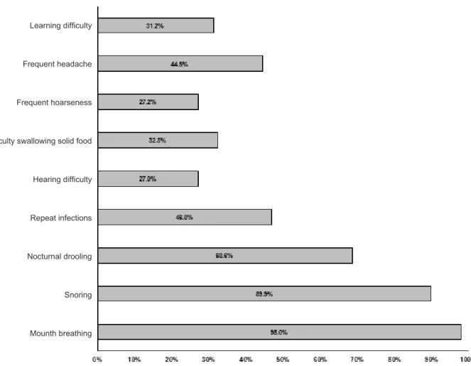
According to the medical history data, the most prevalent complaints of the children seen at the HC-UFMG MBOC were a persistent open mouth posture, snoring, and nocturnal drooling (Figure 1).
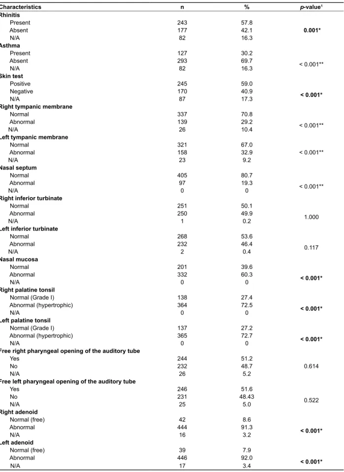
The allergological evaluation (Table 1) indicated rhinitis and positive skin tests among the study children, while no case of asthma was reported. The
otolaryngological examination (Table 1) revealed
compromised nasal mucosa, tonsillar hypertrophy (right and left), adenoidal hypertrophy (right and left), normal tympanic membranes, and normal nasal septum.
observed as they were contracted and inlated,
while the soft palate movements were tested with the production of the sounds “a” and “ã” alternately. Mandibular range of motion was evaluated based on movements of opening, lateralization, protrusion
and maximum mouth opening with the tip of the tongue on the papilla. To determine the nasal airlow,
a Glatzel mirror was used; discrepancy between nares was visually assessed.
Mastication was assessed with a piece of bread; only the swallowing of saliva was observed; speech was evaluated during a guided talk, and the voice was assessed using auditory-perceptual analysis.
The data obtained from the evaluation of the children were digitized; the analyses were done with the aid of the PASW Statistics 18 software (SPSS). For comparisons between proportions, some data were pooled in order to allow for dichotomous variables-namely, abnormal and adequate. This
classiication took into account the presence of at
least one abnormality. Data concerning functional assessments were not pooled, as we prioritized an analysis based on greater detail.
Note: 1binomial test; *p < 0.05 indicates signiicant prevalence of abnormal aspects; **p < 0.05 indicates signiicant prevalence of
adequate aspects
Figure 1 - Medical history data
Learning dificulty
Frequent headache
Frequent hoarseness
Dificulty swallowing solid food
Hearing dificulty
Repeat infections
Nocturnal drooling
Snoring
Table 1 - Data from the allergological and otolaryngological evaluations
Characteristics n % p-value1
Rhinitis
Present 243 57.8
0.001*
Absent 177 42.1
N/A 82 16.3
Asthma
Present 127 30.2
< 0.001**
Absent 293 69.7
N/A 82 16.3
Skin test
Positive 245 59.0
< 0.001*
Negative 170 40.9
N/A 87 17.3
Right tympanic membrane
Normal 337 70.8
< 0.001**
Abnormal 139 29.2
N/A 26 10.4
Left tympanic membrane
Normal 321 67.0
< 0.001**
Abnormal 158 32.9
N/A 23 9.2
Nasal septum
Normal 405 80.7
< 0.001**
Abnormal 97 19.3
N/A 0 0
Right inferior turbinate
Normal 251 50.1
1.000
Abnormal 250 49.9
N/A 1 0.2
Left inferior turbinate
Normal 268 53.6
0.117
Abnormal 232 46.4
N/A 2 0.4
Nasal mucosa
Normal 201 39.6
< 0.001*
Abnormal 332 60.3
N/A 0 0
Right palatine tonsil
Normal (Grade I) 138 27.4
< 0.001*
Abnormal (hypertrophic) 364 72.5
N/A 0 0
Left palatine tonsil
Normal (Grade I) 137 27.2
< 0.001*
Abnormal (hypertrophic) 365 72.7
N/A 0 0
Free right pharyngeal opening of the auditory tube
Yes 244 51.2
0.614
No 232 48.7
N/A 26 5.2
Free left pharyngeal opening of the auditory tube
Yes 246 51.6
0.522
No 231 48.43
N/A 25 5.0
Right adenoid
Normal (free) 42 8.6
< 0.001*
Abnormal 444 91.3
N/A 16 3.2
Left adenoid
Normal (free) 39 7.9
< 0.001*
Abnormal 446 92.0
N/A 17 3.4
Note: 1binomial test; *p < 0.05 indicates signiicant prevalence of abnormal aspects; **p < 0.05 indicates signiicant prevalence of
mixed dentition, 161 (32.1%), deciduous, and 29
(5.8%) children had permanent dentition.
The notes of the speech-language pathology evaluation concerning facial symmetry and type (Figure 3) indicated facial asymmetry.
The analysis of the data regarding the assessment of the orofacial structures (Table 2) revealed that the following features were abnormal: lip tone/tension, resting lip posture, nasolabial angle, and tongue tone/tension.
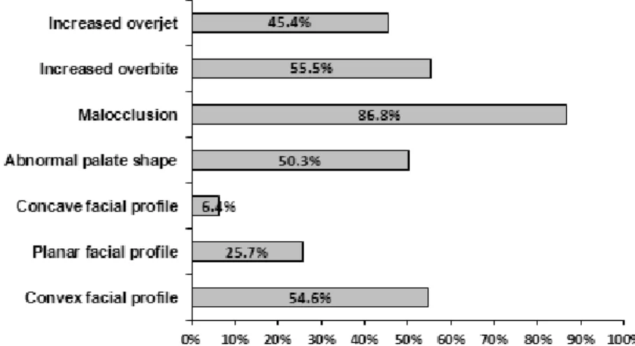
The orthodontic evaluation (Figure 2) revealed the following abnormalities: increased overbite,
presence of malocclusions, and convex facial proile. Among the most frequent malocclusions,
we found Angle class I malocclusion in 168 (33.5%) cases; class II, division 1 in 163 (32.5%); class III in 46 (9.2%), and class II, division 2 in 6 cases (1.2%). One (0.2%) case was noted as “atypical” and no
classiication was documented in 60 (12%) charts.
With regard to dentition, 361 (52.0%) children had
Note: 1binomial test; *p < 0.05 indicates signiicant prevalence of abnormal aspects
Figure 2 - Data from the orthodontic evaluation
Note: 1binomial test; *p < 0.05 indicates signiicant prevalence of abnormal aspects; **p < 0.05 indicates signiicant prevalence of
adequate aspects
Figure 3 - Data from facial type and symmetry evaluation
The nasolabial angle of 212 (42.2%) children was between 90ºand 110º, 175 (34.9%) had an angle > 90º , and for 106 (21.1%) of them the nasolabial angle was < 90º. There was no response to this item in 9 (1.8%) charts.
Table 2 - Data from the orofacial structure evaluation
Structure n % p-value1
Lips
Aspect
Adequate 263 53.4
0.149 Abnormal 230 46.6
Total 493 100.0
Tone/tension
Adequate 162 34.6
<0.001*
Abnormal 306 65.4 Total 468 100.0
Mobility
Adequate 442 91.9
< 0.001**
Abnormal 39 8.1 Total 481 100.0
Habitual posture
Adequate 144 29.5
< 0.001*
Abnormal 344 70.5 Total 488 100.0
Nasolabial angle
Adequate (90 º –110º) 212 43.0
Abnormal 281 57.0 0.002*
Total 493 100.0
Mentolabial angle
Adequate 252 53.7
0.116 Abnormal 217 46.3
Total 469 100.0
Cheeks
Aspect
Adequate 314 64.2
< 0.001**
Abnormal 175 35.8 Total 489 100.0
Tonus/tension
Adequate 256 55.2
0.029**
Abnormal 208 44.8 Total 464 100.0
Mobility
Adequate 393 83.8
< 0.001**
Abnormal 76 16.2 Total 469 100.0
Tongue
Aspect
Adequate 455 95.0
< 0.001**
Abnormal 24 5.0 Total 479 100.0
Tonus/tension
Adequate 164 35.6
< 0.001*
Abnormal 297 64.4 Total 461 100.0
Mobility
Adequate 427 89.3
< 0.001**
Abnormal 51 10.7 Total 478 100.0
Habitual posture
Adequate 193 45.6
0.080 Abnormal 230 54.4
Total 423 100.0
Frenulum
Adequate 383 84.2
< 0.001**
Abnormal 72 15.8 Total 455 100.0
Note: 1binomial test; *p < 0.05 indicates signiicant prevalence of abnormal aspects; **p < 0.05 indicates signiicant prevalence of
The palate was categorized as high/deep in 227 (45.2%) children, narrow in 137 (27.3%), wide in 13 (2.6%), and low in 3 (0.6%) cases. No evaluation was documented in 224 (44.6%) cases.
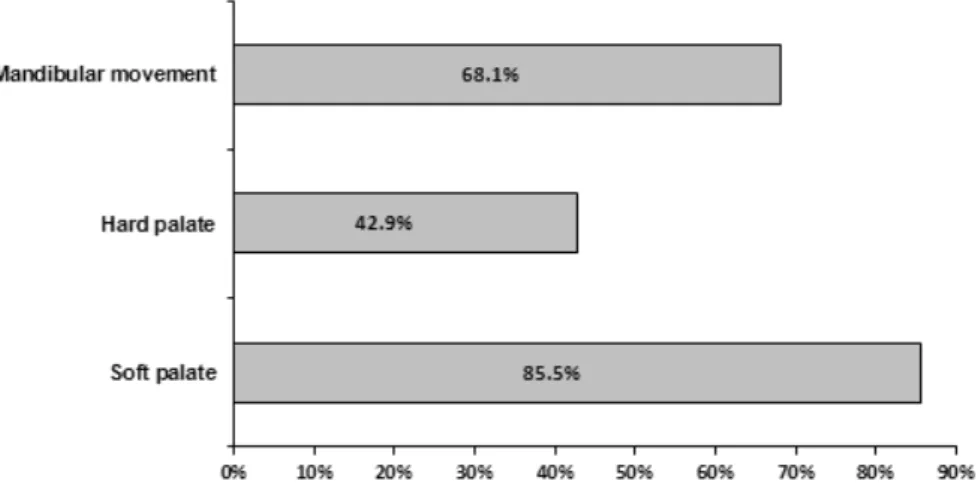
Mandibular range of motion (Figure 4) was found to be adequate. The evaluation of the palate (Figure 4) showed that soft palate mobility was adequate while hard palate mobility was impaired.
Note: 1binomial test; *p < 0.05 indicates signiicant prevalence of abnormal aspects; **p < 0.05 indicates signiicant prevalence of
adequate aspects
Figure 4 - Data from mandibular and palatal evaluation
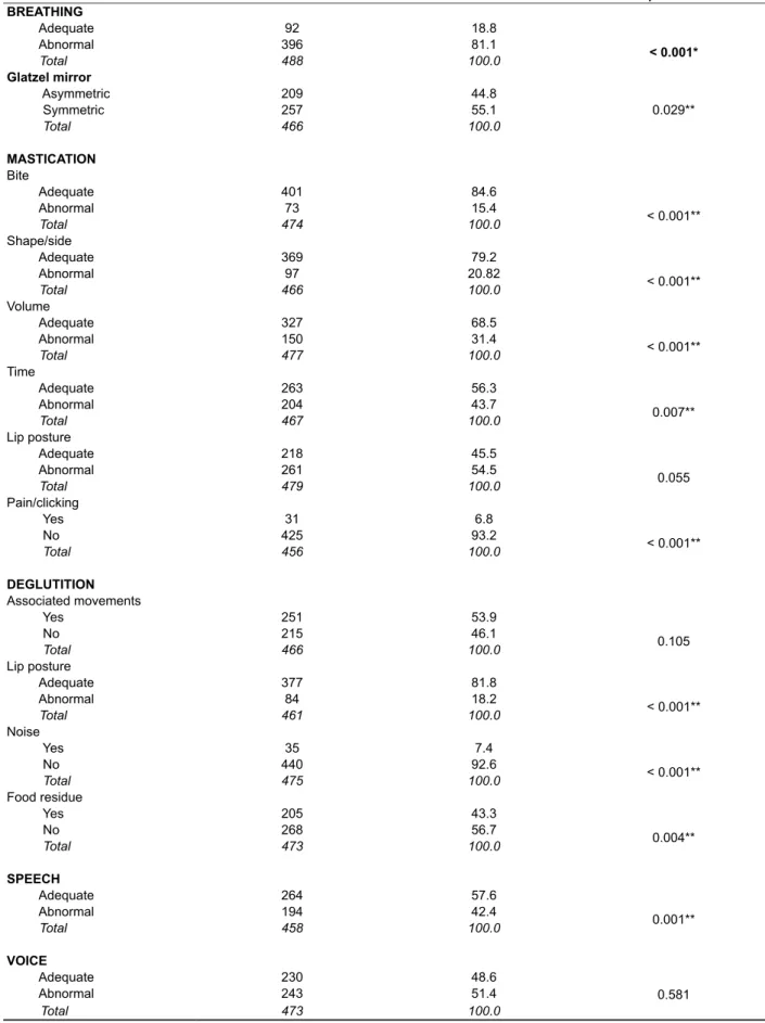
According to the stomatognathic function assessment (Table 3), only the breathing mode was dysfunctional. The following aspects were adequate: 1) breathing: symmetric air escape on the Glatzel mirror; 2) mastication: bite, shape/ side, volume, time, and absence of pain and/or clicking; 3) deglutition: lip posture, absence of noise and/or food residue; 4) speech: adequate in most subjects. Deviations in speech were found in 23.9% of the children, phone substitutions in 13.3%, and
omissions in 11% of cases. Trismus and excessive
salivation occurred in < 1% of cases each.
From the speech-language pathology evaluations derived the following approaches to management: referral to speech therapy in 195 (38.8%) cases, postoperative re-evaluation in 191 (38.0%), or wait for the approach recommendation by other profes-sionals in 32 (6.4%). Only guidance was provided in 4 (0.8%) cases, and the patient was uncooperative in another 4 (0.8%) cases, with the evaluation being completed at a later time. In addition, there were 38 (7.6%) children who did not need speech therapy, 17 (3.4%) who were in therapy elsewhere, and 21 (4.2%) records made no reference to management.
DISCUSSION
Multiple abnormalities can be observed in a mouth breather, such as changes in posture and in the stomatognathic system structures leading
to disturbances in dentofacial growth, suction, mastication, deglutition, and speech, which may progress to cardiorespiratory and endocrinological disease; sleep disturbances; mood disorders, and
achievement deicits in school3.
According to the children’s medical history, signif-icant complaints in this sample included nocturnal drooling, snoring, and open-mouth
breathing-indings in agreement with the literature4,5,7-9. The
reports of dificulty swallowing solid foods and frequent hoarseness were not statistically signiicant. Learning dificulties were not identiied in our study,
although this complaint has been documented in the literature5,8. This discrepancy could be due to one of two reasons: either the age range of the evaluated children, as many participants were at the stage of developing literacy, or the fact that both studies
reporting learning dificulties investigated a much
smaller sample than that of our study, with 142 cases
in the irst of those studies5 and 42 in the second8. The complaint of frequent headaches was reported in 222 cases (44.6%) in our sample; however, this
symptom reached no statistical signiicance.
Of note, one of the previous studies5 showed that some symptoms are more frequent depending on the cause of mouth breathing; however, we did not establish these relationships in the present study.
Table 3 - Data from functional evaluation
Function n % p-value1
BREATHING
Adequate 92 18.8
< 0.001*
Abnormal 396 81.1
Total 488 100.0
Glatzel mirror
Asymmetric 209 44.8
0.029**
Symmetric 257 55.1 Total 466 100.0
MASTICATION
Bite
Adequate 401 84.6
< 0.001**
Abnormal 73 15.4
Total 474 100.0
Shape/side
Adequate 369 79.2
< 0.001**
Abnormal 97 20.82
Total 466 100.0
Volume
Adequate 327 68.5
< 0.001**
Abnormal 150 31.4
Total 477 100.0
Time
Adequate 263 56.3
0.007**
Abnormal 204 43.7
Total 467 100.0
Lip posture
Adequate 218 45.5
0.055 Abnormal 261 54.5
Total 479 100.0
Pain/clicking
Yes 31 6.8
< 0.001**
No 425 93.2
Total 456 100.0
DEGLUTITION
Associated movements
Yes 251 53.9
0.105
No 215 46.1
Total 466 100.0
Lip posture
Adequate 377 81.8
< 0.001**
Abnormal 84 18.2
Total 461 100.0
Noise
Yes 35 7.4
< 0.001**
No 440 92.6
Total 475 100.0
Food residue
Yes 205 43.3
0.004**
No 268 56.7
Total 473 100.0
SPEECH
Adequate 264 57.6
0.001**
Abnormal 194 42.4
Total 458 100.0
VOICE
Adequate 230 48.6
0.581 Abnormal 243 51.4
Total 473 100.0
Note: 1binomial test; *p < 0.05 indicates signiicant prevalence of abnormal aspects; **p < 0.05 indicates signiicant prevalence of
inluenced the data analysis. Thus, the team needs
further instruction in this regard.
The structural evaluation indicated facial
asymmetry and altered nasolabial angle, indings that
have not been described in other studies, although they have been mentioned in the literature25,26.
The functional evaluation showed that only the breathing mode was abnormal. It is worth noting
that not all patients in the sample were classiied
as having abnormalities on the speech-language pathology evaluation because a proportion of those children only had nocturnal mouth breathing,
and the breathing mode classiication was based
solely on what the patient manifested at the time of
examination. However, it should be stressed that
the multidisciplinary diagnosis of all the patients included in the present study was indeed of mouth breathing. No changes were noted with respect to mastication, in contrast to other studies, which detected food residue, noise during mastication, separate lips, premature escape, and reduced mastication time9,27. We believe that no abnormal-ities were detected in our study probably because the patients were monitoring themselves, as they knew they were being evaluated. Hence, they made an effort to keep their lips sealed, avoided chewing
too fast, and prevented extraoral escape. The other
abnormalities found by those authors were not
examined in our study.
In another study, conducted with patients in orthodontic treatment, despite the high preva-lence of masticatory abnormalities, there was no
signiicant association when related to breathing
mode21. However, there are reports in the literature of changes in mastication time, lip posture, and other parameters of mastication9,27.
The absence of a lip seal during mastication
showed a signiicance level that was very close to
the established cut-off point. We believe that some of the children were able to monitor their performance during the assessment, which is in fact commonly observed in clinical practice. A lip seal can be noted in mouth breathing children, although this frequently
occurs at the expense of mentalis muscle strain15.
The evaluation of deglutition identiied no noises,
food residue, changes in lip posture, or associated
movements. The last two of those indings are in
contrast to the reports in the literature20.
The literature reports a relationship between abnormal breathing mode and the presence of speech disorders18,28. A study showed that 31.2% of mouth breathers have speech disorders, but the
signiicance of that inding was not ascertained29 . Another study found that mouth breathing is a risk factor for lisp in speech30. However, a study comparing speech disorders in mouth and nasal though they had met the MBOC inclusion criteria.
Therefore, it is clear that in a few cases the family of a mouth breathing child may not notice important signs of this abnormality.
Studies have shown a variety of causes for mouth breathing5,7,10,11. The children whose charts were reviewed in the present study had rhinitis and a positive skin test, as well as nasal mucosa abnor-malities and hypertrophic tonsils and adenoids.
With regard to the indings of the orthodontic evaluation, the convex facial proile observed in
the present study was found by other authors both among mouth and nasal breathers12. The rate of malocclusions was similar to that of other studies evaluating children in general13,14. The prevalence of increased overbite in the general population of children is lower13,14 than that in the present study; this as, therefore, the only characteristic that distin-guished the group of mouth breathers.
Mouth breathing can cause facial deformities and dental alignment problems3. Reports of maloc-clusion5,6,15,16, long face6, excessive height of the
lower third of the face and of the maxillary arch5 can also be found in the literature. Although anterior open bite can be considered the most prevalent malocclusion in mouth breathers17, the literature
reports that there is no one speciic type of maloc -clusion characteristic of mouth breathing18. The most prevalent facial type was the medium face, which
corroborates the indings of another study19. In the
literature, we ind reports of a vertical facial growth
pattern in mouth breathers 6,20, with increased height of the lower third of the face5,21,22. However, facial type categorization is subjective and inter-rater divergence may occur.
Abnormal lip and tongue tone as found in our sample was also shown in other studies 5,11,20,21.
Another inding in our study was inadequate resting
lip posture, with most children showing a lips-apart posture, which corroborates other studies3,10,15,20,23. However, changes in cheek tension and resting tongue posture, mentioned in the literature20, were
not identiied in our study.
Regarding mandible and palate assessments, changes were noted in the hard palate only, as found in other studies15. More speciically, increased depth18 and reduced width18,24 of the hard palate were noted. It should be noted that the angle of the mandible was not assessed in the present study. Therefore, we were unable to note an obtuse gonial angle as documented by some authors5,21. It is suggested that further studies should address this particular aspect.
CONCLUSION
Among the mouth breathing children in our study, we noted complaints of an open mouth posture, snoring, and nocturnal drooling. We also observed the following: rhinitis, positive skin test, abnormal nasal mucosa, adeno-tonsillar hypertrophy, deep
anterior overbite, malocclusion, convex facial proile,
facial asymmetry, reduced lip and tongue tone, incompetent resting lip posture, altered nasolabial angle, high hard palate, and mouth breathing.
Mouth breathing can impact several dimensions of a child’s life, as it compromises several aspects of health. It is of paramount importance that these patients receive multidisciplinary evaluation and treatment. Our study is relevant in that it compiles
the characteristics identiied in mouth breathing
children, thereby assisting in the establishment of evaluation guidelines and in the proposition of health promotion and prevention initiatives in orofacial myology.
In forthcoming studies, the data will be stratiied
according to facial type and occlusion class so that these relationships can be analyzed. We suggest that studies be conducted including a control group to allow more precise data to be obtained.
ACKNOWLEDGMENTS
We are grateful to the FUNDEP/Santander partnership for the undergraduate research grants
awarded to the irst author.
breathers16 found no signiicant difference, which is consistent with our study results. Among the
speech disorders identiied in the present study, distortions were the most prevalent—likely as a
result of alterations in the stomatognathic system3, such as changes in the tension and position of the phonatory/articulatory organs.
Finally, it was found that the most frequent speech-language pathology approach to treatment was an indication to speech therapy-a relevant aspect, since speech therapy can contribute to breathing mode correction, facilitate the control of mouth breathing and of allergic rhinitis, and assist in the management of asthma28. The second most frequent approach was postoperative re-evaluation, which is related to the high prevalence of adeno-tonsillar hypertrophy in our sample.
Although the initial proposition of the present study was to discuss the abnormalities that had been found, it should be stressed that some data
presented in the literature-hence, expected to be found in our study as well-were not conirmed, whether because no signiicant differences were
found or even because there was a greater frequency of adequate parameters in our sample. This might have occurred due to the sample size of
the studies. Another limitation that could have inlu -enced the results was the fact that the assessments were conducted by different students, even though they were all trained by the same teacher.
8. Neiva PD, Kirkwood RN, Godinho R. Orientation and position of head posture, scapula and thoracic spine in mouth breathing children. Int J Pediatr Otorhinolaryngol. 2009;73:227-36.
9. Cunha DA, Silva GAP, Motta MEFA, Lima CRL, Silva HJ. A respiração oral em crianças e suas repercussões no estado nutricional. Rev Cefac. 2007;9(1):47-54.
10. Farid MM, Metwalli N. Computed tomographic evaluation of mouth breathers among pediatric
patients. Dentomaxillofac Radiol. 2010;39(1):1-10.
11. Junqueira P, Marchesan IQ, Oliveira LR, Ciccone E, Haddad L, Rizzo MC. Speech language
pathology indings in patients with mouth breathing:
multidisciplinary diagnosis according to etiology. Int J Orofacial Myology. 2010;36:27-32.
12. Frasson JMD, Magnani MBBA, Nouer DF, Siqueira VCV, Lunardi N. Estudo cefalométrico comparativo entre respiradores nasais e predominantemente bucais. Rev Bras Otorrinolaringol. 2006;72(1):72-82. 13. Britto DI, Dias PF, Gleiser F. Prevalência de más oclusões em crianças de 9 a 12 anos de idade da cidade de Nova Friburgo (Rio de Janeiro). Dental Press. 2009;14(6):118-24.
14. Boeck EM, Pizzol KEDC, Navarro N, Chiozzini NM, Foschini ALR. Prevalência de maloclusão em escolares de 5 a 12 anos de rede municipal de ensino de Araraquara. Rev Cefac. 2013;15(5):1270-80. 15. Cattoni DM, Fernandes FD, Di Francesco RC, Latorre MRDO. Características do sistema
REFERENCES
Muñoz ICL, Orta PB. Comparison of cephalometric patterns in mouth breathing and nose breathing children. Int J Pediatr Otorhinolaryngol. 2014;78(7):1167-72.
2. Lione R, Buongiorno M, Franchi L, Cozza P.
Evaluation of maxillary arch dimensions and palatal
morphology in mouth-brathing children by using digital dental casts. Int J Pediatr Otorhinolaryngol. 2014;78(1):91-5.
3. Abreu RR, Rocha RL, Lamounier JA, Guerra AF. Prevalência de crianças respiradoras orais. J Pediatr. 2008;84(5):467-70.
4. Felcar JM, Bueno IR, Massan ACS, Torezan RP, Cardos JR. Prevalência de respiradores bucais em crianças de idade escolar. Ciênc Saúde Coletiva. 2010;15(2):437-44.
5. Di Francesco RC, Passerotti G, Paulucci B, Miniti A. Respiração oral na criança: repercussões diferentes de acordo com o diagnóstico. Rev Bras Otorrinolaringol. 2004;70(5):665-70.
6. Menezes VAM, Tavares RLO, Granville-Gracia AF. Síndrome da respiração oral: alterações clínicas e comportamentais. Arq Odontol. 2009;45(3):160-5. 7. Abreu RR, Rocha RL, Lamounier JA, Guerra AF. Etiologia, manifestações clínicas e alterações presentes nas crianças respiradoras orais. J Pediatr. 2008;84(6):529-35.
RESUMO
Objetivo: descrever os achados miofuncionais orofaciais, bem como os principais problemas otorrino-laringológicos, alergológicos e ortodônticos encontrados em crianças com respiração oral. Métodos:
análise de prontuários de 502 crianças do Ambulatório do Respirador Oral do Hospital das Clínicas da Universidade Federal de Minas Gerais. Os participantes tinham idades entre 2 e 12 anos (mediana
de 6,0 anos), sendo 289 (57,6%) do sexo masculino e 213 (42,4%) do sexo feminino. Foram cole -tados dados dos prontuários referentes à anamnese geral, avaliação fonoaudiológica, bem como as partes relevantes das avaliações otorrinolaringológica, alergológica e ortodôntica. Os dados foram submetidos à análise estatística. Resultados: na anamnese, observou-se prevalência signiicante de
permanência de boca aberta (98,0%), ronco (89,9%) e sialorreia noturna (68,6%). Na avaliação
aler-gológica, veriicou-se teste cutâneo positivo (59%) e rinite (57,8%) e na otorrinolarinaler-gológica, hiper
-troia de adenoide (91,7%) e amígdalas (72,6%), além de mucosa nasal alterada (60,3%). A avaliação ortodôntica indicou presença de má oclusão (86,8%), peril facial convexo (62,9%) e trespasse verti -cal aumentado (55,5%). Os dados da avaliação fonoaudiológica indicaram inadequação da posição habitual de lábios (70,5%), tensão de lábios (65,4%) e de língua (64,4%) alteradas, palato duro alto
(57,1%), ângulo nasolabial alterado (57,0%) e assimetria facial (55,0%). Conclusão: veriicaram-se alterações nas avaliações realizadas por todos os proissionais, conirmando o grande impacto da
respiração oral na qualidade de vida e, portanto, a necessidade de tratamento multidisciplinar para esses pacientes.
26. Siqueira VCV. O crescimento craniofacial e o respirador bucal. In: Coelho-Ferraz MJP (org). Respirador bucal: uma visão multidisciplinar. Lovise: São Paulo; 2005. P. 119-26.
27. Silva MAA, Natalini V, Ramires RR, Ferreira LP. Análise comparativa da mastigação de crianças respiradoras nasais e orais com dentição decídua. Rev CEFAC. 2007;9(2):190-8.
28. Campanha SMA, Fontes MJF, Camargos PAM, Freire LMS. O impacto do tratamento fonoaudiológico no controle da asma e da rinite alérgica em crianças e adolescentes respiradores orais. J Pediatr. 2010;86(3):202-8.
29. Hitos SF, Arakaki R, Solé D, Weckx LLM. Oral
breathing and speech disorders in children. J Pediatr. 2013;89(4):361-5.
30. Monteiro VR, Brescovici SM, Delgado SE. A ocorrência de ceceio em crianças de oito a 11 anos em escolas municipais. Rev Soc Bras Fonoaudiol. 2009;14(2):213-8.
31. Tavares JG, Silva EHAA. Considerações teóricas sobre a relação entre respiração oral e disfonia. Rev Soc Bras Fonoaudiol. 2008;13(4):405-10.
32. Martinelli RLC, Fornaro EF, Oliveira CJM, Ferreira LMDB, Rehder MIBC. Correlações entre alterações de fala, respiração oral, dentição e oclusão. Rev CEFAC.2011;13(1):17-26.
33. Souki BQ, Lopes PB, Veloso NC, Avelino RA, Pereira TB, Souza PE et al. Facial soft tissues
of mouth-breathing children: Do expectations
meet reality? Int J Pediatr Otorhinolaryngol. 2014;78(7):1074-9.
34. Nishimura CM, Gimenez SRML. Peril da fala
do respirador oral. Rev CEFAC. 2010;12(3):505-8. 35. Sígolo C, Silveira M, Quintal M, Sakano E, Tessitore A. Ocorrência de movimentos primários de língua em crianças respiradoras oronasais. Rev CEFAC. 2008;10(1):51-7.
36. Hennig TR, Silva AMT, Busanelo AR, Alemida FL, Berwig LC, Boton LM. Deglutição de respiradores orais e nasais: avaliação clínica fonoaudiológica e
eletromiográica. Rev CEFAC. 2009;11(4):618-23.
estomatognático de crianças respiradoras orais: enfoque antroposcópico. Pró-Fono R Atual
Cientiica. 2007;19(4):347-51.
16. Lemos CM, Wilhelmsen NSW, Mion OG, Mello Junior JF. Alterações funcionais do sistema estomatognático em pacientes com rinite alérgica: estudo caso-controle. Braz J Otorhinolaryngol. 2009;75(2):268-74.
17. Ribeiro F, Bianconi CC, Mesquita MCM, Assencio-Ferreira VJ. Respiração oral: alterações oclusais e hábitos orais. Rev Cefac. 2002;4(3):187-90.
18. Feres MFN, Enoki C, Sobreira CR, Matsumoto MAN. Dimensões do palato e características oclusais de crianças respiradoras nasais e bucais. Pesq Bras Odontoped Clin Integr. 2009;9(1):25-9. 19. Bianchini AP, Guedes ZCF, Vieira MM. Estudo da relação entre a respiração oral e o tipo facial. Rev Bras Otorrinolaringol. 2007;73(4):500-5.
20. Valera FCP, Trawitzki LVV, Mattar SEM, Matsumoto MAN, Elias AM, Anselmo-Lima WT. Muscular, functional and orthodontic changes in pré school children with enlarged adenoids and tonsils. Int J Pediatr Otorhinolaryngol. 2003;67(7):761-70. 21. Branco A, Ferrari GF, Weber SAT. Alterações orofaciais em doenças alérgicas de vias aéreas. Rev Paul Pediatr. 2007;25(3):266-70.
22. Cattoni DM, Fernandes FD, Di Francesco RC, Latorre MRDO. Quantitative evaluation of the orofacial morphology: anthropometric measurements in healthy and mouth-breathing children. Int J Orofacial Myology. 2009;35:44-54. 23. Rodrigues HOSN, Faria SR, Paula FSG, Motta AR. Ocorrência de respiração oral e alterações orofaciais em sujeitos em tratamento ortodôntico. Rev CEFAC. 2005:7(3):356-62.
24. Berwig LC, Silva AM, Côrrea EC, Moraes AB, Montenegro MM, Ritzel RA. Hard palate dimensions in nasal and mouth breathers from different etiologies. J Soc Bras Fonoaudiol. 2011;23(4):308-14.
25. Junqueira P. Avaliação miofuncional. In: Marchesan IQ. Fundamentos em Fonoaudiologia: aspectos clínicos da motricidade orofacial. Guanabara Koogan: Rio de Janeiro; 1998. p. 13-21.
Received on: April 14, 2014 Accepted on: October 05, 2014
Mailing address: Mariana da Costa
Rua Sambeatiba, 201 casa 19 – Cachoeirinha Belo Horizonte – MG – Brasil
CEP: 31150-220
APPENDIX 1
MOUTH BREATHING OUTPATIENT CLINIC SPEECH-LANGUAGE PATHOLOGY EVALUATION PROTOCOL
No __________
1) IDENTIFICATION
Patient: ______________________________________________________________________________ Informant: ___________________________Evaluated by: _____________________________________
2) COMPLAINT _______________________________________________________________________ ____________________________________________________________________________________
3) COMPLEMENTARY HISTORY Sleep
a) drools: yes ( ) no ( ) sometimes ( ) doesn’t know ( ) during the day ( ) b) wakes up with his/her mouth dry: yes ( ) no ( ) sometimes ( ) doesn’t know ( ) c) sleeps on his/her: stomach ( ) back ( ) sideways ( ) doesn’t know ( )
Eating
a) breastfeeding: yes ( ) no ( ) until ________ exclusively until ________
b) if he/she is still on the bottle: number/content ______________________________________________ c) introduction of foods: _________________________________________________________________ d) consistency of foods: _________________________________________________________________ e) taste/smell: _________________________________________________________________________ f) chewing: fast ( ) normal ( ) slow ( ) open lips ( ) closed lips ( ) unilateral
( ) bilateral ( ) doesn’t know ( ) noisy ( ) TMJ pain ( ) liquids at meal ( )
g) swallowing: ______________________________________________________________________
Oral habits
a) paciier: yes ( ) no ( ) started ________ until ________ type: orthod. ( ) regular ( )
b) bottle: yes ( ) no ( ) started _______ until ________ type: orthod. ( ) regular ( )
c) inger: yes ( ) no ( ) started________ until________ which/how ________________________________
____________________________________________________________________________________ d) teeth grinding: yes ( ) no ( ) started ________ until ________
e) teeth clenching: yes ( ) no ( ) started ________ until ________ f) nail biting: yes ( ) no ( ) started ________ until ________
g) rests his/her face on the hand to sleep: yes ( ) no ( ) sometimes ( ) doesn’t know ( ) h) bites objects: yes ( ) no ( ) which _____________ started ______ until _______
i) other (which, started and until):__________________________________________________________
Speech
a) changes: yes ( ) no ( )
b) which: ____________________________________________________________________________
Additional information:__________________________________________________________________
_____________________________________________________________________________________
4) SPEECH-LANGUAGE PATHOLOGY EVALUATION
STRUCTURES 1st Assessment Date: ___/___/___
1 postoperative month Date: ___/___/___
3 postoperative months Date: ___/___/___
Eyes
Eye vs. right and left commissure
Nose Morphology Nasolabial angle Philtrum
Lips Morphology Habitual posture Tension
Mobility
Mentolabial angle Cheeks
Morphology Tension Mobility Mental Morphology Tension Mandible Opening Lateralization Protrusion
Maximum opening with tongue on incisive papilla
Masseter muscle Temporalis muscle Tongue
Morphology Habitual posture Tension
Mobility Frenulum Palate Hard Soft Facial Type Facial height
Thirds (upper/middle/lower) Proile
FUNCTION 1st Assessment 1 postoperative month
3 postoperative months
Breathing Mode
Glatzel mirror Mastication Bite Pattern
Mandibular movement Volume
Lip posture
Associated movements/noises Pain/clicking
Temporalis/masseter Deglutition
Associated movements Lip posture
Noises Food residue Speech Abnormalities Voice
Vocal quality Resonance Pitch Loudness Velocity
CPFA1
MPT2 /a/ ; /i/ ; /u/
MPT /s/ ; /z/ S/Z ratio
1CPFA: pneumo-phonic and articulatory coordination; 2MPT: maximum phonation time
5) Otolaryngological evaluation: _________________________________________________________ _____________________________________________________________________________________
6) Orthodontic evaluation: ______________________________________________________________ _____________________________________________________________________________________