example, lack of manpower in agrarian societies may lead to reductions in agri- cultural production. The health systems will surely face major increases in the de- mand for services and may face this de- mand with reduced manpower and im- paired infrastructures. Any of these outcomes will have major implications for food, nutrition, and health planning, and will increase the need for capital
funding while reducing the ability to re- pay debts. The Subcommittee therefore recommended that governments and the United Nations agencies monitor not only the development of the AIDS epi- demic but also the evolution of its struc- tural effects, so that national and interna- tional actions can be set in motion to compensate for the wide-ranging impacts on health and development.
Measurement of Antibodies to Human
Immunodeficiency Virus: An International
Collaborative Study to Evaluate WHO
Reference Sera
A.J. GARRETT,*
V. SEAGROATT,~
E.M.SUPRAN,*
K.O.
HABERMEHL,~
H.HIAMPL,~&G.C.SCHILD~
Human immunodeficiency virus (HIV), the causative agent of the acquired im- munodeficiency syndrome (AIDS) (I, 2, 3), is transmitted primarily through sex- ual contact or the injection of contami- nated blood or blood products such as anti-hemophilic factors (4). Since 1985, the screening of blood donations for anti-
Source: Bulletin of the World Healfh Organimfion 66(2):197-202,1988. 0 World Health Organization. i WHO Collaboratine Center on AIDS, National In-
stitute for Biologycal Standards and Control, South Mimms, Potters Bar, Hertfordshire, EN6 3QG, England:
a WHO Collaborating Center on AIDS, Division of Microbiological Reagents and Quality Control, Central Public Health Service Laboratory, Colin- dale, London, England.
3 WHO Collaborating Center on AIDS, Institute for Clinical and Experimental Virology, Free Univer- sity of Berlin, Berlin (West).
HIV has been instituted in many coun- tries in order to minimize the risk of transmission of AIDS via blood transfu- sions or treatment with blood products. The detection of antibodies to HIV is also of major importance as a relatively simple and rapid determination of the extent and spread of HIV infections (5), and many commercial and “in-house” immu- nochemical tests are now in use through- out the world. At present, the most com- monly used assays are based on enzyme-linked or radioimmunosor- hence, immunofluorescence, immuno- blotting, or immunoprecipitation, and variations in the specificities and sensitiv- ities of the techniques reflect inherent dif- ferences between the principles of the as- says as well as batch-to-batch variations in the preparation of reagents and kits (6,
7). Thus, there is an urgent need for well- characterized reference materials for use in defining the reliability and sensitivity of the tests, for quality control of batches of kits or reagents, and as common refer- ences between laboratories.
This report presents an assessment of two proposed reference preparations of sera, one reactive and the other unreac- tive to HIV in a collaborative study in- volving 21 laboratories in 11 countries.
MATERIALS AND METHODS
Proposed Reference Materials
The preparations of antibody-positive and antibody-negative human sera, freeze-dried in sealed glass ampules, were supplied by Professor K. 0. Haber- mehl (Institute for Clinical and Experi- mental Virology, Berlin (West)). Each preparation was derived from a single donor; one an asymptomatic carrier of HIV and the other a donor with no known risk factors. Each serum was un- reactive when tested for hepatitis B sur- face antigen (HBsAg) by a standard im- munoassay and was heated at 56 “C for one hour before freeze-drying. When re- constituted as recommended (in 0.2 ml water) these preparations had concentra- tions one-twentieth of the original sera; this dilution factor is not included in the calculations presented in this report.
Coded Preparations
Seven freeze-dried serum prepara- tions, coded A to G, were supplied to each participant. Preparations A and C were duplicate samples of the reactive proposed reference material, preparation E was the nonreactive proposed refer- ence material. Samples D and F were pre- pared by the Central Public Health Serv- ice Laboratory, Colindale, London.
Sample D, derived from a single donor, showed weak reactivity in immunosor- bent assays. Sample F, derived from sera pooled from several donors, was highly reactive. Samples B and G, which were National Institute for Biological Stand- ards and Control (England) reference preparations freeze-dried from pooled sera in 1973 and 1967, respectively, were both unreactive.
Design of the Study
The study was designed to identify the coded preparations that reacted with HIV antibodies and to ascertain the minimum amount of the reactive samples that could be detected in the methods rou- tinely used by the participants.
The 21 participating laboratories (see Annex) were supplied with duplicate sets of the coded preparations and requested to assay them by the procedures usually employed in their laboratories.
Assay Methods
All but three participants assayed the preparations by indirect or competitive ELISA. Nine different commercial kits for ELISA were used: Abbott, DuPont, ENI, Genetic, Organon, Ortho, Pasteur (“or- dinary” and “Rapide”), Travenol, and Wellcome. Two laboratories carried out ELISAs using their own “in-house” methods and one included in its series of assays the Abbott “confirmatory” ELISA, a competitive ELISA based on en- velope and core antigens derived from recombinant DNA.
Immunoblots were carried out by 15 laboratories. All except two, whose tech- niques involved the use of a mouse monoclonal antibody specific for human IgG and labelled with i=I, or protein A labeled with lZI, used peroxidase-linked anti-human IgG for the identification of antigen-antibody complexes. Eight used
biotin-avidin amplification of the enzyme system.
One participant used the Karpas method (8) and one used an assay based on particle agglutination (PA).
Method of Analysis
For each test the reactivities of the co- ded samples A to G and the end-point titers for samples A, C, and F were taken to be those recorded by the participants. End-points were defined as the recipro- cals of the highest dilutions of the re- constituted original materials in nomzal serum (not the final dilutions in the assay wells) that gave positive responses in the assays.
Potency ratios of C and F were ex- pressed as ratios of their titers to that of A in the same assays.
RESULTS
Classification
ofSera by Reactivity in
ELBA and Immunoassays
Samples A, C, and F were found reac- tive in all tests and samples B, E, and G
were reported as negative in all but one test. The exception was a test based on particle agglutination (PA) in which sam- ple E was judged to be weakly reactive. Sample D was found to be reactive or weakly reactive in 40 of the 48 ELISAs performed, in both the PA and Karpas test, and in two of the four fluorescence microscopy (FM) tests (Table 1). In the eight ELISAs in which sample D was identified as unreactive, it had a higher optical density (OD) than the negative control (although not, of course, as high as the OD of the cut-off limit), and in all but one the ODs were at least twice that of the negative control.
Titration of Samples A, C, and F
Participants carried out single or dupli- cate assays for individual manufacturers’ ELISAs or by their local methods. Some laboratories used several manufacturers’ ELISAs; in particular, laboratory 1 used seven different kits. For each kit, the geo- metric means of the titers for A, C, and F and of the potency ratios for C and F ob- tained by individual laboratories were calculated. The frequency distributions of
Table 1. Assessment of reactivity of sample D by immunoassays.
No. with stated
No. of No. of reactivity
Assay method laboratories assays + 4 -
ELISA kits:
Abbott 9 I.2 9 2 1
DuPont 2 3 3 -
ENI 1 2 - 2
Genetic 1 2 - 2 -
Organon 3 4 1 1 2
Ortho 2 3 3 -
PasteuP 5 8 6 1 1
Travenol 1 1 - 1
Wellcome 6 10 8 2 1
“In-house” ELISA 2 3 1 1 1
All ELISAs 18 48 31 9 8
Fluorescence microscopy 4 4 1 1 2
Karpas method 1 1 1 - -
Particle agglutination 1 2 2 -
“Includes assays using the “Rapid&’ version.
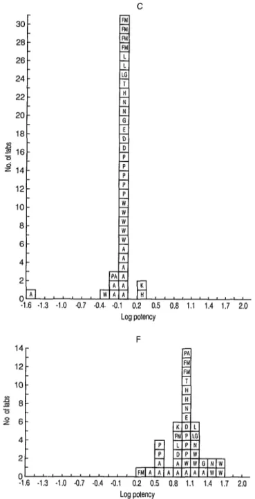
these values are shown in Figures 1 and 2. One of the participants using the Ab- bott kit obtained much higher titers for A and F than did the other participants us- ing both this and other kits. Further, its
titers for C were ten-fold and loo-fold lower than those for A. The results from
this participant were, therefore, consid- ered atypical and excluded from the sub- sequent analyses,
lo-
a-
-5.0 -4.6 -4.2 .3.8 -3.4 -3.0 2.6 -2.2 -1.8 -1.4 -1.0 -0.6 .0.2 Log end point
0
-5.0 -4.6 -4.2 -3.8 .3.4 -3.0 -2.6 -2.2 -1.8 -1.4 .l 0 .0.6 .0.2 Log end point
F
8
0
4.0 -4.6 .4.2 -3.8 -3.4 -3.0 -2.6 -2.2 -1.8 .i.4 -1.0 .0.6 -0.2 Log end point
Figure 1. Frequency distributions of the end-point dilutions obtained for samples A, C, and F. Each square denotes an estimate from one test; the let- ters in the squares refer to the type of assay. A, D, E, G, H, N, I’, T, and W denote the following commercial kits of ELISA: A, Abbott; D, DuPont; E, ENI; G, Genetic; H, Ortho; N, Organon; P, Pasteur; T, Travenol; and W, Wellcome. L and LG denote “in-house” versions of ELISA. Letters FM, K, and PA denote the following methods other than ELISA: FM, fluorescence microscopy; K, Karpas; and PA, particle agglutination test.
30- 28- 26- 24
t
-
Logpotency
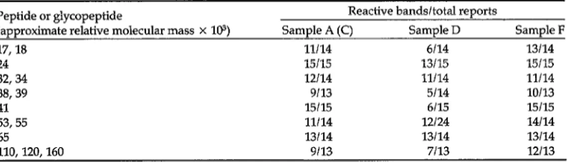
-1.6 -1.3 -1.0 -0.7 -0.4 -0.1 0.2 0.5 0.8 1.1 1.4 1.7 2.0 Log potency
Figure 2. Frequency distributions of the potency ratios of samples C and F in terms of A obtained from individual tests. (See legend in Figure 1 for explanation of the letters in the squares.)
The laboratory mean titers varied con- siderably over about a 20-fold range. There were, however, no obvious differ- ences between the titers from different kits: for instance, the ranges of titers for Abbott, Pasteur, and Wellcome over- lapped with each other.
The laboratory mean potency ratios of C, a coded duplicate of A, were mostly unity. All but one laboratory (mentioned above) found the titers of A and C to be within one dilution step (Figure l), al- though sometimes these dilution steps were as large as five- and tenfold. The laboratory mean potency ratios for F were more variable than those for C. However, the potency ratios were less variable than the titers (Figures 1 and 2). One laboratory’s results gave a potency ratio, based on a single assay, of 256, ten- fold higher than the other estimates, and had the highest titer for F (16,384). This titer and potency ratio were, therefore, considered atypical and excluded from the subsequent analysis.
Differences between the laboratories’ estimations of titers were found to be sig- nificant by analyses of variance, even be- tween those using kits from the same manufacturer. Expressing the reactivities of samples C and F relative to A showed that the differences between laboratories using the same commercial kit were no longer statistically significant. However, there were still significant differences be-
Table 2. Detection of anti-HIV by immunoblot.a
tween the potency ratios from different tests. For example, the overall mean po- tency ratio, i.e., the geometric mean of the laboratory mean potencies, for Wellcome was 22, about three times higher than those for Abbott and Pasteur kits (6 and 7, respectively). The overall mean potency ratios for the other kits fell within this range.
Immunoblots
Fifteen participants tested samples A to G by immunoblot techniques. The results are given in Table 2. The use of control antigens (“mock” antigens) was re- ported from only two laboratories. Faint reactive bands in the regions of relative molecular mass (M,) 24 x lo3 and 64 x
lo3 were detected consistently by one participant using “mock” antigen from H9 cells; more information is required on this important aspect of the assays.
The relative molecular masses recorded in Table 2 are those assigned by the indi- vidual participants. For convenience of presentation, bands reported within nar- row ranges of M, are not differentiated and are classified in groups (e.g., (32-34)
x lo3 and (110-160) x 103).
Samples A and C were duplicates of the proposed reactive reference serum. As was hoped, they produced identical results for each immunoblot system and are considered together. One participant
Peptide or glycopeptide Reactive bands/total reports
(approximate relative molecular mass X 103) Sample A (C) Sample D Sample F
17,18 11114 6114 13114
24 15115 13115 15115
32,34 12114 11114 11114
38,39 9113 5114 10113
41 15115 6115 15115
53,55 11114 12/24 14114
65 13114 13114 13114
110,120,160 9113 7113 12113
“All participants did not report the presence or absence of each peptide or glycopeptide.
reported on the detection of antibodies to peptides p24 and gp41 only. All partici- pants, except one who reported a weak reaction in the p65 region for sample E and another who observed a reaction to p24 antigen in sample G, detected no an- tibodies to HIV in samples B, E, and G. Of the positive samples, F reacted most strongly in all immunoblots but, except for less frequent detection of antibodies to the envelope antigens gpllO-~160 in A and C than in F, samples A, C, and F were qualitatively identical
All participants detected antibodies to ~24, gp41, and p53/55 in A, C, and F; three reported no antibodies to ~17118; and only one reported no antibodies to ~65.
Additional Results
Essex and colleagues included in their study an investigation of the reactivity of samples A to G in immunoblots in which the recently isolated strain HTLV-IV was used as antigen. No reactions with any viral antigens were reported for Western blots and only two reactive regions, gp160 for sample A and p24 for sample F, were detected in radioimmunoprecipita- tion using %-labelled HTLV-IV
DISCUSSION
ELISA kits from nine manufacturers were used in this study. The sensitivities of the various kits were assessed by com- paring end-point titers for the highly re- active samples A and F and by whether or not a weakly reactive serum was found “positive” in the tests. This sensitivity varied between laboratories, even be-
tween those using the same commercial kit. Expressing the reactivity of sample F relative to A reduced the variation be- tween tests and resulted in agreement between the laboratories using the same kit. Nevertheless, there were still consist-
ent differences between the kits in the comparison of the reactivity of A and F. This possibly reflected the differences in specificities of the ELISA systems.
Overall, immunoblots revealed reac- tions of sera A (C) and F to all the ex- pected HIV antigens. The presence of an- tibodies to ~24, gp41, and p55 is considered by most workers an impor- tant indication of infection with HIV (9, 10, 11). However, for reproducible results the source of antigens, the standardiza- tion of electroblotting procedures, and the provision of “control” antigens re- quire careful attention (9); variations of these factors may well explain the differ- ences shown in Table 2.
The results for HTLV-IV confirm earlier findings (21, 12, 23) and emphasize the urgent need for information on the re- sponses of immunoassays to sera from AIDS patients from different geographi- cal areas and for characterization of the
genetic and immunological differences between viral isolates. The proposed ref- erence preparation, A, reacted strongly in all ELISAs and related immunoassays and reacted with all the major HIV anti- gens in immunoblots. Its use will depend on individual requirements, but it may be of value as a qualitative check on the spe- cificity of the assays, to calibrate positive controls included in kits and other assays in arbitrary units, to calibrate detection limits (cut-off) in arbitrary units (as done for HBsAg), and to calibrate immuno- blots, particularly for defining the opti- mal amounts of antigen and for deter- mining the relative mobilities of the major peptides and glycopeptides from HIV Because the unreactive preparation (E), reconstituted in 0.2 ml water as recommended, represents diluted se- rum, its use as a reference material will be limited.
The WHO Expert Committee on Bio- logical Standardization reviewed the re- port of this collaborative study in Decem-
ber 1986 and agreed that preparations A and E would be of value as reference preparations for, respectively, positive and negative anti-HIV sera.
The preparations, under code num- bers 86/6302 (reactive) and 86/6238 (unreactive) are available from the Direc- tor, National Institute for Biological Standards and Control, South Mimms, Potters Bar, Herts., EN6 3QG, England.
Acknowledgment.
We thank Jane Bruce for assisting in the statistical analysis of the data from the study.REFERENCES 1. 2. 3. 4. 5. 6. 7. 8. 9.
Barre-Sinoussi, F. et al. Isolation of a T- lymphotropic retrovirus from a patient at risk for acquired immune deficiency syn- drome (AIDS). Science 220:868-871, 1983. Levy, J. A. et al. Isolation of lymphocyto- pathic retroviruses from San Francisco pa- tients with AIDS. Science 225:840-842, 1984.
Popovic, M. et al. Detection, isolation and continuous production of cytopathic ret- roviruses (HTLV-III) from patients with AIDS and pre-AIDS. Science 224:497-500, 1984.
Curran, J. W. et al. The epidemiology of AIDS: Current status and future pros- pects. Science 229:1352-1357,1985.
Biggar, R. J. The AIDS problem in Africa. lmcet 1:79-83, 1986.
Mortimer, P. I? et al. Which anti-HTLV-1111 LAV assays for screening and confirma- tory testing? Lancet 2~873-877, 1985. Petricciani, J. C. et al. An analysis of se- rum samples positive for HTLV-III anti- bodies. N Eng J Med 313:47-48,1985. Karpas, A. et al. Lytic infection by British AIDS virus and development of rapid cell test for antiviral antibodies. Lancet 2:695- 697,1985.
Essex. M. et al. Antigens of human T- lymphotropic virus “type IIIllymph- adenopathy associated virus. Ann Intern Med 103:700-703, 1985.
10.
11.
12.
13.
Schupbach, J. et al. Antibodies to HTLV- III in Swiss patients with AIDS and pre- AIDS and in groups at risk for AIDS. N Eng 1 Med 312:265-270,1985.
Biberfeld, G. et al. Findings in four HTLV- IV seropositive women from West Africa. Lancet 2:1330-1331, 1986.
Clavel, F. et al. Isolation of a new human retrovirus from West African patients with AIDS. Science 233:343-346, 1986.
Kanki, l? J. et al. New human T- lymphotropic retrovirus related to simian T-lymphotropic virus type III (STLV-III AGM). Science 232:238-243,1986.
ANNEX
Participating Laboratories
National HIV Reference Laboratory, Fair- field Hospital, Fairfield, Victoria, Aus- tralia
Red Cross Blood Transfusion Service, Adelaide, Australia
Laboratory Centre for Disease Control, Ottawa, Ontario, Canada
Laboratoire National de la Sante, De- partement de Controle des Vaccins a Virus et des Produits Derives du Sang, Paris, France
Institut Pasteur, Paris, France
Institute for Clinical and Experimental Virology, Free University of Berlin, Berlin (West)
Max von Pettenkofer Institute, Munich, Federal Republic of Germany
Institute for Virus Research, Kyoto Uni- versity, Kyoto, Japan
Central Laboratory of the Netherlands Red Cross Blood Transfusion Service, Amsterdam, Netherlands
Blood Transfusion Service, Department of Haematology, Singapore General Hospital, Singapore
Centro National de Microbiologia, Viro- logfa e Immunologfa Sanitarias, Maja- dahonda, Madrid, Spain
National Bacteriological Laboratory, Solna (Stockholm), Sweden
North London Blood Transfusion Centre, Edgware, Middlesex, England
Scottish National Blood Transfusion Service, Edinburgh, Scotland
Department of Microbiological Reagents and Quality Control, Central Public Health Laboratory, Colindale, London, England
Department of Haematological Medicine, University of Cambridge, Cambridge, England
Regional Blood Transfusion Centre, Royal Infirmary, Edinburgh, Scotland
National Institute for Biological Stand- ards and Control, Holly Hill, Hamp- stead, London, England
Department of Cancer Biology, Harvard School of Public Health, Boston, Mas- sachusetts, USA
Division of Virology, Center for Drugs and Biologics, Food and Drug Admin- istration, Rockville Pike, Bethesda, Maryland, USA
AIDS Program, Center for Infectious Dis- eases, Centers for Disease Control, At- lanta, Georgia, USA
Animal Models for HIV Infection and AIDS
HlV is a member of the lentivirus sub- family of the retroviruses. Members of the Retroviridae family, or retroviruses, possess enveloped virions containing an RNA genome. The distinctive feature of these viruses, which gave the name to the family, is the presence in the virus particle of a virus-coded RNA-dependent DNA polymerase, or reverse transcrip- tase; upon infection, this enzyme tran- scribes the RNA genome into a DNA pro- virus, which then becomes integrated into the host chromosomal DNA where it may complete the replication cycle by di- recting the synthesis of infectious vi- rions, or it may express none or only part of its genetic information in a covert in- fection. Retroviruses are widely distrib-
Source: World Health Oraanization. Animal models for HIV infection and XIDS: Memorandum from a WHO Meeting. Bulktin of the World Health Organization 66(5), 1988. 0 World Health Organiza- tion. The Memorandum is based on the report of an informal WHO Consultation held in Geneva, Swit- zerland, on 28-30 March 1988. The names ‘of the participants are given on pages 232-233.
uted in nature and for many years have been known to infect numerous verte- brate species. Human retroviruses have only been recognized since the late 197Os, and now include the human T- lymphotropic virus types I and Il (HTLV- I, HTLV-II) and the human immunodefi- ciency virus (HIV) (2).
The Retroviridae family is presently subdivided into three subfamilies (On- covirinae, Spumavirinae, and Lentiviri- nae), according to their different biologi- cal characteristics, which also coincide with different genomic organization. The Oncovirinae subfamily (onto = Greek “tumor”), the largest one, includes vi- ruses most commonly associated with lymphoproliferative disorders in many animal species. The Oncovirinae genome consists of the structural genes, gag, pal, and env. The gag gene (for group-specific antigen) codes for the internal proteins that constitute the “core” of the virion; the poI gene (for polymerase) codes for the reverse transcriptase; and the env gene codes for the glycoproteins found in