Use of microsurgical laps for the treatment of
burn patients: a literature review
Uso de retalhos microcirúrgicos em pacientes queimados: revisão da literatura
ABSTRACT
Patients with severe burns complicated by joint involvement and exposure of noble struc-tures require immediate local cover, which can be achieved using a variety of surgical procedures. Local laps are the irst choice due to the simplicity of their preparation and the resulting acceptable cover. However, the tissue adjacent to the burned area is often of low quality as a consequence of local changes that mainly affect blood circulation. When local laps cannot be used, distant and/or microsurgical laps can be applied. However, distant laps generally require reconstructions performed in separate surgical procedures, which can prolong bed rest and immobilization of the patient. For more than 3 decades, micro-surgery has been used to repair signiicant tissue losses in a single surgical procedure. This technique enabled the use of tissue transplantation for the repair of burn sequelae. Often in association with other established surgeries, such as skin graft or tissue expansion, these procedures provide better functional and aesthetic results. In the present study, aspects of this therapeutic strategy, as well as the indications, contraindications, and technical details of tissue transplantation are discussed.
Keywords: Burns. Microsurgery. Tissue transplantation.
RESUMO
Pacientes com queimaduras graves, em casos de acometimento articular e de exposição de estruturas nobres, necessitam de cobertura local o mais breve possível. Em ambas as situações referidas, faz-se necessária a realização de procedimentos que proporcionem cobertura adequada de tais tecidos e estruturas. Retalhos locais são a primeira escolha, em decorrência da simplicidade de sua confecção e da boa cobertura propiciada por eles. Entre -tanto, no universo dos pacientes queimados, as áreas contíguas à área lesionada geralmente apresentam-se queimadas ou com tecidos de baixa qualidade, em decorrência de alterações locais, principalmente na circulação. Quando não é possível o emprego de retalhos locais, utilizam-se retalhos à distância e/ou microcirúrgicos. Entretanto, retalhos à distância geral-mente necessitam de reconstruções em tempos diversos e, em alguns casos, imobilização prolongada no leito. Com a introdução da técnica microcirúrgica para reparo de grandes perdas de substância em tempo único, há mais de três décadas, em nosso meio, o transplante de tecido passou a ser uma realidade no arsenal técnico do cirurgião para reparo dessas graves sequelas, proporcionando resultados funcionais e estéticos mais aceitáveis, associado ou não a outros métodos consagrados, como expansão tecidual ou enxertia cutânea. Neste trabalho, são discutidos os aspectos relativos a esse arsenal terapêutico, suas indicações e contraindicações, e os aspectos técnicos relativos a cada região.
Descritores: Queimaduras. Microcirurgia. Transplante de tecidos. Study conducted at the
Associação Beneicente Santa Casa de Campo Grande (Beneicent Association of Campo Grande Santa Casa), Campo Grande, MS, Brazil. Submitted to SGP (Sistema de Gestão de Publicações/Manager Publications System) of RBCP (Revista Brasileira de Cirurgia Plástica/Brazilian Journal of Plastic Surgery). Article received: October 2, 2011 Article accepted: December 1st, 2011
1. Aspirant member of the Sociedade Brasileira de Cirurgia Plástica (Brazilian Society for Plastic Surgery) – SBCP, plastic surgery resident at the Associa -ção Beneicente Santa Casa de Campo Grande (Beneicent Association of Campo Grande Santa Casa), Campo Grande, MS, Brazil.
2. Full member of SBCP, member of the Sociedade Brasileira de Microcirurgia (Brazilian Society of Microsurgery), assistant professor of Disciplina de Morfoisiologia da Universidade Federal de Mato Grosso do Sul (Morphophysiology at the Federal University of Mato Grosso do Sul), Preceptor of the Residency Program of Plastic Surgery at the Associação Beneicente Santa Casa de Campo Grande (Beneicent Association of Campo Grande Santa Casa), Campo Grande, MS, Brazil.
3. Specialist member of SBCP, plastic surgeon at the Hospital Regional de Mato Grosso do Sul (Regional Hospital of Mato Grosso do Sul), Campo Grande, MS, Brazil.
Bruno Barrosde azevedo
Coutinho1
Marina Buainain BalBuena1
tatyanne Ferreirada
silva1
FáBio taCla saad1
Kleder GoMes de alMeida2
Paulete yuri nuKariya
INTRODUCTION
Patients with severe burns, in particular those presenting with joint involvement with major integumentary losses, consistently evolve with signiicant cicatricial retractions that lead to aesthetic and functional disorders. Moreover, po tentially exposed noble structures such as bones, tendons, blood vessels, and central and peripheral nervous system components require immediate local cover. To provide ade -quate cover, these defects can be repaired using local, distant, or microsurgical laps, discarding the possibility of using a graft because noble structures require additional protec tion and prevention from scarring sequelae.
Based on the assumption that the best reconstruction is the simplest procedure that can restore form and function without resulting in sequelae in the donor area, Mathes and Nahai proposed the principle of the reconstructive ladder. This allows selection of the indicated surgical technique based on its simplicity and on the specific features of each lesion. Taking this into account and considering the cases mentioned above, it is necessary to exclude a priori primary
synthesis and grafts due to the specificity of the lesion in question1.
Local laps are the irst choice because they are simple to prepare and provide adequate cover. However, in burn patients, the tissues adjacent to the injured area are often damaged because of the burn or are of low quality as a conse-quence of local changes that mainly affect blood circula tion. The production of a local flap is therefore often difficult and its viability may be compromised.
When local laps are not a viable option, distant and/or microsurgical laps are used. However, reconstructions with distant laps generally involve different surgical procedures, which often prolongs the period of bed rest and immobi -lization. This is undesirable in burn patients because of the possibility of thromboembolisms due to extensive trauma and the inconvenience of multiple surgical procedures.
Microsurgery has been in use for more than 3 decades for the repair of signiicant tissue losses in a single surgical procedure. This technique enables the surgeon to repair se -rious burn sequelae with improved functional and aesthetic results. Microsurgical procedures can be associated with other established methods, such as skin graft or tissue expansion2.
Initially, microsurgical transplants were performed in pa -tients with burn sequelae. The procedure was later modiied, and the indications for microsurgery increased as surgeons acquired more experience with the procedure, resulting in the application of vascular microsurgery for the repair of more complex cases and acute burns. Currently, the tendency is to perform microsurgical reconstruction in the acute phase in an attempt to prevent sequelae3.
Microsurgical laps are beneicial because they enable the surgeon to close deep lesions in a single procedure,
resulting in lower infection rates, and decreased morbidity and hospitalization time. This leads to earlier rehabilitation of the patient and preservation of the function of the affec ted region. In tissue retractions, which are common in burn sequelae, free laps provide suficient vascularized tissue with features similar to those of the receiving area, resulting in reconstitution of the local anatomy and improved aesthe tic and functional results3.
INDICATIONS AND CONTRAINDICATIONS OF MICROSURGERY
The indications for microsurgical laps include limb in ju ry sequelae caused by extensive electrical and deep ther -mal burns, for which local laps cannot be employed.
Further, microsurgical laps are generally used in cases of exposure of noble structures requiring immediate cover. In keeping with this notion, early sequential debridements are performed in all the devitalized tissues, and microsur-gical transplantations are carried out within a maximum of 3 weeks. Although adopted by many surgical services, this procedure is not recommended as a standard treatment.
The criteria for the exclusion of patients (contraindica -tions) are systemic or localized vascular disease, diabetes, arterial hypertension, previous radiotherapy, severe heart di -sease with or without arrhythmia, comorbidities that might lead to signiicant clinical-surgical alterations, sepsis, and/or local infection.
Microsurgical transplants, in particular those applied to the treatment of electrical burns or ionizing radiation4,5,
should be carefully planned in advance, as the vasculariza -tion (receiving vessels and/or donors) in the area in which the microanastomosis is performed might be compromised, increasing the risk of vessel thrombosis.
The presence of pulses, routinely assessed by Doppler
ul trasonography or angiotomography of the receiving vessels, should be veriied to establish an appropriate preoperati ve plan3.
Free laps used in burn reconstructions can be divided into myocutaneous, muscular, cutaneous, and fasciocutaneous. Myocutaneous and muscular laps, which are mainly used to ill cavities, provide better control of the infection rate and are preferred for the repair of areas undergoing radical debridements, as in the case of electric trauma. Cutaneous and fasciocutaneous laps show eficient local adaptation and provide a thin and eficient cover, which is often required, when applied to the limbs3.
Taking into consideration the extension, location, and the structures or tissues requiring repair, the speciic indications for microsurgical transplants are as follows:
• skull: musculocutaneous and muscular laps; • face: cutaneous, fasciocutaneous, and
• neck: cutaneous, fasciocutaneous, and muscular laps; • hand and foot: fasciocutaneous, cutaneous, and mus
-cular laps;
• exposure of osteotendinous structures (long bones): muscular laps.
ANATOMICAL AREA INVOLVED
Areas in which the skin is thinner, such as the upper limbs (dorsal hand and ingers), lower limbs (anterior portion of the leg and dorsum of the foot), face, and the cervical re gion, require a matching skin cover, and in these cases the use of fasciocutaneous laps is indicated. Burns caused by elec-tricity, combustion, or contact may be the cause of damage in these regions3.
Areas showing more extensive tissue damage, such as tho se undergoing radical debridement with possible bo ne exposure, require large amounts of donor tissue. In these si tua -tions, muscular or myocutaneous laps are often suggested3.
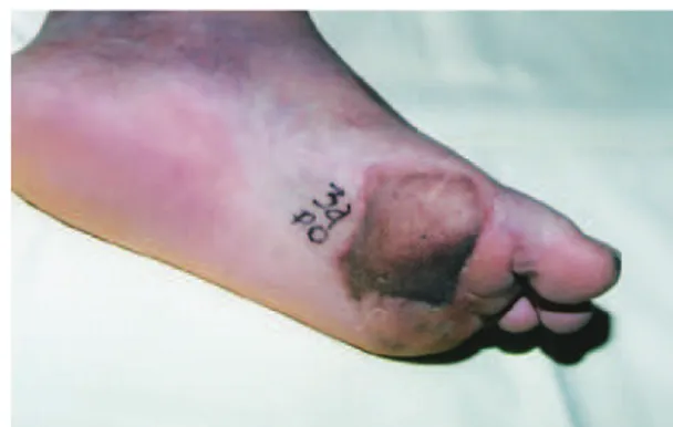
For the repair of hands and feet, the quality of the cover should meet the functional requirements. In the repair of hand defects, the aim is to create a pincer that, when the pa -tient attempts to grip an object, provides a good opening of the interdigital space and preserves the length and sensitivi ty of the digital pulp. This enables the patient to regain thumb opposition and grasp objects. Regarding the foot, especially the sole, the preservation of stability and sensitivity are the main goals when the repair involves areas of plantar support. Lateral arm and forearm fasciocutaneous laps are preferred for the repair of palms and soles because they are thin, and when transferred to support areas, they increase stability and provide satisfactory aesthetic results without causing ulcers or persistent edema. Figure 1 shows the appearance of the plantar cover of an electrical burn area 3 years after surgery. In this case, 2 surgical procedures were performed to decrease the subcutaneous cellular tissue.
Head and Neck Covers
The major challenges in the repair of head and neck burns are to obtain sufficient cover of scalp and skull tissue losses and to repair severe cervicothoracic adhesions while preserving the aesthetic and functional properties of the face6. Microsurgical flaps have been used to replace areas showing retractions, which may present with adhesions to deep planes (late), or those that require cover of noble struc tures (early).
In the surgical treatment of cervicothoracic adhesions, which lead to signiicant limitations related to neck extension at varying degrees, the use of skin grafts or local laps to replace the scar tissue has been questioned after consi deration of the short- and long-term aesthetic and functional results. This procedure thus requires improvements, and new strate-gies aimed at increasing cervical extension in the presence
of late scar retraction are essential. The ideal lap should be thin and long enough to cover a large region and prevent the development of late retractions.
To repair these large areas, the use of thin and/or pre -viously expanded skin laps, which can be associated with bone tissue in jaw reconstructions, is necessary. The most frequently used laps are the scapular, rectus abdominis, and osteomyocutaneous laps with inclusion of the ibula.
The so-called “pre-molded” laps, which are expanded in the donor area7, might undergo secondary retractions when transferred to the receiving region, requiring additional sur -geries. However, in cases involving the cervical contour, for which a more elastic tissue is needed, the microsurgical lap has provided excellent functional results, reducing the time requirement for the use of orthoses and compressive meshes, and allowing earlier physical therapy intervention.
Upper Limb and Hand Covers
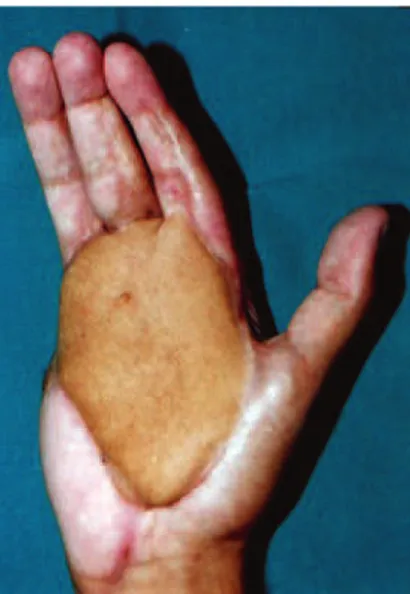
The repair of upper limb injuries, mainly those invol-ving the middle and distal thirds of the posterior or anterior forearm; those showing exposure of vessels, nerves, or ten dons; and defects involving the palm and dorsum of the hand, require immediate cover. In keeping with this notion and considering the preservation of the lexo-extension function of the forearm, a muscular lap of the latissimus dorsi or gracilis muscles should be used. For the repair of the dorsum and palm of the hand, a lateral arm lap is preferred. Figure 2 shows a hot oil burn in the palmar region that was covered with a lateral arm lap anastomosed to the radial artery and vein.
Although electrical burns constitute only a small propor -tion of thermal-related injuries, most electric burns affect the lexo-extensor apparatus as well as the median and ulnar nerves. Whenever possible, in late reconstructions, deep structures should be repaired in a single surgical procedure. In primary repairs, priority should be given to the skin to enable the subsequent reconstruction of deep structures such as muscles and tendons. Following the complication period Figure 1 – An electric current burn in the plantar region covered
after the transplant, the functional reconstruction of the gra -cilis muscle (muscle-tendon) should be planned, followed by the reconstruction of the lexor or extensor apparatus of the forearm.
The use of several proximal flaps in different surgical pro cedures can result in a delay of the functional rehabi li tation of the affected area. Extensive areas could be co -ve red in a single surgical procedure using microsurgical flaps, which allows an early functional rehabilitation of the limb.
The choice of microsurgical lap depends on the expe -rience of the speciic surgical service and on the complexity of each case.
The requirement of bridge grafts for arterial or venous anastomoses is due to the presence of vessels near the injury site that are not patent or reliable for anastomosis. Impor -tantly, the preservation of the function of the hand or upper limb is the irst priority in the repair of injuries involving these regions, and this consideration should determine the choice of treatment.
Figure 3 shows a patient with an electric burn that evolved to transmetacarpal amputation that was covered with lateral arm lap.
Lower Limb Covers
Functional sequelae resulting from burns in the lower limb are less common. However, retractions in the popliteal fossa, dorsal region of the foot, toes, and ankle cause severe functional deiciencies in the lower limb, which limit ambu -lation and musculoskeletal development. In these regions, conventional procedures such as zetaplasty, grafts, and local laps are effective for the repair of most retractions.
The challenges in the lower limb are associated with plantar defects and injuries causing joint movement limitations, which may or may not be repaired using conventional me -thods. For the repair of ankle burns, the use of the muscular lap of the gracilis is preferred, whereas in cases of major losses of the dorsum of the foot and lower-middle third of the leg, the latissimus dorsi lap is the irst choice, especially in the presence of bone exposure or losses. Figure 4 shows the covering of an area with extensive tissue loss in the dorsum of the foot and ankle and bone exposure.
The use of a lateral arm flap to cover the plantar region provided stability and satisfactory aesthetic and functional re sults, without the appearance of a plantar ulcer. However, the presence of a significant scar in the donor area was ob served.
CONCLUSIONS
Microsurgical laps are indicated for complex recons -tructions in burn patients, especially when the lesions in- vol ve noble structures and joints. Clearly, this is an excep-tional pro cedure adopted only when simpler techniques,
Figure 4 – Dorsum of the foot and ankle covered with a latissimus dorsi lap.
Figure 2 – Palmar region covered with a lateral arm lap presenting suficient digital extension.
Figure 3 – Transmetacarpal amputation covered with
such as local laps, cannot be employed due to the morbidi ty asso ciated with surgery. However, if indicated properly, microsurgical laps provide excellent results when used for the reconstruction of noble structures, with preservation of functional and aesthetic properties.
REFERENCES
1. Mathes SJ, Nahai F. Reconstructive surgery: principles, anatomy, and technique. London: Churchill Livingstone; 1997. p. 617-42.
2. Horibe EK, Pinto WS, Yamaguchi CT. Uso de expansores de tecidos
no tratamento de sequelas de queimaduras. Rev Bras Cir. 1988;78(2): 117-24.
3. Milcheski DA, Busnardo F, Ferreira MC. Reconstrução microcirúrgica em queimaduras. Rev Bras Queimaduras. 2010;9(3):100-4.
4. Hunt JL, McManus WF, Haney WP, Pruitt BA Jr. Vascular lesions in acute electric injuries. J Trauma. 1974;14(6):461-73.
5. Tuma Junior P, Faria JCM, Fontana C, Goldenberg DC, Ferreira MC. Queimaduras elétricas dos membros superiores. Rev Hosp Clin Fac Med Univ São Paulo. 1995;50 Supl:13-6.
6. Pitanguy I, Lima PV, Muller P, Persichetti P. Considerações sobre queimadura elétrica do lábio. Rev Bras Cir. 1986;76(4):231-42. 7. Morais-Besteiro J, Aki FE, Mendes JA. Retalhos livres pré-moldados.
Rev Soc Bras Cir Plást. 1992;7(1):82-9.
Correspondence to: Bruno Barros de Azevedo Coutinho