Article
J. Braz. Chem. Soc., Vol. 26, No. 2, 273-281, 2015. Printed in Brazil - ©2015 Sociedade Brasileira de Química 0103 - 5053 $6.00+0.00
A
*e-mail: [email protected]
Preparation, Characterization, Cytotoxicity and Antioxidant Activity of DOPA
Melanin Modified by Amino Acids: Melanin-Like Oligomeric Aggregates
Thiago G. Costa,a Mateus J. Feldhaus,a Felipe S. Vilhena,a Melina Heller,b
Gustavo A. Micke,b Aldo S. Oliveira,c Inês M. C. Brighente,c Fabiola B. F. Monteiro,d
Tânia B. Creczynski-Pasad and Bruno Szpoganicz*,a
aLaboratório de Equilíbrio Químico and bLaboratório de Eletroforese Capilar, Departamento de Química, Universidade Federal de Santa Catarina (UFSC), CP 476, 88040-900 Florianópolis-SC, Brazil
cDepartamento de Química, Universidade Federal de Santa Catarina (UFSC), Campus Universitário, Bairro Trindade, CP 5069, 88040-970 Florianópolis-SC, Brazil
dDepartamento de Ciências Farmacêuticas, Universidade Federal de Santa Catarina (UFSC), CP 476, 88040-900 Florianópolis-SC, Brazil
Two new synthetic melanin-like oligomers inspired by the synthesis of pheomelanin (cys-DOPA) were prepared using L-DOPA as a precursor and the amino acids serine (ser-DOPA) and threonine (thr-DOPA) for modifications. The products obtained exhibited appreciable antioxidant activity. The compounds were characterized by liquid chromatography-mass spectrometry (LC-MS), elemental analysis and infrared spectroscopy. The pKa values of the groups present in each melanin suspension were determined in aqueous solution by potentiometric and spectrophotometric titration. Cytotoxicity assays showed an IC50 > 500 mg L
-1 resulting in non-toxic compounds for living cells. Finally, cys-DOPA, ser-DOPA and thr-DOPA exhibited antioxidant activity determined by the DPPH scavenging assay, with EC50 = 7.69, 1.02 and 27.60 mg L
-1, respectively.
Keywords: synthetic melanins, amino acid modification, antioxidant activity
Introduction
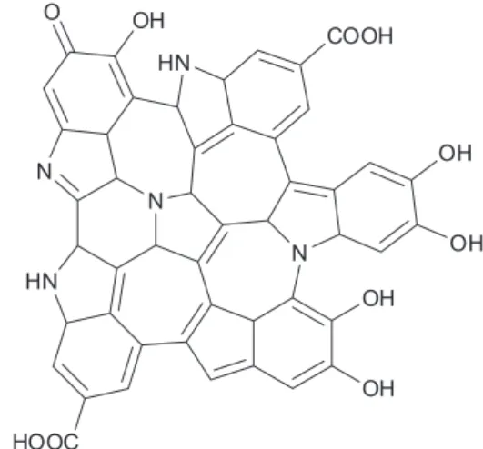
Melanin is a natural pigment produced through the oligomerization of 5,6-dihydroxyindole (DHI) and 5,6-dihydroxyindole-2-carboxylic acid (DHICA) catalyzed by tyrosinase. It contains between 3 to 9 monomer units.1
It can be found in different species in nature including fungus,2 humans,3 vegetables4 and marine life.5 These
organisms produce melanin for protective application in biological systems. In the literature, two major types of melanin pigments are described: eumelanin and
pheomelanin, which exhibit dark and brown colors,
respectively.6 Figure 1 shows the most accepted melanin
chemical structure described in the literature.7
Besides their biological functions, melanins have a wide range of applications, including technological, dermatological, medicinal and pharmacological, and they are also being used in food products.8-10 Recently, Ye et al.11 described a method for fungal melanin extraction and
modification with amino acids, and the sample obtained showed antioxidant activity. Other authors have described the antioxidant efficiency of other types of melanin11,12 and
other oligomeric compounds with phenolic groups.13,14 The
antioxidant activity can be attributed to the presence of catechol groups in the structure,7,9 as shown in Figure 1.
OH
HN
COOH
N
OH
OH N
N
HN
HOOC
OH
OH O
solubility, biological compatibility, providing availability for important applications in bioinorganic materials, medicinal products and food chemistry. The determination of antioxidant activity is an extremely necessary and useful tool in the initial selection of substances that can be used as drugs. Due to the formation of different reactive oxygen species (ROS) and their various mechanisms of action in living organisms, there is no simple and universal method available to measure the antioxidant activity accurately and quantitatively.
The aim of this research was to prepare, purify and characterize 3,4-dihydroxyphenylalanine DOPA-melanin derivatives chemically modified through oxidation with cysteine (synthetic pheomelanin), serine and threonine. In addition, their antioxidant activity was evaluated using two methods, free radical scavenging and reducing power assays. To assess the biological compatibility of these new antioxidants the cytotoxicity was evaluated in cell lines. This study is an advance in the field of bio-inspired synthetic oligomers, and the results obtained may contribute to the development of new dermatological products, which can be applied to skin protection.
Experimental
Preparation of DOPA melanin modified with cysteine, serine and threonine
The modified DOPA melanins were prepared based on the formation of pheomelanin described by Deibel and Chedekel15 as detailed below. Firstly, 280 mg (1.44 mmol)
of L-DOPA (Sigma-Aldrich) were homogenized in phosphate buffer pH 7.4 at 37 oC and to this solution 1 mg
of tyrosinase originating from mushrooms as a lyophilized powder (Sigma-Aldrich) was added; when the solution became pink, 2.88 mmol of L-cysteine (cys, 349 mg), L-serine (ser, 302 mg) and L-threonine (thr, 343 mg) (Sigma-Aldrich) were added to each reaction mixture. The reaction was carried out under vigorous stirring for 48 h, and then 0.1 mol L-1 HCl (Vetec) was added until
pH 2. The final solution was centrifuged at 4000 rpm at 0 oC and the solid was collected and washed with
1:1 ethanol:water several times to remove any precursor contamination (L-DOPA dimers and amino acids). Lastly, the solid was placed in 1:1 ethanol:water mixture and heated to 40 oC, and centrifuged again for purification
of the cys-DOPA, ser-DOPA and thr-DOPA melanin. In each centrifugation process, one drop of AgNO3 was
added to the supernatant for chloride testing and when the supernatant became clear, the purification process
and characterized. Scheme 1 summarizes the process and shows the proposed structures.
Characterization
Elemental analysis
Measurements to determine the percentage of carbon, hydrogen, nitrogen and sulfur were carried out in a Carlo Erba CHNS elemental analyzer E-1110.
Spectrophotometric titration
The titration was carried out by monitoring the changes in the electronic spectrum of each modified melanin sample in the region between 200 and 400 nm with an increase in pH until 14.5. The experiment was carried out in a Varian CARY 50BIO spectrophotometer.
All experiments were performed in aqueous solution, under inert atmosphere and at 25 oC using one thermostatic
cell. For each experiment, a solution containing 3 mg L-1
of melanin was used. The solutions were titrated with 4 mol L-1 KOH. The aliquots of the base were calculated
for each pH value.
Potentiometric titration
The potentiometric studies were carried out in 30 mL of an aqueous solution containing 50 mg of each melanin and titrated using a Methrom TITRINO PLUS 350 automatic burette with a combined Ag/AgCl electrode. The experimental samples were analyzed in a closed 50 mL thermostatic cell which was maintained at 25 oC, and argon
was bubbled into the cell to ensure an inert atmosphere. The pH of the experimental solution was adjusted to 2.5 with 0.100 mol L-1 HCl and titrated with 0.100 mol L-1 KOH.
The time to reach the equilibrium in each experimental procedure was around 20 min. The pKa values were calculated using the Best7 program.16
Infrared (IR) studies
The analysis was carried out in KBr pellets with approximately 5-10 mg of the melanin sample in a Perkin-Elmer FT-IR 1600 spectrometer with a computerized detection system, in the region of 500 to 4000 cm-1.
Scanning electron microscopy (SEM)
Liquid chromatography-mass spectrometry (LC-MS)
The liquid chromatography instrument (Agilent Technologies 1200) was coupled to a mass spectrometer with an electrospray ionization source (Applied Biosystems/ MDS Sciex QTRAP 3200) and the analysis were carried out in a ZIC-HILIC column (150 mm × 2.1 mm i.d. × 3.5 µm
particle size) (Merck) with the temperature set at 30 °C. The mobile phase consisted of 0.1% formic acid and methanol, and the linear gradient elution method was applied. The flow rate of mobile phase was 100 mL min-1. In each
analysis, the injected volume was 2.0 µL and total analysis time was 35 min, with preconditioning of 10 min. The mass analysis was carried out in a positive ionization mode with the following source parameters: temperature at 350 °C; ion spray voltage of 5500 V, curtain gas at 10 psi. The Analyst software (version 1.5.1, Applied Biosystems) was used for the LC-MS/MS system control and data analysis.
Cytotoxicity assays
NIH/3T3 cells were seeded into 96-well plates at a density of 1 × 10⁴per well (100 µL) and treated with the
following: vehicle control (dimethylsulfoxide, DMSO),
cys-DOPA, ser-DOPA, and thr-DOPA at concentrations of 10 to 500 mg L-1. The cells were treated for 24 h. After
incubation, the cells were washed with fresh culture medium and 5 mg mL-1 of
3-(4,5-dimethylthiazol-2-yl)-2,5-diphenyltetrazolium bromide (MTT) were added, followed by incubation for 2 h at 37 °C. The precipitated produced was dissolved in 100 µL of DMSO and the absorbance was measured at 540 nm using a micro-well system reader (Biotek microplate reader elx800), n = 3.
Antioxidant activity
DPPH scavenging assay
The test for the determination of antioxidant activity using the free radical 2,2-diphenyl-1-picryl-hydrazyl (DPPH) assay was carried out with an ethanolic solution containing 0.004% of DPPH (freshly prepared), and solutions with different concentrations of the samples.17 An
aliquot of the DPPH solution was mixed with an aliquot of the test solution (2:1). The mixture was stirred and after 30 min the reading was performed on a spectrophotometer at 527 nm, correcting the absorbance for each sample
HO
HO
OH O
NH2
L-Dopa
+
HS OH
O
NH2 cys
HO OH
NH2 O
ser
HO OH
CH3
NH2 O
thr
1) phosphate buf f er 0.1 mol L-1pH 7.4 2) tyrosinase / O2
3) vigorous stirring 37 °C - 48 h 4) centrif ugation 4000 rpm
5) purif ication
cys-DOPA
ser-DOPA
thr-DOPA
N
COOH HO
N
S HO
HO
cys-DOPA
N
COOH HO
N
O HO
HO
ser-DOPA N
COOH HO
N
O HO
HO
thr-DOPA CH3
the DPPH solution in the absence of test solution. The percentage of DPPH bleaching was determined for the various concentrations of test solution. The graph of the percentage decrease in absorbance of DPPH as a function of the sample concentration provides the 50% of effective concentration (EC50) value, which indicates the
concentration of the compound required to obtain 50% of antioxidant activity.
Reducing power
The assay for evaluating antioxidant activity by determining the reducing potential consisted of adding the sample (at a concentration of 1000 mg L-1) to 8.5 mL
of distilled water and 1.0 mL of a 0.1 mol L-1 solution of
FeCl3 in a 0.1 mol L
-1 HCl solution. After 3 min, 1.0 mL
of 0.008 mol L-1 potassium ferricyanide was added. After
15 min, the absorbance of each solution was read on a spectrophotometer at 720 nm.18-20 As the background, a
solution prepared according to the above procedure, in the absence of the sample, was used. The appearance of Prussian blue staining is indicative of the reducer potential. The calibration curve was obtained with standard solutions of ascorbic acid (AA) at concentrations of 100-1000 mg L-1
(in triplicate). The results were obtained in equivalents of ascorbic acid (AA), i.e., mg of ascorbic acid per g of sample.
Fe2+ chelating activity
Solutions of the compounds (1 mL) in different concentrations were evenly mixed with 0.05 mL FeCl2
(2 mmol L-1), and 0.2 mL ferrozine solution (5 mmol L-1)
were added. The mixtures were shaken and left standing at room temperature for 20 min, the absorbance values
(Asample) of the mixtures were measured at 562 nm.
Methanol was used instead of the sample solution as blank control (Ablank) and Na2EDTA was used as positive control.
Fe2+ chelating rate (%) × 100 × [(A
blank – Asample) / Ablank]).21
Total antioxidant capacity
Total antioxidant capacities were evaluated by the phosphomolybdenum method. To 0.1 mL of varying concentration of compounds, 1 mL of the reagent solution (0.6 mol L-1) sulfuric acid, 28 mmol L-1 sodium phosphate
and 4 mmol L-1 ammonium molybdate were added and
incubated at 95 °C for 90 min. After cooling to room temperature, the absorbance was measured at 695 nm. The antioxidant activity was expressed as the number of equivalents of ascorbic acid.22
The statistical analysis of all triplicate assay results was performed using STATISTICA software. The averages, standard deviations and linear regression (R2) were
determined. The differences between the samples were established through analysis of variance (ANOVA). The level of significance was determined at p < 0.05 for all experiments.
Results and Discussion
Characterization of DOPA melanin modification by amino acids
Residual amino acid analysis
Aiming to ensure the purity of the modified synthetic melanins, the samples were analyzed to determine the residual amino acids. The fragmentation pairs were monitored in multiple reaction monitoring (MRM) mode and other parameters relating to cone voltage and collision energy are listed in Table S1.
In all samples the amount of free amino acids in the solution were below the limit of detection, evidencing the high purity of the three synthesized melanin samples in relation to possible residues of reactive precursors and the effectiveness of the purification method used to obtain products with high purity. Interference from the reaction precursors can cause errors in the characterization techniques applied in this study, generating false positive results for cytotoxicity and antioxidant assays.
CHNS analysis, electron microscopy and IR spectroscopy
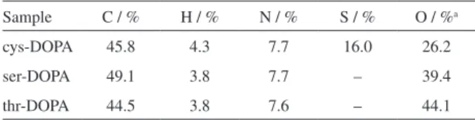
According to the elemental analysis shown in Table 1, the presence of sulfur in cys-DOPA (originating from cysteine) and the absence of this element in the other two synthesized melanin samples can be observed. The structure of the cys-DOPA obtained coincides with that described for this oligomer by Ito and Wakamatsu,23 wherein the sulfur
atom can be attached to the 1,4-benzothiazine intermediates of the pheomelanin precursors. Conversely substituting the heteroatoms for oxygen can characterize the other
Table 1. Carbon, hydrogen, nitrogen and sulfur contents of the three synthetic melanin samples
Sample C / % H / % N / % S / % O / %a
cys-DOPA 45.8 4.3 7.7 16.0 26.2
ser-DOPA 49.1 3.8 7.7 – 39.4
thr-DOPA 44.5 3.8 7.6 – 44.1
two melanins, ser-DOPA and thr-DOPA. An extensive comparison of the elemental composition with other natural and synthetic melanins can be seen in Table S2.
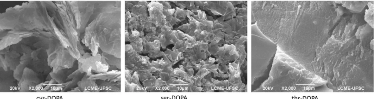
The morphology of the melanins after oligomerization is another important parameter in the characterization, since the melanin samples can then be compared with those extracted from different sources. The SEM images of the three melanins synthesized in this study are shown in Figure 2. Simon and co-workers24 performed the first electron microscopy
studies on samples of skin melanin and others extracted from marine animals. Comparing their results with those for the melanin samples synthesized from L-DOPA reported herein, a notable difference was observed, the morphologies of the natural melanins exhibiting the form of pellets or spheres. In our study, the three melanin samples presented amorphous form without self-organization, similarly to synthetic melanins derived from 3,4-diacetoxyindol recently described in the literature.25 To the best of our knowledge, a
synthetic approach to synthetic melanins that mimic natural morphology has not yet been reported.
An alternative characterization technique for melanins, used by some authors,26-28 is infrared spectroscopy, and
the respective spectra for the modified melanins are given in Figure S1. The spectra for the two novel melanins have characteristic bands at 3422 cm-1 attributed to the
–O–H stretching of carboxylic acids, and protonated catechol and quinone-imine groups, along with another characteristic band at 1620 cm-1 related to the –C=O
stretching, attributed to carboxylic groups present in quinone-imine and carboxylic acid. Two bands with weak intensity are also observed nearby at 1490 and 1395 cm-1,
characteristic of aromatic –C=C and imine –C=N present in the heterocyclic rings and also in molecules derived from
o-hydroxyquinoline27 here characterized by quinone-imine
groups. Additionally the spectra display a band of medium intensity at 1070 cm-1, as also observed by Bilińska29 and
attributed to aromatic rings in substituted macromolecular systems, which are also present in humic substances, as well as overlapping –C–O stretching.28
From these results, differences between the infrared spectra of the three melanins can be noted, especially in the carbonyl stretching region where ser-DOPA and thr-DOPA show a band at 1620 cm-1 and the cys-DOPA spectrum
presents two other bands at 1720 and 1503 cm-1, which
can be attributed to carboxylic acid groups and quinone-imine containing adjacent sulfur groups, respectively. The latter have a lower degree of freedom, shift the –C–O stretching to 1000 cm-1. The proposed structure of
cys-DOPA corresponds to previous descriptions in the literature and based on this model, together with the experimental structural data, the structure of the novel modified melanins, ser-DOPA and thr-DOPA, are proposed in Figure 2.
Equilibrium and UV-Vis spectroscopy
Equilibrium studies of the melanins in aqueous solution are fundamental in order to understand properties such as the chelation of metal ions,9,30,31 and structural
characteristics, and most importantly here, to correlate the results with the antioxidant properties. Szpoganicz et al.9
have determined the constants for the acid/base equilibrium of melanins derived from 5,6-diacetoxyindole, and correlated the results with their interactions with divalent metals. The acid/base equilibrium of melanins, especially the phenolic groups responsible for the antioxidant activity, is of great interest in this study, in order to identify the proton-donating capacity, which is directly linked to the reducing power of the samples. The three groups present in eumelanins (black pigments) are: carboxylic, quinone-imine and catechol,9,23 and the catechol group is largely
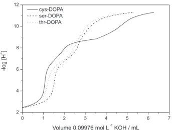
responsible for the antioxidant properties. The titration curves of the three modified melanins are shown in Figure 3.
The corresponding curves for thr-DOPA and ser-DOPA have small differences, although both have three buffered regions. At acidic pH values, there is a buffered region due to deprotonation of the carboxylic groups, at neutral and alkaline pH values there is the acid/base equilibrium of the quinone-imine and catechol groups in basic pH values. The titration curve of cys-DOPA shows the same buffered
regions observed for the thr-DOPA and ser-DOPA titration curves, but the presence of an additional buffer region was also noted, which can be assigned to deprotonation of the –SH group, referred to as thiol in this paper, which might be present in the structure. These groups are important because they can also act as antioxidants due to the formation of R–S–S–R disulfide bridges. This is the first report demonstrating that pheomelanins have another ionized group, however, it needs additional structural characterization studies to clearly elucidate which is the mechanism that leads to the free-thiol formation. The pKa values for each equilibrium observed for each type
of melanin in this study were calculated using the Best7 program and the results are presented in Table 2.
The pKa values for the deprotonation of the second ionized hydrogen atom present in the catechol group are very important as they can be correlated with the antioxidant activity, and they were determined by spectrophotometric titration. The melanins modified with cysteine, serine and threonine had λ
max values of 235, 283 and 290 nm,
respectively. These wavelength values were detected based
addition of base. The UV-Vis spectra, absorbance vs. pH curves and their derivatives are shown in Figure S2.
The absorbance values for ser-DOPA and thr-DOPA were very similar, but cys-DOPA had a λ
max value of
235 nm and this finding may be associated with a more energetic process than the resonance occurring in the other two melanin samples. This melanin has sulfur groups in the thiol form, which might be free or cyclized as benzothiazinones.6 This can be explained by the presence
of a greater number of electrons in sulfur compared with the oxygen present in the two other amino acids and this could also be the reason for the hypsochromic shift compared with the other two melanins. Additionally, the increase in absorbance is proportional to the pH increase, which may be associated with the fact that the susceptibility toward the formation of a catecholate group, which exhibits resonance effect when deprotonated, as was previously observed by Szpoganicz et al.9 in synthetic
DHI melanins, derived from diacetoxy-indol. Scheme 2 shows the equilibria associated with the three chemically modified melanins.
Cytotoxicity
For the use of the modified melanins as antioxidant agents in biological applications, dermatological systems and medicinal in general, the evaluation of possible cytotoxic effects in healthy cells in vitro is necessary to evaluate possible toxic effects. Urabe et al.31 reported
experiments of this type using several precursors, catalysts and other species formed during melanogenesis. They observed higher cytotoxicity for melanins derived from diacetoxyindol (DAI) than for those derived from L-DOPA. Moreover, a high level of cell death was observed in the presence of products derived from both synthetic melanins and dihydroxyindol (DHI) and dihydroxyindol-2-carboxylate (DHICA). However, the L-DOPA and the tyrosinase free samples showed no cytotoxic effects.
Another important aspect that must be taken into consideration is the formation of H2O2 during melanin
polymerization and a small amount after exposure to light and other chemical agents.32 Thus, the cys-DOPA,
ser-DOPA and thr-DOPA melanin samples synthesized in this study were submitted to cytotoxicity assays using murine NIH/3T3 fibroblasts. The results of the cytotoxicity assay are shown in Figure 4.
It can be observed that synthetic melanins modified with the three amino acids showed no cytotoxicity even at high concentrations, and with an IC50 > 500 mg L
-1. The low
cytotoxicity shows that even synthetic melanins modified
0 1 2 3 4 5 6 7
2 4 6 8 10
-l
o
g
[H
+ ]
Volume 0.09976 mol L-1KOH / mL
ser-DOPA thr-DOPA
Figure 3. Titration curves of each modified melanin under inert atmosphere. T = 25 °C, µ = 0.1 mol L-1 KCl, combined glass electrode.
Table 2. Log of dissociation constants of the major groups present in each melanin
Equilibriuma cys-DOPAb ser-DOPAb thr-DOPAb
[Ac–][H+] / [HAc] 3.33 3.36 4.25
[QI–][H+] / [HQI] 6.83 6.96 6.94
[R–S–][H+] / [R–SH] 8.29 – –
[HCat–][H+] / [H
2Cat] 10.99 10.85 11.06
[Cat2–][H+] / [HCat–] 14.28c 14.07c 14.45c
aAc: acetate, QI: quinone-imine, Cat: catechol groups; bvalues obtained by
the average of three titrations; cpKa values obtained by spectrophotometric
with amino acids have good cell compatibility and can be used as potential natural antioxidants.
Antioxidant activity analysis and correlations
For the three modified melanins synthesized in this study, the same groups were observed: carboxylic acid, quinone-imine and catechol. There was, however, a difference in terms of the acidity of the protons of the hydroxyl of the catechol groups that can be seen by their pKa values (Table 2).
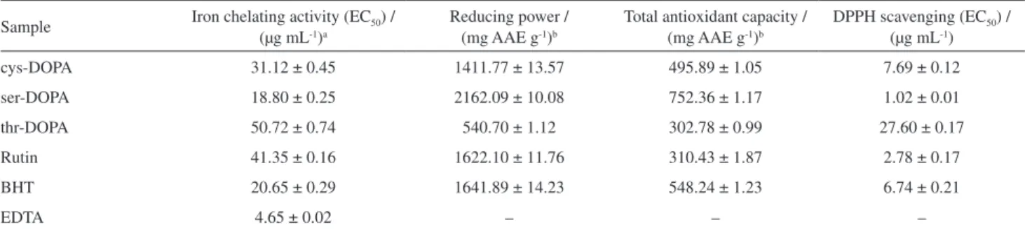
There was a linear correlation between the EC50 values
obtained in the DPPH scavenging assay and the acidity of the compounds for both the first and second deprotonations. This demonstrates that the acidity of the phenolic compounds is also correlated with the antioxidant activity. All melanin samples showed EC50 values (Figure 5) close to those of
the standards used (rutin and butylatedhydroxytoluene (BHT), notably ser-DOPA, which showed an EC50 value
of 1.02 µg mL-1, that is half that of the most active standard
rutin tested using this method (Table 3). Likewise, when the compounds were analyzed using the method of reducing power it was observed that ser-DOPA showed superior activity and the other two melanins presented antioxidant activity comparable to the standards used in the same order of activity observed for the DPPH assays.
It is noteworthy that when reporting the values for the antioxidant activity obtained by the two methods, that is, the reducing power and DPPH scavenging assays, a correlation of 97.2% was observed, which demonstrates the effectiveness of the methods used to determine the antioxidant activity of these compounds (Table 3).
Total antioxidant capacities were evaluated by the phosphomolybdenum method. This assay is based on the reduction of Mo(VI) to Mo(V) by the antioxidant compounds and the subsequent formation of a green phosphate/Mo(V) complex at acidic pH with a maximal absorption at 695 nm.20 The obtained results, expressed
1.0 1.5 2.0 2.5 3.0 0 20 40 60 80 100
cysDOPA serDOP A thrDOP A C e ll v ia b ili ty / %
logC / ppm
Figure 4. Cell viability in the presence of each melanin.
N H C O OH R
pKa 1 pKa 2
pKa 3* pKa 4 HO pKa 5 HO Protonated species
R = Oligomeric chain
N O HO HS N H C O O -R HO HO N O HO HS N H C O O -R HO HO N O -O HS N H C O O -R HO HO N O -O -S N H C O O -R HO -O N O -O -S N H C O O -R -O -O N O -O -S HAc Ac -HQI QI --S -
HS-H2Cat
HCat -HCat
-Cat
Table 3. Antioxidant activity of modified melanins and standards
Sample Iron chelating activity (EC50) /
(µg mL-1)a
Reducing power / (mg AAE g-1)b
Total antioxidant capacity / (mg AAE g-1)b
DPPH scavenging (EC50) /
(µg mL-1)
cys-DOPA 31.12 ± 0.45 1411.77 ± 13.57 495.89 ± 1.05 7.69 ± 0.12
ser-DOPA 18.80 ± 0.25 2162.09 ± 10.08 752.36 ± 1.17 1.02 ± 0.01
thr-DOPA 50.72 ± 0.74 540.70 ± 1.12 302.78 ± 0.99 27.60 ± 0.17
Rutin 41.35 ± 0.16 1622.10 ± 11.76 310.43 ± 1.87 2.78 ± 0.17
BHT 20.65 ± 0.29 1641.89 ± 14.23 548.24 ± 1.23 6.74 ± 0.21
EDTA 4.65 ± 0.02 – – –
aEC
50 value of iron(II) ions that were chelated by 50%; bresults in mg of ascorbic acid equivalents per g of compound. Calculated for sample concentrations
of 100 µg mL-1. Each value is expressed as mean ± standard deviation.
Figure 5. Antioxidant activities: (a) DPPH scavenging assay; (b) chelating rate of Fe2+ of cys-DOPA, ser-DOPA and thr-DOPA.
as ascorbic acid equivalents (AAE), are presented in Table 3. All investigated samples were active in a concentration-dependent manner and their potencies were high. Among the tested compounds, ser-DOPA showed the highest total antioxidant capacity with a value of 752.36 ± 1.17 mg AAE per gram of compound. Total antioxidant capacities are also well correlated with other activities, 76 and 92% with reducing power and DPPH, respectively, which demonstrates the effectiveness of the analysis to investigate the antioxidant activity of compounds.
The antioxidant activities of compounds are also attributed to their ability to chelate transition metal ions, such as those of iron and copper, which have been proposed as catalysts for the initial formation of reactive oxygen species. Chelating agents may stabilize pro-oxidative metal ions in living systems by complexating them.33 Iron(II)
ion is known as a potent inducer of lipid peroxidation. Ferrozine can quantitatively form complexes with Fe2+. In
the presence of chelating agents, the complex formation is disrupted resulting in a decrease in the red color of the complex. Measurement of the reduction in color intensity at the 562 nm wavelength allows estimation of the metal chelating activity of the coexisting chelator.34
All compounds demonstrated an ability to chelate Fe2+. Among the compounds analyzed ser-DOPA proved
to be the most potent chelator, more active than one of the standard used and only 4 times less potent than the most active of standards, EDTA. The values found for the standards are in agreement with literature data.33 As before,
the percentage of chelated Fe2+ is directly correlated with
the other activities with excellent correlation percentages: 84, 91 and 72% for reduction power, total antioxidant capacity and DPPH scavenging, respectively.
Conclusions
In this study two new types of melanin chemically modified with amino acids were synthesized. The molecules were inspired by the synthesis of pheomelanin
The pKa values of the groups present in all synthesized
melanins were determined in solution. The results were found to correlate with the antioxidant activity of each melanin sample. ser-DOPA showed better antioxidant activity and lower pKa values for the catechol group. None
of the melanins studied showed significant cytotoxicity and all had high antioxidant activity, indicating that these melanins have attractive characteristics for potential medicinal applications.
Supplementary Information
Supplementary data are available free of charge at http://jbcs.sbq.org.br as PDF file.
Acknowledgments
The authors thank the Central Laboratory of Electron Microscopy at the UFSC and CNPq (Brazil) for scholarship.
References
1. Raper, H. S.; J. Biochem.1926, 20, 89.
2. Gómez-Marín, A. M.; Sánchez, C. I.; J. Non-Cryst. Solids2010,
356, 1576.
3. Novelino, L.; Napolitano, A.; Prota, G.; Biochim. Biophys. Acta
2000, 1475, 295.
4. de Angelis, F.; Arcadi, A.; Marinelli, F.; Paci, M.; Botti, D.; Pacioni, G.; Miranda, M.; Phytochemistry1996, 43, 1103. 5. Boyle, P.; Rodhouse, P.; Cephalopods: Ecology and Fisheries;
Blackwell Science: Oxford, 2005, pp. 32.
6. Simon, J. D.; Peles, D. N.; Acc. Chem. Res.2010, 43, 1452. 7. Gidanian, S.; Farmer, P. J.; J. Inorg. Biochem.2002, 89, 54. 8. Zhou, J.; Shang, J.; Ping, F.; Zhao, G.; J. Ethnopharmacol.
2012, 143, 639.
9. Szpoganicz, B.; Gidanian, S.; Kong, P.; Farmer, P.; J. Inorg. Biochem.2002, 89, 45.
10. Takahashi, A.; Tsuchiya, K.; Yamanome, T.; Amano, M.; Yasuda, A.; Yamamori, K.; Kawauchi, H.; Peptides2004, 25, 1613.
11. Ye, M.; Wang, Y.; Guo, G.; He, Y.; Lu, Y.; Ye, Y.; Yang, Q.; Yang, P.; Food Chem.2012, 135, 2490.
12. Hung, Y. C.; Savab, V. M.; Makan, S. Y.; Chen, T. H. J.; Hong, M. Y.; Huang, G. S.; Food Chem.2002, 78, 233.
13. Abdel-Hamid, R.; Newair, E. F.; J. Electroanal. Chem.2011,
657, 107.
14. Giacomelli, C.; Ckless, K.; Galato, D.; Miranda, F. S.; Spinelli, A.; J. Braz. Chem. Soc.2002, 13, 332.
15. Deibel, R. B.; Chedekel, M. R.; J. Am. Chem. Soc. 1982, 104, 7306.
16. Motekaitis, R. J.; Martell, A. E.; Can. J. Chem.1982, 60, 2403. 17. Brighente, I. M. C.; Dias, M.; Verdi, L. G.; Pizzolatti, M. G.;
Pharm. Biol.2007, 45, 156.
18. Moresco, H. H.; Queiroz, G. S.; Pizzolatti, M. G.; Brighente, I. M. C.; Rev. Bras. Farmacogn. 2011, 22, 319.
19. Dos Santos, M. H.; Batista, B. L.; Quim. Nova2007, 30, 604. 20. Waterman, P. G.; Analysis of Phenolic Plant Metabolites;
Blackwell Scientific: Oxford,1994, pp. 238.
21. Wang, J.; Zhang, Q.; Zhang, Z.; Li, Z.; Int. J. Biol. Macromol.
2008, 42, 127.
22. Prieto, P.; Pineda, M.; Aguilar, M.; Anal. Biochem.1999, 269, 337.
23. Ito, S.; Wakamatsu, K.; Photochem. Photobiol. 2008, 84, 582. 24. Liu, Y.; Kempf, V. R.; Nofsinger, J. B.; Weinert, E. E.;
Rudnicki, M.; Wakamatsu, K.; Ito, S.; Simon, J. D.; Pigm. Cell Res.2003, 16, 355.
25. Costa, T. G.; Younger, R.; Poe, C.; Farmer, P. J.; Szpoganicz, B.; Bioinorg. Chem. Appl.2012, 712840, DOI: 10.1155/2012/712840.
26. Krohn, J.; Xu, C. T.; Svenmarker, P.; Khoptyar, D.; Andersson-Engels, S.; Exp. Eye Res.2010, 90, 330.
27. Bilińska, B.; Spectrochim. Acta, Part A1996, 52, 1157. 28. Pierce, J. A.; Rast, D. M.; Phytochemistry1995, 39, 49. 29. Bilińska, B.; Spectrochim. Acta, Part A2001, 57, 2525. 30. Stainsack, J.; Mangrich, A. S.; Maia, C. M. B. F.; Machado,
V. G.; dos Santos, J. C. P.; Nakagaki, S.; Inorg. Chim. Acta
2003, 356, 243.
31. Urabe, K.; Aroca, P.; Tsukamoto, K.; Biochim. Biophysica Acta
1994, 1221, 272.
32. Beer, R. J. S.; Broadhurst, T.; Robertson, A.; J. Chem. Soc.
1954, 1947.
33. Shahidi, F.; Liyana-Pathirana, C. M.; Wall, D. S.; Food Chem.
2006, 99, 478.
34. Gülçin, İ.; Toxicology2006, 217, 213.