A
RTIGOO
RIGINAL Revista Brasileira de FisioterapiaEvaluation of acute radiation effects on
mandibular movements of patients with head
and neck cancer
Avaliação dos efeitos agudos da radioterapia sobre os movimentos mandibulares
de pacientes com câncer de cabeça e pescoço
Karoline C. Bragante1, Daniela M. Nascimento1, Neiro W. Motta2
Abstract
Objectives: To evaluate radiotherapy effects (RT) on mandibular movements of patients with head and neck cancer (H&NC) and associate them to the variables: functional capacity, radiation field, disease staging, type of feeding, concomitant chemotherapy and total dose of RT. Methods:
Twenty-six patients with H&NC were followed up at the RT service. Physical examination was performed in 3 follow up time points: before the beginning of RT (T0), between 14th and 17th session of RT (T1) and after the last session of RT (T2). The physical examination consisted of the
assessment of the following variables: mouth opening without pain (MO), maximum mouth opening (MMO), right lateral excursion (RLE), left lateral excursion (LLE) and protrusion (PR) of the jaw. The feeding type and the Karnofsky Performance Status Scale (KPS) were evaluated in each follow up time point. Data with regards to the tumor lesion and the RT were collected from the patient’s clinical notes. Results: There was a statistical significant reduction in the values of MO (p=0.006), MMO (p=0.001), LLE (p=0.006) and KPS (p=0.001). There was significant a statistical association among the reduction in KPS and decreased measure of MO (r=0.390, p=0.048) and MMO (r=0.435, p=0.026). The mouth and oropharynx radiation fields when combined showed a significant reduction for both the measure of MO (p=0.005) and for MMO (p=0.004). Patients who used nasoenteric tube feeding (NTF)had greater reduction in the measurement of MMO (p=0.031). The remaining variables showed no statistically significant difference. Conclusion: Patients with H&NC present reduction of the measures of MO and MMO during the RT, especially if they present reduced functional capacity, have radiation in the mouth and oropharynx fields and used NTF.
Keywords: radiotherapy; trismus; head and neck neoplasms; joint range of motion.
Resumo
Objetivos: Avaliar os efeitos da radioterapia (RT) sobre os movimentos mandibulares de pacientes com câncer de cabeça e pescoço (CCeP) e associá-los às variáveis: capacidade funcional, campo de radiação, estadiamento da doença, tipo de alimentação, quimioterapia concomitante e dose total de RT. Métodos: Vinte e seis pacientes com CCeP foram acompanhados em um serviço de RT. O exame físico ocorreu em três momentos: antes do início da RT (M0), entre a 14º e 17º sessão (M1) e após a última sessão de RT (M2) para verificação de variáveis, como: abertura bucal sem dor (AB), abertura bucal máxima (ABm), excursão lateral direita (EXd), excursão lateral esquerda (EXe) e protrusão (PR) da mandíbula. O tipo de alimentação e a Escala de Karnofsky (EK) foram reavaliados em cada momento. Dados a respeito da lesão tumoral e RT foram coletados do prontuário do paciente. Resultados: Houve redução significativa nos valores de AB (p=0,006), ABm (p=0,001), EXe (p=0,006) e EK (p=0,001). Houve associação estatisticamente significativa entre a redução na EK e a diminuição de AB (r=0,390; p=0,048) e de ABm (r=0,435; p=0,026). Os campos de radiação da boca e orofaringe, quando agrupados, apresentaram redução significativa tanto para a medida de AB (p=0,005) quanto para ABm (p=0,004). Os pacientes que utilizaram sonda nasoentérica (SNE) apresentaram maior redução da medida de ABm (p=0,031). As demais variáveis não apresentaram diferença estatisticamente significativa.
Conclusão: Os pacientes com CCeP apresentam redução das medidas de AB e ABm no decorrer da RT, principalmente se apresentarem
redução da capacidade funcional, tiverem irradiação para os campos da boca e orofaringe e fizerem uso de SNE.
Palavras-chave: radioterapia; trismo; neoplasias de cabeça e pescoço; amplitude de movimento articular.
Received: 07/15/2011 – Revised: 10/02/2011 – Accepted: 10/25/2011
1 Physical Therapy Department, Methodist University, Instituto Porto Alegre (IPA), Porto Alegre, RS, Brazil
2 Department of Radiotherapy, Santa Rita Hospital, Irmandade Santa Casa de Misericórdia de Porto Alegre (ISCMPA), Porto Alegre, RS, Brazil
Correspondence to: Karoline Camargo Bragante, Rua Coronel Corte Real, 827/402, CEP 90630-080, Porto Alegre, RS, Brasil, e-mail: [email protected]
1 1
Introduction
Head and neck cancer (H&NC) presents a worldwide esti-mated incidence of 780.000 new cases per year1. In Brazil, the
National Institute of Cancer (INCA) estimated that in 2010 10.330 new cases of cancer involving the head and neck areas among men and 3.790 among women occurred2. he
anatomi-cal sites that are included in this group of neoplasms are the oral cavity, which comprises oral mucosa, lips, gums, hard palate, tongue, mouth loor and retromolar trigone; pharynx, which includes oropharynx (base of tongue and soft palate), nasopharynx and hypopharynx (pyriform sinus, pharyngeal wall and post-cricoid area); nasal cavity and paranasal sinuses; glottis and supraglottic larynx; and salivary glands1,3.
Approxi-mately 40% of H&NC occur in the oral cavity, 25% in the larynx, 15% in the pharynx, 7% in the salivary glands and 13% in other areas3,4. he most frequent histological type is the squamous
cell carcinoma, present in more than 90% of cases1,3,5.
Several factors are involved in the genesis of head and neck cancer, including genetic predisposition1,6; professional activity
such as exposure to textile ibers, leather and nickel7; social
conditions and habits, being the consumption of tobacco and alcohol the most signiicant risk factors1,3,8,9. Variables such as
age over 40 years-old, male gender and white race may be also considered as predisposing factors for its development3.
herapeutic modalities for the treatment of head and neck cancer include surgical, procedures, radiotherapy and chemo-therapy, which may be used separately or in combination8-10.
Radiotherapy (RT) is a modality used in the treatment of malignant tumors which the therapeutic agent is the ionizing radiation, i.e. that one which promotes ionization in the en-vironment where charged, turning it electrically unstable11,12.
By creating unstable atoms whose free electrons join to other adjacent atoms, which also become unstable with increased negative charges, damage to the cellular deoxyribonucleic acid (DNA) occurs, preventing the replication of the neoplastic cell13.
he ionizing radiation treatment, however, is not selective, as it does not have the capacity to diferentiate the normal cells from the malignant ones, which turns it toxic for the body13.
Adverse reactions due to RT depend on the volume and the area that will be irradiated, whether the exposure will be unilateral or bilateral, total dose, dose fractionation, age, pa-tient clinical condition, social habits, such as alcoholism and smoking and the associated treatments12-15. Most of patients
undergoing RT for head and neck receive a total dose from 50 to 70 Gray (Gy) as a curative dose. hese doses are fractionated in a period from ive to seven weeks, once a day, ive days a week, with daily dose of approximately 2 Gy8,11,15.
he side efects of RT established for the treatment of patients with H&NC signiicantly interfere on the quality of
life of these individuals16. Among the efects located in the
head and neck area it may be mentioned: mucositis, derma-titis, dry mouth, hypogeusia, osteoradionecrosis, ibrosis and trismus4,11,13,15. Radiation-induced trismus, a reduced mobility
of the jaw17,18 due to the ibrosis that occurs in the masticatory
muscles when they are in the radiation ield5,17-19 has a negative
impact on the quality of life of the patients5,13, as it causes
alter-ations in the facial appearance18, diiculty in food intake5,10,20, in
the use of dental prostheses10 and in the speech5, compromises
the oral hygiene5,19 and it may lead to depression5.
Some authors9,21 suggest that radiation-induced trismus
may only be established months after the end of radiation therapy and if the patient underwent to high doses, while other authors5,22 state that, during the course of radiation therapy
may occur restriction of the mandibular movements even after low doses of irradiation. Several studies17,21-24 reported the
rela-tionship of trismus with speciic anatomical structures in the radiation ield. In addition, some authors17,25 conirm the direct
inluence of this therapy associated with chemotherapy in the emergence of this complication, while other authors5 disagree.
Due to the contradictory data in the scientiic literature with regards to the appearance of radiation-induced trismus in the course of irradiation therapy and the high rate of patients with H&NC undergoing high doses of RT in extensive radiation ields11, there is a need to better investigate the occurrence of
possible changes in the mandibular mobility in irradiated pa-tients, as well as which associated factors would be involved in the onset of these complications, so that after this investiga-tion, data for prevention programs and/or reduction of such dysfunctions may be provided.
herefore, the objectives of this study were to evaluate the efects of RT on the mandibular movements of patients with H&NC and, secondarily to investigate the association of these efects with the following variables: functional capacity, radia-tion ield, disease staging, type of feeding, use of concomitant chemotherapy and total dose of RT.
Methods
Study design and sample
of Grandi et al.26 for a signiicance level of 5%, a power of 80%
and an efect size of at least 0.6 among the evaluations, and it resulted a minimum number of 22 patients to contemplate the study design.
he assessment of eligibility for participation in the study followed the criteria: 1) inclusion criteria: Male and female over 18 years-old, with diagnosis of H&NC undergoing cura-tive RT alone or in combination with chemotherapy, with one or more masticatory muscles on the radiation ield and who had percentile higher than 40% in the Karnofsky Performance Status Scale (KPS)27. 2) exclusion criteria: individuals who had
performed surgical intervention to remove the tumor in any of the mastication muscles, presenting facial palsy, trigemi-nal neuralgia or herpes zoster, patients in treatment with brachytherapy, patients who were receiving physical therapy, who have not performed any of the stages of the study and who refused to participate in the study.
Data from 32 patients were collected, being ive patients lost to follow-up due to interruption of the radiotherapy as requested by the physician (n=1) and for abandonment of the treatment with irradiation therapy in the above-mentioned institution (n=4). One patient was excluded from the study for not being assessed in the last follow up. his study had a inal sample of 26 patients. he data collection was concluded in July 2010.
Data collection
All patients who started the RT in HSR received informa-tion, in group, about the treatment by the nursing staf. In these groups, patients who met the inclusion criteria of the study were identiied and the informed consent form was presented to them. After signing the consent form the data collection was initiate.
Each patient individually underwent to a brief initial inter-view and variables such as age, family history of cancer, smok-ing and alcoholic habits, as well as how long they have besmok-ing smoking and/or drinking, classiication by KPS scale, which is a scale of functional performance used in the prognosis of can-cer therapy27, and type of feeding28,29 were collected. Data such
as tumor location, histological type of tumor, tumor stage30,
associated chemotherapy, radiation ield and dose per session of RT were collected from the patient’s chart for future correla-tions and characterization of sample.
he data collection from the physical examination aimed to assess the variables mouth opening without pain (MO), maxi-mum mouth opening (MMO), right lateral excursion (RLE), left lateral excursion (LLE) and protrusion (PR) of the jaw followed the instructions and speciications of the clinical examination of the Protocol of Diagnostic Criteria of Temporomandibular
Disorders (RDC/TMD)31. he physical examination was always
carried out by the same examiner, who was familiarized with this protocol, in three diferent time points: before the begin-ning of the RT, time 0 (T0); between 14th and 17th session, time 1
(T1) and immediately after the last session of the RT, time 2 (T2). Variables such as type of feeding and KPS scale were also evaluated at each time points.
All patients were treated with irradiation therapy by pho-tons in parallel pairs or wedge angle with daily dose from 1.8 to 2 Gy until the end of treatment of approximately seven weeks. he total dose of the RT ranged from 50 to 70 Gy.
he present study was approved by the Ethics in Research Committee of the Centro Universitário Metodista do Insti-tuto de Porto Alegre (IPA) (Porto Alegre, RS, Brazil, number 393/2009), and by the Ethics in Research Committee of the ISCMPA, protocol 031/10.
Statistical Analysis
he collected data were stored in the Microsoft Excel 2003. To characterize the sample, the categorical variables of this study were described through absolute and relative frequen-cies; the continuous variables, through means and standard deviations for those that presented a normal distribution, and median and interquartile ranges in case of asymmetric distri-bution variables. he Shapiro-Wilk test was used to verify the normality of data. he comparison between physical examina-tion variables in the three diferent time points was veriied by the ANOVA for repeated measures followed by Bonferroni test or, when appropriate, by its correspondent non-parametric Friedman test followed by Wilcoxon test. he comparison among the physical examination variables and disease stage, type of feeding and radiation ield was obtained by the one-way ANOVA test followed by Tukey test, and the variable chemotherapy was compared with the variables of physical ex-amination by the Student t test. Correlations were performed through Pearson correlation test. he statistical program used was SPSS, version 17.0. he signiicance level was set at α=0.05 for all statistical analyses.
Results
In relation to the variable gender, all 26 (100%) participants of this study were male. he volunteers’ age ranged between 45 and 74 years old, with a mean age of 59.0 (SD=8.8) years. Re-garding the distribution by race, there were 20 (76.9%) Cauca-sians, ive (19.2%) Afrocaucasian and one (3.9%) melanoderm. Regarding social habits, 26 (100%) were smokers, with smoking mean time duration of 37.4 (SD=12.7) years, and 19 (73.1%)
participants reported regular intake of alcohol, with mean time duration of 29.5 (SD=13.0) years. With regards to the family his-tory of cancer, 13 (50%) have reported family hishis-tory of cancer and 13 (50%) were not sure or had no cases of cancer in the family. Patients’ distribution according to the tumor location, tumor histological type and tumor stage is shown in Table 1.
Regarding the comparison of the variables of the physical examinaton in the three diferent time points, there was a sta-tistically signiicant reduction on the values of MO, MMO and LLE. here was also observed a statistically signiicant reduc-tion in relareduc-tion to KPS scale (Table 2).
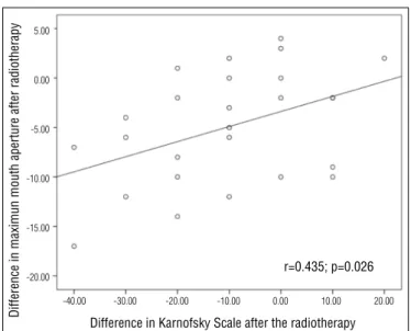
here was a moderate positive statistically signiicant corre-lation among the reduction of the KPS scale and the decreased of MO (r=0.39; p=0.048) and MMO measurements (Figure 1).
he analysis of the radiation ields shows that there was a tendency of the oropharynx and mouth ields present a greater reduction of mouth opening (Table 3). If oropharynx and mouth ields are combined and compared with the other radiation ields, it can be observed a statistically signiicant reduction in both, MO (p=0.005) and MMO (p=0.004) measurements. here were no sta-tistically signiicant diferences (p>0.05) among tumor staging and both MO and MMO measurements.
With regards to the type of feeding in T0 all patients reported oral food intake being the food in the normal consistency; in
T1, seven (26.9%) patients remained with normal feeding; 11 (42.3%) reported eating only food in pasty consistency and eight (30.8%) patients informed feeding exclusively for nasoen-teric tube feeding (NTF). In T2, data about the type of feeding were the same as in T1.
Comparison between the variation of MO and MMO mea-surements according to the type of feeding showed values sig-niicantly lower of MMO measurement in patients who used NTF (Table 4).
Regarding the use of chemotherapy (cisplatin), 13 (50%) participants had used it combined with the RT. When the pa-tients who underwent chemotherapy are compared with those who did not realize this therapy there was no statistically sig-niicant diference (p>0.05) on the variation of MO and MMO measurements.
here was no statistically signiicant association between the total dose of radiation and the reduction of MO (r=-0.111; p=0.591) and MMO (r=-0.164; p=0.423) measurements.
Table 2. Comparison of the variables from the physical examination at
three follow up times points.
The values of MO: Mouth opening; MMO: maximun mouth opening and KPS: Karnofsky Performance Status scale, are expressed in means and standard deviations. The values of RLE: right lateral excursion; LLE: left lateral excursion e PR: protrusion are expressed as medians and interquartile ranges *ANOVA for repeated measurements **Friedman test. a,b letters are not significantly different by the Bonferroni test or Wilcoxon p<0.05.
Figure 1. Pearson’s correlation.
Difference in maximun mouth aperture after radiotherapy
5.00
0.00
-5.00
-10.00
-15.00
-20.00
Difference in Karnofsky Scale after the radiotherapy r=0.435; p=0.026
-40.00 -30.00 -20.00 -10.00 0.00 10.00 20.00
Table 1. Patients distribution by tumor location, tumor histological
type and tumor stage.
Variables n (%)
Larynx 12 (46.2)
Oropharynx 10 (38.9)
Mouth 8 (30.8)
Hypopharynx 4 (15.4)
Nasopharynx 1 (3.8)
Squamous cell carcinoma 25 (96.2)
Undifferentiated Carcinoma 1 (3.8)
Stage I 1 (3.8)
Stage II 4 (15.4)
Stage III 5 (19.2)
Stage IVA 12 (46.2)
Stage IVB 4 (15.4)
Variables T0 T1 T2 p
MO 36.7±10.4b 34.2±11.4a,b 32.2±9.8a 0.006*
MMO 40.9±10.6b 38.7±12.1b 35.9±10.2a 0.001*
RLE 8 (5-10) 8 (4.8-10) 5.5 (3.8-8.5) 0.133**
LLE 9 (6.8-10)b 8 (5.0-10)a 8 (3-10)a 0.006**
PR 5 (2.8-8.3) 4 (2.0-5.0) 4 (2-6.3) 0.154**
KPS 83.1±13.5b 75.8±13.3a 72.7±14.8a 0.001*
Table 3. Mouth opening variation according to the radiation field.
The values of MO: Mouth Opening and MMO: Maximum Mouth Opening are expressed as means and standard deviations. *Anova one-way.
Variables ∆ MO=M0-M2 p* ∆ MMO=M0-M2 p*
Oropharynx -13±4.2 -11.5±7.8
Larynx -5.2±7.4 0.059 -5.3±6.3 0.059
Hypopharynx 0.0±0.0 -2.0 ±0.0
Mouth -10.0±4.7 -11.0±1.7
Drainage areas -2.1±5.2 -2.8±4.5
The values of MO: Mouth Opening e MMO: Maximun Mouth Opening are expressed as means and standard deviations.*Anova one-way. a,b: same letters do not differ by the Tukey test (p<0.05).
Table 4. Mouth opening variation according to the field of feeding.
Variables ∆MO=M0-M2 p* ∆MMO=M0-M2 p*
Normal -0.6±3.8 -0.6±3.6b
Pasty -4.2±5.8 0.057 -5.6±5.0a,b 0.031
Discussion
In relation to gender, age and race, the present results support the results existent in the literature, which showed a higher incidence of H&NC in males3,7-9,12,32, with predominance
of individuals between the 6th and 7th decade of life3,4,8,9,33 and
Caucasian3,12,26.
Regarding the social habits all participants of this study declared themselves smokers and 73.1% and consumed alco-hol, which strengthens the association between the intake of alcohol and tobacco for the development of H&NC3,7,9,34. Half
of sample of the present study reported to have family history of cancer indicating that genetic predisposition may also be a risk factor for H&NC as shown in recent studies1,34-36.
he predominant histological type in this study was the squamous cell carcinoma, which is in agreement with the sta-tistics of INCA2 and with other studies3,5,9,26. With regards to the
anatomical site larynx tumor was the most prevalent in this study disagreeing with epidemiological studies that indicate predominance of cancers of the oral cavity3,4,34. Such
disagree-ment may be explained due to the previous studies surgical patients were also included and in the present study patients with mouth cancer who require surgical procedures that em-brace the masticatory muscles37 were excluded of the study. In
relation to the patients’ distribution by tumor staging, 46.2% presented IVA stage. he literature shows high incidence of ad-vanced stage H&NC at the moment of the clinical diagnosis3.
Our results demonstrate that the RT signiicantly reduces the measure of MO and MMO over the course of the radio-therapy as reported in the study of Goldstein et al.22. Some
au-thors, however, obtained diferent results, inding the presence of trismus only after nine weeks and six months of the end of the RT5,21. hese discrepancies in the literature may be related
with bias of retrospective data collections and with the vari-ability in the evaluation form and deinitions of trismus. Many authors21,38 have adopted as a measure of trismus a MO lower
than 35 mm, while other authors19,39 have adopted the measure
of 40 mm. Another study5 considered as criterion for trismus a
MO lower than 35 mm in dentulous patients and a MO lower than 40 mm in edentulous patient. here is also no consensus on how to evaluate the mouth opening. It can be performed with a millimeter ruler, Will´s compasses or calipers40.
he present study used a millimeter ruler for the measure-ments, as recommended by RDC/TMD31 and had not established
a cutof point for trismus. We consider to be more accurate to evaluate the variation of measurement of the mandibular move-ments at diferent time points, thus if there were any restrictions of these movements already (inherent to the individual or due to the tumor) it was identiied before the beginning of the RT allowing therefore, that only the alterations in consequence
of the irradiation were analyzed. Many patient with head and neck cancer already present before the beginning of RT limited MO due to tumor invasion in the masticatory muscles or due to spasm relex of these muscles18,21,41,42. his symptom tends to
relieve or disappear during the course of the irradiation therapy, but it may reappear gradually in case of radiation-induced ibro-sis in the masticatory muscles21.
he LLE measure showed statistically signiicant reduc-tion. Due to the patients in this study had received radiation therapy in parallel pairs or in angled wedge, this reduction could be attributed to the unequal application of irradiation on parallel ields in patients who received bilateral radiation, as the uneven application on parallel opposed ields results in radiation-induced trismus on the side that received more irradiation according to the study of Wollin, Gilbert and Kagan43. Since in the present study the application method of
the RT was not controlled and the radiation doses were not calculated for the speciic muscle groups, it is not possible to airm that such outcome is due to uneven application of radiation on parallel ields, being this diference occurring by chance. Prospective studies controlling the application of the RT on parallel ields should be carry out to determine whether the unequal application results in radiation-induced trismus on the side that have received more irradiation.
Technological advances in radiotherapy have been increas-ing the survival of patients with tumors15 demonstrating the
importance of taking in consideration the impact of RT on functional and cosmetic aspects44. Our results demonstrated
a signiicant reduction on the patients’ functional status in the middle of the treatment, as have been reported in other study45.
here was a positive correlation among the reduction in KPS scale and reduction of MO and MMO measurements, i.e. the patients that have presented a greater reduction the MO mea-surement were the ones who had the worst level of physical performance, which corroborate with the literature18. here is a
need for a global physical rehabilitation in order to beneit the functionality of oncologic patients.
he region where the tumor is irradiated is known as ra-diation ield. he core of the tumor receives the dose prede-termined by the radiotherapist and the surrounding tissues receive lower doses of irradiation15. Our results showed that the
radiation ields of oropharynx and mouth presented a greater reduction of the MO and MMO measurements. his occurs be-cause in these ields the main jaw elevator muscles (masseter and medial pterygoid)46 are in the path of the radiation beam
receiving higher doses of radiation, which may lead to ibrosis of these muscles and generate radiation-induced trismus, cor-roborating with the literature5,17,19,22,42.
In the study of Johnson et al.18, patients who presented
greater MO restriction were the patients with more advanced
disease staging. his inding conlicts with the results of the present study, which demonstrate no statistically signiicant diference between the variation of the MO and MMO mea-surements and the tumor stages. According to some studies1,47,
tumors that have the same clinical staging may demonstrate diferent patterns of evolution, suggesting that adverse re-actions of RT require the analysis of other complementary factors.
According to some studies29,41,48,49 from 50 to 60% of patients
with H&NC have a relatively impaired feeding due to factors such as sore throat, dysphagia, xerostomy, ageusia, among others, and a signiicant involuntary weight loss becoming extremely necessary the enteral feeding tube. In this study, 30.8% of the patients fed through NTF since the middle of ra-diotherapy, and 42.3% had to change the consistency of food at the same period. Our results demonstrated that patients who fed trough NTF had greater MMO restriction compared to patients who remained feeding orally. he mandibular hy-pomobility caused by the use of NTF accelerates the degenera-tion of the muscles and the temporomandibular joint caused by RT50. Several studies have reported the use of chewing gum
for the treatment of trismus19,26. hus, it may be supposed that
the chewing had a preventive function on the MO restriction in patients who remained feeding orally.
In the organ preservation therapy, chemotherapy combined with RT is frequently used nowadays17. In this study, half of
patients had chemotherapy combined with RT. Researchers re-port that the combination of these treatments presents greater beneit for the tumor control, on the other hand, it increases the toxicity to the body resulting in excessive side efects17,29. In
our results, however, chemotherapy combined with RT did not increase the incidence of MO and MMO restrictions, conirm-ing the indconirm-ings of Louise’s Kent et al.5.
In the present results there was no association between to-tal dose of RT and MO restriction. Individuals who received the same total dose of RT showed diferent MO pattern, corrobo-rating with some studies51,52. Factors as radiation ield, patient’s
functionality and type of feeding showed greater inluence on the patient’s MO compared to the total dose of radiation in the short-term evaluation of trismus.
Several studies24,42,53,54 have reported that the prevention of
trismus instead of its treatment is the most desirable objective since the radiation-induced trismus is of diicult to recover. herefore, early intervention of a physiotherapist, who is in-serted in the multidisciplinary team working in the care of cancer patient, becomes extremely necessary to prevent or to reduce this frequent complication, with is occasionally ignored but with signiicant negative impact on the quality of life of patients with H&NC. It is suggested that clinical trials to verify which physical therapeutic exercises would be more efective in the prevention of MO limitation should be carried out.
he main limitation of this study was that, when creating subgroups for analysis of the secondary objectives a type II error might have occurred due to the small sample size. Due to the small sample size, the anatomical subsites of the tumor lesion were not analyzed with regards to the MO restriction, which should be investigated in future researches. Longer follow up period is also necessary for understanding the progression and severity of trismus over time. Prospective cohort study with a longer follow up time is suggested.
Conclusion
he results of this study suggest that patients with H&NC, without physical therapeutic intervention, present restriction of mandibular movements during radiotherapy indicating that this physical complication should be evaluated during the course of RT, mainly if the patients present a reduced functional capacity, have radiation to mouth and oropharynx ields and used of NTF. he variables disease staging, total dose of RT and combined chemotherapy had not represented statistically signiicant risk factors in relation to the occur-rence of mandibular hypomobility.
References
1. Colombo J, Rahal P. Alterações genéticas em câncer de cabeça e pescoço. Rev Bras Cancerol. 2009;55(2):165-74.
2. Instituto Nacional de Câncer (Brasil). Estimativa 2010: Incidência de câncer no Brasil. Acesso em 27 set. 2011. Disponível em: <http://www.inca.gov.br/estimativa/2010/index.asp>.
3. Alvarenga LM, Ruiz MT, Pavarino-Bertelli EC, Ruback MJC, Maniglia JV, Goloni-Bertollo EM. Avaliação epidemiológica de pacientes com câncer de cabeça e pescoço em um hospital universitário do noroeste do estado de São Paulo. Rev Bras Otorrinolaringol. 2008;74(1):68-73.
4. Cardoso MFA, Novikoff S, Tresso A, Segreto RA, Cervantes O. Prevenção e controle das seqüelas bucais em pacientes irradiados por tumores de cabeça e pescoço. Radiol Bras. 2005;38(2):107-15.
5. Louise Kent M, Brennan MT, Noll JL, Fox PC, Burri SH, Hunter JC, et al. Radiation-induced trismus in head and neck cancer patients. Support Care Cancer. 2008;16(3):305-9.
6. Choi S, Myers JN. Molecular pathogenesis of oral squamous cell carcinoma: implications for therapy. J Dent Res. 2008;87(1):14-32.
7. Ridder T. Orofacial physiotherapy after radiotherapy in the head and neck region. Cranio. 1993;11(3):242-4.
8. Caccelli EMN, Rapoport A. Para-efeitos das irradiações nas neoplasias de boca e orofaringe. Rev Bras Cir Cabeça Pescoço. 2008;37(4):198-201.
10. Shenoy VK, Shenoy KK, Rodrigues S, Shetty P. Management of oral health in patients irradiated for head and neck cancer: A review. Kathmandu Univ Med J (KUMJ). 2007;5(1):117-20.
11. Jham BC, Freire ARS. Complicações bucais da radioterapia em cabeça e pescoço. Rev Bras Otorrinolaringol. 2006;72(5):704-8.
12. Rocha RCA, Lehn CN, Oliveira JX, Marcucci M. Incidência de osteorradionecrose em pacientes com câncer de boca tratados com radioterapia exclusiva ou em associação com cirurgia. Rev Bras Cir Cabeça Pescoço. 2008;37(2):91-4.
13. Salazar M, Victorino FR, Paranhos LR, Ricci ID, Gaeti WP, Caçador NP. Efeitos e tratamento da radioterapia de cabeça e pescoço de interesse ao cirurgião dentista: revisão da literatura. Rev Odonto (São Bernardo do Campo). 2008;16(31):62-8.
14. Vier FV, Cherubini K, Figueiredo MAZ, Yurgel LS. Manejo da osteorradionecrose em pacientes submetidos à radioterapia de cabeça e pescoço. Rev Odonto Ciênc. 2005;20(47):23-8.
15. Sassi LM, Machado RA. Protocolo pré-radioterapia de cabeça e pescoço. Rev Bras Cir Cabeça Pescoço. 2009;38(3):208-10.
16. Lazarus C. Tongue strength and exercise in healthy individuals and in head and neck cancer patients. Semin Speech Lang. 2006;27(4):260-7.
17. Teguh DN, Levendag PC, Voet P, van der Est H, Noever I, de Kruijf W, et al. Trismus in patientes with oropharyngeal cancer: relationship with dose in structures of mastication apparatus. Head Neck. 2008;30(5):622-30.
18. Johnson J, van As-Brooks CJ, Fagerberg Mohlin B, Finizia C. Trismus in head and neck cancer patients in Sweden: incidence and risk factors. Med Sci Monit. 2010;16(6):CR278-82.
19. Dhanrajani PJ, Jonaidel O. Trismus: aetiology, differential diagnosis and treatment. Dent Update. 2002;29(2):88-94.
20. Angelo AR, Medeiros AC, Biasi RCCG. Qualidade de vida em pacientes com câncer na região de cabeça e pescoço. Rev Odontol UNESP (Online). 2010;39(1):1-7.
21. Ichimura K, Tanaka T. Trismus in patients with malignant tumours in the head and neck. J Laryngol Otol. 1993;107(11):1017-20.
22. Goldstein M, Maxymiw WG, Cummings BJ, Wood RE. The effects of antitumor irradiation on mandibular opening and mobility: a prospective study of 58 patients. Oral Surg Oral Med Oral Pathol Oral Radiol Endod. 1999;88(3):365-73.
23. Dijkstra PU, Kalk WW, Roodenburg JL. Trismus in head and neck oncology: a systematic review. Oral Oncol. 2004;40(9):879-89.
24. Vissink A, Burlage FR, Spijkervet FK, Jansma J, Coppes RP. Prevention and treatment of the consequences of head and neck radiotherapy. Crit Rev Oral Biol Med. 2003;14(3):213-25.
25. Oppenheimer R, Finkel R, Brennan A. Treatment of radiation-induced fibrosis of the face with manual compression therapy. Ear Nose Throat J. 2004;83(7):478-80.
26. Grandi G, Silva ML, Streit C, Wagner JC. A mobilization regimen to prevent mandibular hypomobility in irradiated patients: an analysis and comparison of two techniques. Med Oral Patol Oral Cir Bucal. 2007;12(2):E105-9.
27. Schag C, Heinrich RL, Ganz PA. Karnofsky performance status revisited: reliability, validity and guidelines. J Clin Oncol. 1984;2(3):187-93.
28. Unamuco MRDL, Marchini JS. Sonda nasogástrica/nasoentérica: cuidados na instalação, na administração da dieta e prevenção de complicações. Medicina (Ribeirão Preto). 2002;35(1): 95-101.
29. Chasen MR, Bhargava R. A descriptive review of the factors contributing to nutritional compromise in patients with head and neck cancer. Support Care Cancer. 2009;17(11):1345-51.
30. União Internacional Contra o Câncer. TNM Classificação de Tumores Malignos. 6ª Ed. Rio de Janeiro: INCA; 2004.
31. Research Diagnostic Criteria for Temporomandibular Disorders Consortium. Acesso em 26 set 2010; Disponível em: <http://www.rdc-tmdinternational.org>.
32. Dedivitis RA, França CM, Mafra ACB, Guimarães FT, Guimarães AV. Características clínico-epidemiológicas no carcinoma espinocelular de boca e orofaringe. Rev Bras Otorrinolaringol. 2004;70(1):35-40.
33. Cintra AB, Vale LP, Feher O, Nishimoto IN, Kowalski LP, Angelis EC. Deglutição após quimioterapia e radioterapia simultânea para carcinomas de laringe e hipofaringe. Rev Assoc Med Bras (1992). 2005;51(2):93-9.
34. Döbrossy L. Epidemiology of head and neck cancer: magnitude of the problem. Cancer Metastasis Rev. 2005;24(1):9-17.
35. Lothaire P, de Azambuja E, Dequanter D, Lalami Y, Sotiriou C, Andry G, et al. Molecular markers of head and neck squamous cell carcinoma: promising signs in need of prospective evaluation. Head Neck. 2006;28(3):256-69.
36. Thomas GR, Nadiminti H, Regalado J. Molecular predictors of clinical outcome in patients with head and neck squamous cell carcinoma. Int J Exp Pathol. 2005;86(6):347-63.
37. Magrin J, Kowalski LP. Complicações das cirurgias por câncer de boca e de orofaringe. Rev Bras Cir Cabeça Pescoço. 2003;31(2):45-8.
38. Dijkstra PU, Huisman PM, Roodenburg JL. Criteria for trismus in head and neck oncology. Int J Oral Maxillofac Surg. 2006;35(4):337-42.
39. Ribas PF, Savioli C, André M, Dias RB. Avaliação da abertura bucal em pacientes submetidos à radioterapia de cabeça e pescoço. Odonto (São Bernardo do Campo). 2011;19(38):99-104.
40. Al-Ani MZ, Gray RJ. Evaluation of three devices used for measuring mouth opening. Dent Update. 2004;31(6):346-8, 350.
41. Vissink A, Jansma J, Spijkervet FK, Burlage FR, Coppes RP. Oral sequelae of head and neck radiotherapy. Crit Rev Oral Biol Med. 2003;14(3):199-212.
42. Dijkstra PU, Sterken MW, Pater R, Spijkervet FK, Roodenburg JL. Exercise therapy for trismus in head and neck cancer. Oral Oncol. 2007;43(4):389-94.
43. Wollin M, Gilbert HA, Kagan AR. Unequal weighting of given doses in opposed fields in treatment of cancer of the tonsillar region using 60Co, 4-, 8-, 15-, 24-Mvp photons. Med Phys. 1976;3(2):113-6.
44. Araújo SSC, Padilha DMP, Baldisserotto J. Avaliação da condição de saúde bucal e da qualidade de vida de pacientes com câncer de cabeça e pescoço atendidos em um hospital público de Porto Alegre. Rev Bras Cancerol. 2009;55(2):129-38.
45. Minarrini BAS, Diniz TS, Mazzini APB, Verdeiro ACH, Vital FMR, Sleutjes LF. Déficit funcional pós-radioterapia. Proceedings of the III Congresso Internacional de Oncologia e Cirurgia Onco Reparadora do CBOnco; 2005 Oct 28-29; Muriaé – MG. Muriaé: Revista Científica da FAMINAS. 2006;2:14.
46. Rocabado M. A articulação temporomandibular. In: Rocabado M. Cabeça e pescoço: tratamento articular. Seção 2. São Paulo: Oclusivo; 1999. p. 15-47.
47. Thomas GR, Nadiminti H, Regalado J. Molecular predictors of clinical outcome in patients with head and neck squamous cell carcinoma. Int J Exp Pathol. 2005;86(6):347-63
48. Duval PA, Vargas BL, Fripp JC, Arrieira ICO, Lazzeri B, Destri K, et al. Caquexia em pacientes oncológicos internados em um programa de internação domiciliar interdisciplinar. Rev Bras Cancerol. 2010;56(2):207-12.
49. Melo ILP, Dantas MAM, Silva LC, Lima VT, Lima SCV, Sena KCM. Avaliação nutrional de pacientes cirúrgicos com câncer de cabeça e pescoço sob a terapia nutricional enteral. Rev Bras Nutr Clin. 2006;21(1):6-11.
50. Bensadoun RJ, Riesenbeck D, Lockhart PB, Elting LS, Spijkervet FK, Brennan MT, et al. A systematic review of trismus induced by cancer therapies in head and neck cancer patients. Support Care Cancer. 2010;18(8):1033-8.
51. Steelman R, Sokol J. Quantification of trismus following irradiation of the temporomandibular joint. Mo Dent J. 1986;66(6):21-3.
52. Nguyen TD, Panis X, Froissart D, Legros M, Coninx P, Loirette M. Analysis of late complications after rapid hyperfractionated radiotherapy in advanced head and neck cancers. Int J Radiat Oncol Biol Phys. 1988;14(1):23-5.
53. Stubblefield MD, Manfield L, Riedel ER. A preliminary report on the efficacy of a dynamic jaw opening device (dynasplint trismus system) as part of the multimodal treatment of trismus in patients with head and neck cancer. Arch Phys Med Rehabil. 2010;91(8):1278-82.
54. Tang Y, Shen Q, Wang Y, Lu K, Wang Y, Peng Y. A randomized prospective study of rehabilitation therapy in the treatment of radiation-induced dysphagia and trismus. Strahlenther Onkol. 2011;187(1):39-44.