Evaluation of salivary flow in patients during head and
neck radiotherapy
Análise do fluxo salivar em pacientes durante a radioterapia em
cabeça e pescoço
Paulo Rogério Ferreti Bonan* Fábio Ramoa Pires**
Márcio Ajudarte Lopes*** Osvaldo Di Hipólito Jr***
ABSTRACT:Radiotherapy is frequently employed for the treatment of head and neck squamous cell carcinoma. Among the side effects, xerostomia is one of the most important. With the objective of evaluating the role of radiother-apy in salivary flow, we performed three salivary sample collections: at the beginning of, during, and immediately after radiotherapy. The results showed that the salivary flow values of the first collection were very similar to those of the control group. However, during treatment, there was a significant decrease of the salivary flow (p = 0.0008), which con-tinued low immediately after radiotherapy (p = 0.0009). Our study showed that radiotherapy leads to an important re-duction of salivary flow during and after radiotherapy.
DESCRIPTORS:Radiotherapy; Xerostomia; Squamous cell carcinoma.
RESUMO:A radioterapia é um tratamento comumente empregado em pacientes portadores de carcinomas espinocelu-lares em cabeça e pescoço. Entre os efeitos colaterais locais, a xerostomia é um dos mais importantes. Com o objetivo de avaliar o efeito da radioterapia sobre o fluxo salivar, foram feitas 3 coletas salivares: no início, em um período inter-mediário e posteriormente ao tratamento radioterápico. Os resultados obtidos demonstraram médias de fluxo salivar semelhantes entre a coleta inicial e o grupo controle. Com o decorrer da radioterapia, houve diminuição significativa do fluxo salivar na coleta intermediária (p = 0,0008), que se manteve após o término da radioterapia (p = 0,0009). Nos-so estudo enfatiza que há redução significativa do fluxo salivar durante e após a radioterapia.
DESCRITORES:Radioterapia; Xerostomia; Carcinoma espinocelular.
INTRODUCTION
The most important therapies for treatment of head and neck squamous cell carcinomas (HNSCC) are surgery and radiotherapy. Chemo-therapy is restricted for HNSCC, and is generally employed in advanced tumors2
.
Although radiotherapy is effective to control HNSCC, side effects are undesirable and may ag-gravate the patient’s health status19
. Alterations found in irradiated sites occur mainly in skin, mu-cosa, bones, salivary glands and teeth10,11,18
. As a consequence, different grades of dermatitis and mucositis, bone and teeth alterations are obser-ved. In salivary glands, atrophy and acinar dege-neration caused by radiotherapy commonly result in a decreased saliva production, which is a fre-quent complaint of head and neck irradiated pati-ents6,9
.
The severity of reduced salivary flow ranges from a little dryness complaint on a relatively nor-mal mucosa to the total absence of saliva with se-vere mucosa burns6
. With the reduction of salivary flow, which is observed at the beginning of treat-ment, other alterations may occur including an in-crease of salivary viscosity, pH, ions and immunoglobulins levels13
. As a consequence, irra-diated patients often present higher risk for the de-velopment of dental caries and difficulties to swal-low, speak and eat1,4,7,12,17.
Although compensatory salivary production by non-affected salivary glands may exist, permanent salivary flow reduction is frequently found even one year after the end of the radiotherapeutic treatment. Reduction of salivary flow is strongly associated with the involvement of the parotid glands in the radiation field. The total dose is also
important, and the greater harm is produced when doses reach over 44 grays (Gy)14
.
The aim of this study was to evaluate salivary flow alterations during head and neck radiother-apy applied to patients with squamous cell carci-noma and to compare them with the values ob-tained for the control group.
MATERIAL AND METHODS
This study included 47 patients divided in two groups. The research methodology was approved by the Ethical Research Committee, School of Den-tistry of Piracicaba:
a) group I - (study group) - 22 patients attended at the Oral Diagnosis Clinic (OROCENTRO), School of Dentistry of Piracicaba, and at the
Radiotherapeutic Service, Sugar Cane Sup-pliers Hospital, Piracicaba, Brazil. The age of these patients ranged from 34 to 83 years, with an average of 58.5 years. There were 18 males (82%) and four females (18%). The majority of the patients (90.9%) were white, and 9.1% were black. These patients presented squamous cell carcinomas and were submitted to head and neck radiotherapeutic treatment, alone or com-bined with surgery. Three non-stimulated sali-vary collections were performed for 5 minutes each: at the beginning of, during, and at the end of radiotherapy (Table 1).
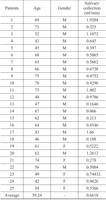
b) group II - (control group) - 25 healthful patients. Of these patients, 20 (80%) were males and 5 (20%) were females. The average age was 59.24 years, ranging from 42 to 78 years. The control
TABLE 1 - Distribution of patients (group I) according to age, gender, skin color, tumor site, clinical stage and
radiotherapeutic total dose (RTD).
Patients Age Gender Skin Color Tumor site Clínical stage RTD (Gy)
1 58 M B Floor of the mouth IV 81
2 61 M W Floor of the mouth III 71
3 75 M W Tongue III 70
4 58 M W Alveolar ridge IV 82.6
5 74 M W Tongue IV 70
6 83 M W Floor of the mouth III 72
7 58 M W Tongue III 71
8 62 M W Pyriform sinus IV 71
9 49 M W Cervical metastasis (unknown primary) IV 60
10 52 M W Tongue II 70
11 47 M W Pyriform sinus IV 80
12 56 F W Tonsil IV 57
13 46 M W Tongue IV 68.4
14 34 F W Retromolar IV 58.8
15 46 M W Pyriform sinus III 70.4
16 64 M W Oropharynx IV 70
17 69 M W Buccal mucosa IV 68.4
18 63 F B Buccal mucosa IV 70
19 53 F W Floor of the mouth II 72.2
20 64 M W Oropharynx IV 71
21 59 M W Hypopharyx IV 60.4
22 54 M W Larynx III 68
group was matched by age, skin color and gen-der. None of the patients had undergone radio-therapy or presented medication-related xerostomia. In the control group, only one sali-vary collection was performed (Table 2).
For the saliva collection, all patients were in-structed to swallow the saliva present in their mouths, and after this step they were asked to put the total saliva produced in glass recipients. The saliva collected was identified and weighed. If pa-tients were wearers of complete dentures or
remov-able partial dentures, they were informed to re-move their prostheses during the collection.
The weight of each collection was measured and adjusted to flow in ml/min for each patient. To classify the salivary flows, values below 0.1 ml/min were indicative of xerostomia16
. Two-tailed tests were used for evaluation of the salivary flow.
RESULTS
The regions more affected by the squamous cell carcinomas were floor of the mouth (18.2%), buccal mucosa (9%) and pyriform sinus (13.6%). The more frequent clinical stages were stage IV (63.5%) and stage III (27.3%). The average dose of radiation used for the tumors was of 69.69 Gy. Radiotherapeutic fields involved the parotid glands in all patients, and total doses were higher than the minimum values for permanent xerostomia (Table 1).
Regarding the salivary flow in the control group, the average was 0.6618 ml/min (Table 2). In irradi-ated patients, the average in the first collection was 0.5827 ml/min, being similar to that of the control group, with no statistical difference (p = 0.5366). However, these patients presented a significant decrease between the initial collection and collection 2 (p = 0.0008). In addition, this re-duction persisted in collection 3. Statistical analy-sis did not show differences between collection 2 and 3 (p = 0.9822). The data about the distribution of salivary flows (group I) are shown in Graph 1 and Table 3.
DISCUSSION
The reduction of salivary flow is an important side effect of head and neck radiotherapy, causing difficulties to chew, swallow, speak and eat5,10,11. It
GRAPH 1 - Salivary flow averages (ml/min) in group I
(three collections) and group II (control).
TABLE 2 -Distribution of patients (group II) according
to age, gender and salivary flow (ml/min).
Patients Age Gender
Salivary collection
(ml/min)
1 69 M 1.9204
2 73 M 0.325
3 52 M 1.1072
4 42 M 0.643
5 45 M 0.397
6 68 M 0.5065
7 65 M 0.5662
8 66 M 0.4738
9 75 M 0.4752
10 78 M 0.4296
11 73 M 1.402
12 48 M 0.9786
13 47 M 0.1646
14 67 M 0.066
15 62 M 0.213
16 64 M 0.8546
17 43 M 1.66
18 46 M 0.108
19 61 F 0.5222
20 62 M 1.2612
21 74 F 0.278
22 56 M 0.5004
23 49 F 0.74432
24 42 F 0.4626
25 54 F 0.5366
also results in alterations in the oral microbiota as well as in an increase of the risk for the develop-ment of dental caries2,9,10,12
. Patients submitted to radiotherapy present reduction of salivary flow in the first weeks of treatment. In many cases, this reduction becomes irreversible15. The sensation of mouth dryness called xerostomia is normally
asso-ciated with salivary flow. Normally, the level of sali-vary flow considered to be xerostomic in non-stim-ulated saliva is below 0.1 ml/min. In our study, 5 out of 22 evaluated patients (22.5%) were classi-fied as xerostomic in collection 2 (during radio-therapy), and 7 patients (32%) were thus classified in collection 3 (end of radiotherapy).
In the first collection, patients from group I (ir-radiated) presented average salivary flows similar to those of patients from group II (control). Regarding the second collection of saliva, a signifi-cant reduction of saliva was observed. This reduc-tion is in agreement with the fact that radiareduc-tion ap-plied to the head and neck promotes a decrease of saliva with doses of 20 Gy. It occurs particularly when parotid glands are included in the radiotherapeutic fields14,15
. However, in our cases, salivary flow did not reach xerostomic levels, which could indicate that, in these cases, xerostomia is more a symptomatic phenomenon, or that the complaints of dry mouth and saliva vis-cosity are more relevant than the actual quantita-tive reduction of salivary flows.
In the third collection (end of treatment), sali-vary flows were low and similar to those of the sec-ond collection (during treatment). Some studies have also demonstrated that the salivary flow re-duction is more significant in the first two weeks of radiotherapy, without important changes in the 13 following weeks3
. However, others showed that there is a linear reduction of non-stimulated
saliva8 .
CONCLUSION
To summarize, our study showed that radio-therapy leads to an important reduction of salivary flow during and after treatment.
ACKNOWLEDGMENT
We wish to thank the Grant from the São Paulo State Research Foundation (FAPESP, No. 99/1364-7), Brazil.
REFERENCES
1. Almstahl A, Wikstrom M. Oral microflora in subjects with reduced salivary secretion. J Dent Res 1999;78:1410-16.
2. Blozis GG, Robinson JE. Oral tissues changes caused by radiation therapy and their management. Dent Clin North Am 1968;643-56.
3. Burlage FR, Coopes RP, Meertens H, Stokman MA, Vissink A. Parotid and submandibular/sublingual salivary flow
during high dose radiotherapy. Radioth Oncol 2001;61:271-4.
4. Carl W. Local radiation and systemic chemotherapy: pre-venting and managing the oral complications. J Am Dent Assoc 1993;124:119-23.
5. Epstein JB, Emerton S, Kolbison DA, Le ND, Phillips N, Stevenson-Moore P,et al. Quality of life and oral function
TABLE 3 -Distribution of patients (group I) according to
salivary collections (ml/min).
Patient Collection 1 Collection 2 Collection 3
1 0.958 0.5908 0.3226
2 0.6508 0.4914 0.4024
3 0.6116 0.0956 0.0936
4 0.2434 0.201 0.1766
5 0.87 0.2848 0.5354
6 0.2676 0.2864 0.4412
7 0.3164 0.0586 0.089
8 1.329 0.1396 0.2164
9 0.8172 0.188 0.0566
10 0.6742 0.2716 0.1335
11 0.2422 0.3218 0.2692
12 0.0924 0.2272 0.0018
13 1.6884 0.1348 0.1986
14 0.2322 0.0158 0.749
15 1.1346 0.1868 0.2796
16 0.1736 0.3248 0.0616
17 0.3449 0.304 0.27
18 0.3756 0.0394 0.0694
19 0.2374 0.0232 0.1044
20 0.3528 0.22316 0.1826
21 0.6632 0.391 0.0988
22 0.544 0.2564 0.2796
following radiotherapy for head and neck cancer. Head and Neck 1999, 21:1-11.
6. Garg A, Malo M. Manifestations and treatment of xerostomia and associated oral effects secondary to head and neck radiation therapy. J Am Dent Assoc 1997;128:1128-33.
7. Gibson G. Identifying and treating xerostomia in restor-ative patients. J Esth Dent 1998;10:253-64.
8. Henson BS, Eisbruch A, D’Hondt E, Ship JA.Two-year lon-gitudinal study of parotid salivary flow rates in head and neck patients receiving unilateral neck parotid-sparing ra-diotherapy treatment.Oral Oncol 1999;35:234-41.
9. Holmes S. Xerostomia: aetiology and management in can-cer patients. Support Care Cancan-cer 1998;6:348-55. 11. Lopes MA, Colleta RD, Alves FA, Abadde N, Rossi Jr A.
Reconhecendo e controlando os efeitos colaterais da radioterapia. Rev Assoc Paul Cirur Dent 1998;52:241-4.
12. Regelink G, Vissink A, Reintsema H, Nauta JM.Efficacy of a synthetic polymer saliva substitute in reducing oral
com-plaints of patients suffering from irradiation-induced xerostomia. Quintessence Int 1998;29:383-8.
13. Rothwell BR. Prevention and treatment of orofacial compli-cation of radiotherapy. J Am Dent Assoc 1987;114:316-22. 14. Schubert MM, Izutsu KT. Iatrogenic causes of salivary
gland dysfunction. J Dent Res 1987;66:680-8.
15. Scully C, Epstein JB. Oral health care for the cancer pa-tient. Eur J Cancer B Oral Oncol 1996;32B:281-92. 16. Sreebny LM. Saliva in healthy and disease: an appraisal
and update. Int Dent J 2000;50:140-61.
17. Weerkamp AH, Wagner K, Vissink A, Gravenmade EJ. Ef-fect of the application of mucin-based saliva substitute on the oral microflora of xerostomic patients. J Oral Pathol 1987;16:474-8.
18. Whitmyer CC, Esposito SJ, Terezhalmy GT. Radiotherapy for head and neck neoplasm. Gen Den 1997;45:363-70, 19. Williams DW, Lewis MA. Isolation and identification of
Candidafrom the oral cavity. Oral Dis 2000;6:3-11.