Osteointegration of porous absorbable bone
sub-stitutes: A systematic review of the literature
Maria Ju´lia Escanhoela Paulo, Mariana Avelino dos Santos, Bruno Cimatti, Nelson Fabrı´cio Gava, Marcelo Riberto, Edgard Eduard Engel*
Departamento de Biomecanica, Medicina e Reabilitacao do Aparelho Locomotor, Faculdade de Medicina de Ribeirao Preto, Universidade de Sao Paulo, Ribeirao Preto, SP, BR.
Biomaterials’ structural characteristics and the addition of osteoinductors influence the osteointegration capacity of bone substitutes. This study aims to identify the characteristics of porous and resorbable bone substitutes that influence new bone formation. An Internet search for studies reporting new bone formation rates in bone defects filled with porous and resorbable substitutes was performed in duplicate using the PubMed, Web of Science, Scielo, and University of Sa˜o Paulo Digital Library databases. Metaphyseal or calvarial bone defects 4 to 10 mm in diameter from various animal models were selected. New bone formation rates were collected from the histomorphometry or micro-CT data. The following variables were analyzed: animal model, bone region, defect diameter, follow-up time after implantation, basic substitute material, osteo-inductor addition, pore size and porosity. Of 3,266 initially identified articles, 15 articles describing 32 experimental groups met the inclusion criteria. There were no differences between the groups in the experimental model characteristics, except for the follow-up time, which showed a very weak to moderate correlation with the rate of new bone formation. In terms of the biomaterial and structural characteristics, only porosity showed a significant influence on the rate of new bone formation. Higher porosity is related to higher new bone formation rates. The influence of other characteristics could not be identified, possibly due to the large variety of experimental models and methodologies used to estimate new bone formation rates. We suggest the inclusion of standard control groups in future experimental studies to compare biomaterials.
KEYWORDS: Biomaterials; Osteointegration; Systematic Review; Bone Substitute.
Paulo MJ, dos Santos MA, Cimatti B, Gava NF, Riberto M, Engel EE. Osteointegration of porous absorbable bone substitutes: A systematic review of the literature. Clinics. 2017;72(7):449-453
Received for publication onDecember 28, 2016;First review completed onMarch 21, 2017;Accepted for publication onMay 5, 2017
*Corresponding author. E-mail: [email protected]
’ INTRODUCTION
The use of autografts for the treatment of bone defects has well-known restrictions, including limited availability and donor site morbidity (1). This fact has sparked an intense search for bone substitutes of different compositions and structural conformations (2). Some are already available on the market, whereas many still await evidence attesting their capacity for osteointegration and consequent commercial viability.
A large number of new materials and material combina-tions have been developed. Structural characteristics have also been improved. The presence of pores significantly increases the osteointegration capacity, whereas solid bio-materials tend to form a fibrosis layer on the surface (3). Furthermore, the presence of pores allows fluid circulation
inside the biomaterial, accelerates absorption of absorbable biomaterials and decreases the peak temperature of cements during setting (4,5). Most authors believe that pore size, porosity and interconnection of pores enhances new bone ingrowth. The ideal magnitude of these characteristics, however, has not yet been established (5-7). Progressive reabsorption and replacement of the biomaterial by nor-mal bone is also considered an advantageous property because inert substitutes affect bone remodeling and can compromise its structure and mechanical resistance (8). The addition of growth factors and other osteoinductive factors seems to increase osteointegration, but conflicting data also exist (9).
Comparing the many combinations of materials is a demanding task, and the use of many different analysis methods makes comparisons even more challenging. Com-puted microtomography (micro-CT) and histomorphometry (HMM) have frequently been used to quantify new bone formation (NBF) in bone defects created in animal models and inside the porous biomaterial (10).
The aim of this study was to identify the chemical and structural characteristics that influence the capacity of new bone formation of porous and absorbable bone substitutes implanted in animal models using a systematic review.
DOI:10.6061/clinics/2017(07)10
Copyright&2017CLINICS–This is an Open Access article distributed under the terms of the Creative Commons License (http://creativecommons.org/licenses/by/ 4.0/) which permits unrestricted use, distribution, and reproduction in any medium or format, provided the original work is properly cited.
Methodology
A systematic review was performed to evaluate how spe-cific characteristics of porous and absorbable bone substi-tutes used to fill bone defects in experimentalin vivostudies affects the capacity of new bone formation using micro-CT or HMM quantifications.
Search strategy
An electronic search was independently performed by two researchers (MJEP and MAS) between July 2014 and February 2015 without restrictions on the publication date. The following databases were used: PubMed, Web of Sci-ence, Scielo, and Theses and Dissertations of the University of São Paulo Digital Library. Only articles written in English or Portuguese were selected. The following search term combinations were used: (bone substitute AND porous) OR (bone substitute AND cancellous) OR (bone substi-tute AND spongy) OR (bone substisubsti-tute AND pore) OR (cement AND porous) OR (cement AND cancellous) OR (cement AND spongy) OR (cement AND pore) OR (bone cement AND porous) OR (bone cement AND cancellous) OR (bone cement AND spongy) OR (bone cement AND pore).
Article selection strategy
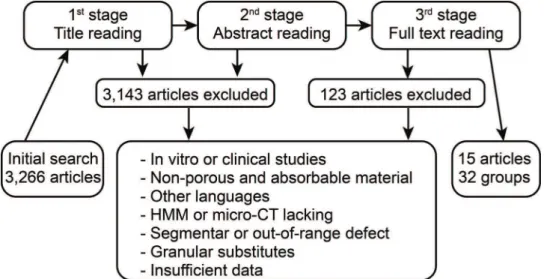
Two researchers (MJEP and MAS) independently per-formed the article selection based on the eligibility criteria by reading the titles, summaries or full text, according to the search strategy (Figure 1). Consensus meetings, with the parti-cipation of a third researcher (EEE), were utilized to resolve conflict situations. Some articles were included from the reference lists of the selected articles.
The experimental groups of each article that met eligibility criteria were analyzed independently; therefore, each article could have more than one group.
Eligibility criteria
Inclusion criteria:
1. Experimental,in vivostudies in animal models;
2. Orificial defects, 4.0 mm to 10.0 mm in diameter, pro-duced by curettage or drilling of holes;
3. Implantation of porous and resorbable bone substitutes in the form of premolded blocks or cement with a clear description of the composition, porosity and pore size; 4. NBF indicated as a rate according to HMM or micro-CT data.
Exclusion criteria:
1. Clinical trials, implantation in humans orin vitrostudies; 2. Insufficient description of the substitute characteristics,
methodology or results;
3. Non-porous or non-absorbable substitutes;
4. Presentation in granular form. This form was excluded due to interference with the porosity and pore size of the substitute.
Variables
The end point variable was the rate of new bone formation (NBF), which was based on the histomorphometry (HMM) or micro-CT analysis data. The remaining dependent vari-ables were as follows: animal model, bone region of the defect, diameter of the defect (in mm), follow-up time after implantation (in weeks), substitute basic material (calcium phosphate, hydroxyapatite, bioglass, etc.), osteoinductor addition (fibroblast growth factor (FGF), BMP or bone marrow mesenchymal stem cells (BMSCs)), maximum pore size and porosity.
Presentation of results
The NBF rates are described as the means, maximum and minimum values. The categorical variables and maximum defect size were grouped and compared using the Kruskal-Wallis test. The defect size, follow-up time and porosity were correlated to NBR using the Spearman’s correlation coeffi-cient. PASW software version 17 (IBM SPSS, Armonk, USA) was used for the data analysis and the level of significance was set as 5%.
’ RESULTS
The initial search identified 3,266 studies. Figure 1 illus-trates the selection flow. A total of 3,143 articles were exclu-ded because the biomaterial characteristics, experimental
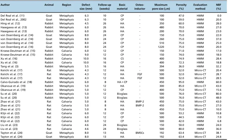
model, NBF measurement or article language were not eligible. Another 108 articles were excluded due to insuffi-cient data or incomplete description of the biomaterial, methodology or defect characteristics. From the remaining 15 articles, 32 experimental groups with different implanted bone substitutes were considered for the analysis (Table 1).
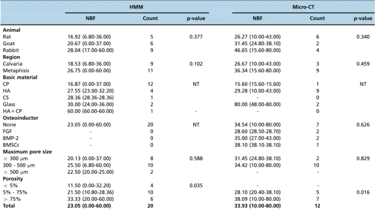
HMM was used more frequently than micro-CT for NBF quantification (20 groups, 62.5%). In both analysis methods, no significant differences were found when the NBF means were grouped according to the animal model and bone region (Table 2). The basic materials of the bone substitutes could not be compared because many of the material groups became too small due to uneven distribution of the experi-mental groups. The addition of osteoinductors to the bone substitute did not result in a significant increase in NBF. In addition, the influence of the pore size on NBF could not be detected. There was no correlation between the defect size and NBF in the HMM analysis (r=–0.181, p=0.446) and micro-CT analysis (r=0.103, p=0.751). NBR showed a moderate correlation to the follow-up time in the HMM analysis (r=0.441, p=0.052) but a weak correlation in the micro-CT analysis (r=0.122, p=0.705). NBF was significantly influenced by porosity (Figure 2).
’ DISCUSSSION
The search for bone substitutes is a contemporary and relevant subject (9). The large number of studies that present new biomaterials or the further development of known biomaterials confirms this statement. Prior to February 2015,
3,266 studies were published describing porous and absorb-able bone substitutes filling bone defects in experimental animals.
A large number of materials have been developed and tested, separately or combined, to increase the capacity of new bone formation. This continuous and effervescent development hinders the classification of these materials (9). In this review, we aimed to classify these materials according to the basic material used in the production of the bone substitute; however, there are overlaps between groups and many materials that are classified into the same group are not always similar. Most of the substitutes contained HA or other types of CPs or a combination of them as the basic material. The accumulated experience on modulat-ing the resorption rate and mechanical resistance of these compounds make this an attractive combination (10,18). The number of other materials was too small to allow a statistical analysis.
The influence of the other chemical and structural characteristics of the biomaterials had to be very large to be detected in such a heterogeneous sample. For this reason, it was not possible to identify differences in the capacity of new bone formation by comparing the different groups of experimental animals, the sizes and locations of bone defects, the association with osteoinducers and the pore sizes. The follow-up time presented a weak to moderate correlation to NBF, depending on the analysis method (HMM or micro-CT). However, the porosity presented significant differences, suggesting that this parameter has a strong impact on the NBF rate.
Table 1-Characteristics of the experimental groups.
Author Animal Region Defect
size (mm)
Follow-up (weeks)
Basic material
Osteo-inductor
Maximum pore size (mm)
Porosity (%)
Evaluation method
NBF (%)
Del Real et al. (11) Goat Metaphysis 6.3 10 CP - 100 47.0 HMM 0.0 Del Real et al., 2002 Goat Metaphysis 6.3 10 CP - 100 59.0 HMM 20.0 Hing et al. (12) Rabbit Metaphysis 4.5 26 HA - 350 60.0 HMM 28.0 Hasegawa et al. (13) Rabbit Metaphysis 6.0 26 HA - 480 70.0 HMM 27.0 Hasegawa et al. (13) Rabbit Metaphysis 6.0 26 HA - 200 70.0 HMM 23.0 von Doernberg et al. (14) Goat Metaphysis 8.0 24 CP - 150 75.0 HMM 22.0 von Doernberg et al. (14) Goat Metaphysis 8.0 24 CP - 260 75.0 HMM 37.0 von Doernberg et al. (14) Goat Metaphysis 8.0 24 CP - 510 75.0 HMM 25.0 von Doernberg et al. (14) Goat Metaphysis 8.0 24 CP - 1220 75.0 HMM 20.0 Kroese-Deutman et al. (15) Rabbit Calvaria 6.0 12 CP - 150 71.0 HMM 17.0 Kroese-Deutman et al. (15) Rabbit Calvaria 9.0 12 CP - 150 74.0 HMM 18.0
Xu et al. (16) Rabbit Calvaria 10.0 16 CS - 400 74.9 HMM 28.4
Xu et al. (16) Rabbit Calvaria 10.0 16 CP - 400 72.3 HMM 18.8
Tang et al. (3) Rabbit Metaphysis 5.5 16 HA - 394 36.0 HMM 32.2 Keiichi et al. (17) Rat Metaphysis 4.3 12 HA - 500 52.0 Micro-CT 20.4 Keiichi et al. (17) Rat Metaphysis 4.3 12 HA FGF 500 52.0 Micro-CT 28.7 Keiichi et al. (17) Rat Metaphysis 4.3 12 HA FGF 500 52.0 Micro-CT 28.5 Calvo-Guirado et al. (18) Rabbit Metaphysis 6.0 8 HA+CP - 450 95.0 HMM 60.0 Okanoue et al. (19) Rabbit Metaphysis 5.0 12 HA - 300 85.0 Micro-CT 43.0 Okanoue et al. (19) Rabbit Metaphysis 5.0 12 CP - 400 75.0 Micro-CT 15.6 Su et al. (20) Rabbit Metaphysis 5.0 12 Bioglass - 500 76.0 Micro-CT 80.0 Su et al. (20) Rabbit Metaphysis 5.0 12 Bioglass - 500 76.0 Micro-CT 48.0 Zhao et al. (21) Rat Calvaria 5.0 8 HA BMP-2 450 75.0 Micro-CT 43.0 Zhao et al. (21) Rat Calvaria 5.0 8 HA BMP-2 450 75.0 Micro-CT 27.0 Zhao et al. (21) Rat Calvaria 5.0 8 HA - 450 75.0 Micro-CT 10.0
Klijn et al. (22) Rat Calvaria 6.0 12 CP - 500 53.2 HMM 10.8
Klijn et al. (22) Rat Calvaria 6.0 12 CP - 500 44.5 HMM 7.0
Klijn et al. (22) Rat Calvaria 6.0 12 CP - 500 42.0 HMM 6.8
Liu et al. (23) Rat Calvaria 4.6 24 Bioglass - 150 50.0 HMM 24.0 Liu et al. (23) Rat Calvaria 4.6 24 Bioglass - 500 80.0 HMM 36.0 Tayton et al. (24) Goat Metaphysis 8.0 13 HA BMSCs 192 63.4 Micro-CT 38.1 Tayton et al. (24) Goat Metaphysis 8.0 13 HA - 192 63.4 Micro-CT 24.8
Despite the large number, few studies could be included due to the significant variation in the presentation of the bone substitutes and experimental models. In addition, the NBF assessment method varied greatly, making comparison unsuitable. In this review, we used the two most cited methods, which, according to some articles, do not present comparable values but are related (10,25,26). Therefore, the HMM and micro-CT results were analyzed separately.
Although a substantial effort has been made by the scien-tific community in recent years to improve bone substitutes, some skepticism still exists regarding the effectiveness of the currently available biomaterials to cure critical size bone
defects (27). The wide variety and geometrical shapes of bone materials indicates that many questions remain to be answered. More research is necessary to better understand how NBF can be increased.
This review demonstrates that the lack of standardization of NBF analyses has hampered the comparison of the various types of porous and absorbable bone substitutes. Even when limiting the evaluation to two types of quantitative anal-yses for NBF, it was not possible to obtain accurate results. A better description of the analyzed region of interest (ROI) by each method could offer more precise data interpretation, and consequently, a more consistent comparison with other Table 2-Median, minimum and maximum values for NBF rates and the experimental group counts according to the experimental model and biomaterial characteristics.
HMM Micro-CT
NBF Count p-value NBF Count p-value
Animal
Rat 16.92 (6.80-36.00) 5 0.377 26.27 (10.00-43.00) 6 0.340
Goat 20.67 (0.00-37.00) 6 31.45 (24.80-38.10) 2
Rabbit 28.04 (17.00-60.00) 9 46.65 (15.60-80.00) 4
Region
Calvaria 18.53 (6.80-36.00) 9 0.102 26.67 (10.00-43.00) 3 0.459
Metaphisis 26.75 (0.00-60.00) 11 36.34 (15.60-80.00) 9
Basic material
CP 16.87 (0.00-37.00) 12 NT 15.60 (15.60-15.60) 1 NT
HA 27.55 (23.00-32.20) 4 29.28 (10.00-43.00) 9
CS 28.36 (28.36-28.36) 1 - 0
Glass 30.00 (24.00-36.00) 2 80.00 (48.00-80.00) 2
HA+CP 60.00 (60.00-60.00) 1 - - 0
Osteoinductor
None 23.05 (0.00-60.00) 20 NT 34.54 (10.00-80.00) 7 0.626
FGF - 0 28.60 (28.50-28.70) 2
BMP-2 - 0 35.00 (27.00-43.00) 2
BMSCs - 0 38.10 (38.10-38.10) 1
Maximum pore size
o300mm 20.13 (0.00-37.00) 8 0.588 31.45 (24.80-38.10) 2 0.829
300 - 500mm 25.50 (6.80-60.00) 10 34.42 (10.00-80.00) 10
4500mm 22.50 (20.00-25.00) 2 -
-Porosity
o5% 11.50 (0.00-32.20) 4 0.035 -
-5% - 7-5% 21.50 (10.80-28.36) 10 28.10 (20.40-38.10) 5 0.016
475% 33.33 (20.00-60.00) 6 38.09 (10.00-80.00) 7
Total 23.05 (0.00-60.00) 20 33.93 (10.00-80.00) 12
NBF, new bone formation; HA, hydroxyapatite; CP, calcium phosphate; CS, calcium silicate; FGF, fibroblast growth factor; BMP-2, bone morphogenetic protein-2; BMSC, bone marrow mesenchymal stem cells; HMM, histomorphometry; NT, not tested. P-values from Kruskal –Wallis test.
similar studies. Additionally, the inclusion of standard con-trol groups with autografts or a commonly used substitute would allow the calibration of the results and the compar-ison of many different study groups.
We conclude that porosity has a high impact on NBF rates and that the current lack of standardized analysis methods and the broad variety of experimental models makes iden-tifying the chemical and structural characteristics that pro-vide a greater capacity for NBF almost impossible. We suggest that standard control groups should always be used to allow for a better comparison of the results.
’ AUTHOR CONTRIBUTIONS
Paulo MJ and dos Santos MA conducted the electronic search and data collection. Cimatti B and Gava NF reviewed the electronic search and data collection. Riberto M revised the manuscript and conducted the data analysis. Engel EE wrote the manuscript, reviewed the data analysis and supervised the study.
’ REFERENCES
1. Summers BN, Eisenstein SM. Donor site pain from the ilium. A compli-cation of lumbar spine fusion. J Bone Joint Surg Br. 1989;71(4):677–80.
2. Fini M, Giavaresi G, Aldini NN, Torricelli P, Botter R, Beruto D, et al. A bone substitute composed of polymethylmethacrylate and alpha-tricalcium phosphate: results in terms of osteoblast function and bone tissue formation. Biomaterials. 2002;23(23):4523–31, http://dx.doi.org/
10.1016/S0142-9612(02)00196-5.
3. Tang PF, Li G, Wang JF, Zheng QJ, Wang Y. Development, characteriza-tion, and validation of porous carbonated hydroxyapatite bone cement. J Biomed Mater Res B Appl Biomater. 2009;90(2):886–93, http://dx.doi.
org/10.1002/jbm.b.31360.
4. Andrade JC, Camilli JA, Kawachi EY, Bertran CA. Behavior of dense and porous hydroxyapatite implants and tissue response in rat femoral defects. J Biomed Mater Res. 2002;62(1):30–6, http://dx.doi.org/10.1002/
jbm.10242.
5. Rentsch C, Rentsch B, Scharnweber D, Zwipp H, Rammelt S. Bone sub-stitute. Transplants and replacement materials--an update. Unfallchirurg. 2012;115(10):938–49, http://dx.doi.org/10.1007/s00113-012-2238-4.
6. Panseri S, Cunha C, D’Alessandro T, Sandri M, Russo A, Giavaresi G, et al. Magnetic hydroxyapatite bone substitutes to enhance tissue regen-eration: evaluation in vitro using osteoblast-like cells and in vivo in a bone defect. PLos One. 2012;7(6):e38710, http://dx.doi.org/10.1371/journal. pone.0038710.
7. Yuan H, Kurashina K, de Bruijn JD, Li Y, de Groot K, Zhang X. A pre-liminary study on osteoinduction of two kinds of calcium phosphate ceramics. Biomaterials. 1999;20(19):1799–806, http://dx.doi.org/10.1016/
S0142-9612(99)00075-7.
8. Kasuya A, Sobajima S, Kinoshita M. In vivo degradation and new bone formation of calcium phosphate cement-gelatin powder composite related to macroporosity after in situ gelatin degradation. J Orthop Res. 2012;30(7): 1103–11, http://dx.doi.org/10.1002/jor.22044.
9. Campana V, Milano G, Pagano E, Barba M, Cicione C, Salonna G, et al. Bone substitutes in orthopaedic surgery: From basic science to clinical practice. J Mater Sci Mater Med. 2014;25(10):2445–61, http://dx.doi.org/
10.1007/s10856-014-5240-2.
10. Park SY, Kim KH, Koo KT, Lee KW, Lee YM, Chung CP, et al. The evaluation of the correlation between histomorphometric analysis and micro-computed tomography analysis in AdBMP-2 induced bone regen-eration in rat calvarial defects. J Periodontal Implant Sci. 2011;41(5): 218–26, http://dx.doi.org/10.5051/jpis.2011.41.5.218.
11. Del Real RP, Ooms E, Wolke JG, Vallet-Regi M, Jansen JA. In vivo bone response to porous calcium phosphate cement. J Biomed Mater Res A. 2003;65(1):30–6, http://dx.doi.org/10.1002/jbm.a.10432.
12. Hing KA, Best SM, Tanner KE, Bonfield W, Revell PA. Mediation of bone ingrowth in porous hydroxyapatite bone graft substitutes. J Biomed Mater Res A. 2004;68(1):187–200, http://dx.doi.org/10.1002/jbm.a.10050.
13. Hasegawa S, Tamura J, Neo M, Goto K, Shikinami Y, Saito M, et al. In vivo evaluation of a porous hydroxyapatite/poly-DL-lactide composite for use as a bone substitute. J Biomed Mater Res A. 2005;75(3):567–79, http://dx.
doi.org/10.1002/jbm.a.30460.
14. von Doernberg MC, von Rechenberg B, Bohner M, Grünenfelder S, van Lenthe GH, Müller R, et al. In vivo behavior of calcium phosphate scaf-folds with four different pore sizes. Biomaterials. 2006;27(30):5186–98,
http://dx.doi.org/10.1016/j.biomaterials.2006.05.051.
15. Kroese-Deutman HC, Ruhé PQ, Spauwen PHM, Jansen JA. Bone induc-tive properties of rhBMP-2 loaded porous calcium phosphate cement implants inserted at an ectopic site in rabbits. Biomaterials. 2005;26(10): 1131–8, http://dx.doi.org/10.1016/j.biomaterials.2004.04.021.
16. Xu HHK, Quinn JB. Calcium phosphate cement containing resorbable fibers for short-term reinforcement and macroporosity. Biomaterials. 2002;23(1):193–202, http://dx.doi.org/10.1016/S0142-9612(01)00095-3.
17. Keiichi K, Mitsunobu K, Masafumi S, Yutaka D, Toshiaki S. Induction of new bone by basic FGF-loaded porous carbonate apatite implants in femur defects in rats. Clin Oral Implants Res. 2009;20(6):560–5, http://dx.
doi.org/10.1111/j.1600-0501.2008.01676.x.
18. Calvo-Guirado JL, Delgado-Ruíz RA, Ramírez-Fernández MP, Maté-Sán-chez JE, Ortiz-Ruiz A, Marcus A. Histomorphometric and mineral degradation study of Ossceram: a novel biphasic B-tricalcium phosphate, in critical size defects in rabbits. Clin Oral Implants Res. 2012;23(6): 667–75, http://dx.doi.org/10.1111/j.1600-0501.2011.02193.x.
19. Okanoue Y, Ikeuchi M, Takemasa R, Tani T, Matsumoto T, Sakamoto M, et al. Comparison of in vivo bioactivity and compressive strength of a novel superporous hydroxyapatite with beta-tricalcium phosphates. Arch Orthop Trauma Surg. 2012;132(11):1603–10, http://dx.doi.org/10.1007/
s00402-012-1578-4.
20. Su J, Cao L, Yu B, Song S, Liu X, Wang Z, et al. Composite scaffolds of mesoporous bioactive glass and polyamide for bone repair. Int J Nano-medicine. 2012;7:2547–55, http://dx.doi.org/10.2147/IJN.S29819.
21. Zhao J, Shen G, Liu C, Wang S, Zhang W, Zhang X, et al. Enhanced healing of rat calvarial defects with sulfated chitosan-coated calcium-deficient hydroxyapatite/bone morphogenetic protein 2 scaffolds. Tissue Eng Part A. 2012;18(1-2):185–97, http://dx.doi.org/10.1089/ten.tea.2011.0297.
22. Klijn RJ, van den Beucken JJ, Félix Lanao RP, Veldhuis G, Leeuwenburgh SC, Wolke JGC, et al. Three different strategies to obtain porous calcium phosphate cements: comparison of performance in a rat skull bone aug-mentation model. Tissue Eng Part A. 2012;18(11-12):1171–82, http://dx.
doi.org/10.1089/ten.tea.2011.0444.
23. Liu H, Hu G, Shang P, Shen Y, Nie P, Peng L, et al. Histological char-acteristics of induced membranes in subcutaneous, intramuscular sites and bone defect. Orthop Traumatol Surg Res. 2013;99(8):959–64, http://
dx.doi.org/10.1016/j.otsr.2013.08.009.
24. Tayton E, Purcell M, Smith JO, Lanham S, Howdle SM, Shakesheff KM, et al. The scale-up of a tissue engineered porous hydroxyapatite polymer composite scaffold for use in bone repair: an ovine femoral condyle defect study. J Biomed Mater Res A. 2015;103(4):1346–56, http://dx.doi.org/
10.1002/jbm.a.35279.
25. Chappard D, Retailleau-Gaborit N, Legrand E, Baslé MF, Audran M. Comparison insight bone measurements by histomorphometry and microCT. J Bone Miner Res. 2005;20(7):1177–84, http://dx.doi.org/
10.1359/JBMR.050205.
26. Müller R, Van Campenhout H, Van Damme B, Van der Perre G, Dequeker J, Hildebrand T, et al. Morphometric analysis of human bone biopsies: a quantitative structural comparison of histological sections and micro-computed tomography. Bone. 1998;23(1):59–66, http://dx.doi.org/10.1016/
S8756-3282(98)00068-4.
27. Afifi AM, Gordon CR, Pryor LS, Sweeney W, Papay FA, Zins JE. Calcium phosphate cements in skull reconstruction: A meta-analysis. Plast Reconstr Surg. 2010;126(4):1300–9, http://dx.doi.org/10.1097/PRS.0b01