Domain Based on X-ray Structure Analyses
Julia Wenger1., Eva Klinglmayr1., Chris Fro¨hlich2
, Clarissa Eibl1, Ana Gimeno1, Manuel Hessenberger1, Sandra Puehringer3,4, Oliver Daumke2, Peter Goettig1*
1Department of Molecular Biology, University of Salzburg, Salzburg, Austria, 2Crystallography, Max-Delbru¨ck-Center for Molecular Medicine, Berlin, Germany,
3Macromolecular Crystallography, Helmholtz-Zentrum Berlin fu¨r Materialien und Energie, Berlin, Germany,4Department of Biology and Chemistry, Freie Universita¨t Berlin, Berlin, Germany
Abstract
Human dynamin-1-like protein (DNM1L) is a GTP-driven molecular machine that segregates mitochondria and peroxisomes. To obtain insights into its catalytic mechanism, we determined crystal structures of a construct comprising the GTPase domain and the bundle signaling element (BSE) in the nucleotide-free and GTP-analogue-bound states. The GTPase domain of DNM1L is structurally related to that of dynamin and binds the nucleotide 59-Guanylyl-imidodiphosphate (GMP-PNP) via five highly conserved motifs, whereas the BSE folds into a pocket at the opposite side. Based on these structures, the GTPase center was systematically mapped by alanine mutagenesis and kinetic measurements. Thus, residues essential for the GTPase reaction were characterized, among them Lys38, Ser39 and Ser40 in the phosphate binding loop, Thr59 from switch I, Asp146 and Gly149 from switch II, Lys216 and Asp218 in the G4 element, as well as Asn246 in the G5 element. Also, mutated Glu81 and Glu82 in the unique 16-residue insertion of DNM1L influence the activity significantly. Mutations of Gln34, Ser35, and Asp190 in the predicted assembly interface interfered with dimerization of the GTPase domain induced by a transition state analogue and led to a loss of the lipid-stimulated GTPase activity. Our data point to related catalytic mechanisms of DNM1L and dynamin involving dimerization of their GTPase domains.
Citation:Wenger J, Klinglmayr E, Fro¨hlich C, Eibl C, Gimeno A, et al. (2013) Functional Mapping of Human Dynamin-1-Like GTPase Domain Based on X-ray Structure Analyses. PLoS ONE 8(8): e71835. doi:10.1371/journal.pone.0071835
Editor:Paul A. Randazzo, National Cancer Institute, United States of America
ReceivedMarch 31, 2013;AcceptedJuly 3, 2013;PublishedAugust 19, 2013
Copyright:ß2013 Wenger et al. This is an open-access article distributed under the terms of the Creative Commons Attribution License, which permits unrestricted use, distribution, and reproduction in any medium, provided the original author and source are credited.
Funding:This study was supported by the Austrian Science Fund project W_01213 and by the Erwin-Schro¨dinger Postdoc Fellowship J3173-N17 for Sandra Puehringer (both http://www.fwf.ac.at/). The funders had no role in study design, data collection and analysis, decision to publish, or preparation of the manuscript.
Competing Interests:The authors have declared that no competing interests exist.
* E-mail: [email protected]
.These authors contributed equally to this work.
Introduction
Members of the dynamin superfamily comprise a family of conserved GTPases, which are mostly found in the eukaryotic kingdom and mediate functions typically related to membrane remodeling [1,2]. A defining feature of dynamin superfamily members is a large GTPase domain of roughly 300 amino acids, which distinguishes it from other signaling GTPases. Despite variations in size, the GTPase domains of most dynamin superfamily members contain five conserved GTP-binding motifs (G1-5), similar to small Ras-like GTPases [3]. The P-Loop (G1) or GXXXXGKS/T motif is also present in ATPases (Walker A motif) and functions as a coordinator of the phosphate groups of the bound nucleotide [4]. A conserved threonine in switch-I (G2) and the conserved residues DxxG of switch-II (G3) are involved in Mg2+
binding and GTP (Guanosine-59-triphosphate) hydrolysis. These regions are rather flexible in the GDP-bound form but are stabilized in GTP-bound state [5]. The nucleotide binding affinity of dynamins is typically low, with specificity for GTP provided by the mostly conserved N/TKxD motif (G4) [2,3]. The G5-motif is involved in binding the ribose moiety.
Although dynamins display a rather high basal GTP turnover rate, additional stimulation (10–100-fold) has been observed for
some superfamily members due to self-assembly and lipid-binding [2,6–8]. GTP hydrolysis results in conformational changes that might be necessary for their function as mechanochemical enzymes [8–12].
Besides the GTPase domain, dynamin superfamily members share at least two more characteristic sequences: a middle domain and a C-terminal GTPase effector domain (GED) [1,2]. These sequences constitute two distinct domains, the stalk and the bundle signaling element (BSE). The latter comprises three helices located at the N- and terminus of the GTPase domain and at the C-terminus of the GED, respectively [8,13]. The BSE was proposed to mediate nucleotide-dependent conformational changes from the GTPase domain to the stalk and to regulate dynamin activity in membrane fission [12,14]. The middle domain and the amino-terminal portion of the GED form an antiparallel four-helix bundle, the stalk of dynamin superfamily proteins [15–17]. This stalk mediates dimerization and tetramerization, and the forma-tion of higher-ordered structures, such as rings or spirals [18–20]. Lipid binding motifs, such as the PH domain in dynamin or the lipid-binding loop L4 in MxA, are situated at the tip of the stalk, at the opposite end of the GTPase domain [20].
domain architecture related to their diverse cellular functions [1]. For example, human dynamin-1-like protein (DNM1L, formerly dynamin-related protein 1, Drp1) and optic atrophy 1 (OPA1) have essential roles in controlling mitochondrial dynamics. DNM1L is targeted from the cytoplasm to the outer mitochondrial membrane (MOM) and is a key player in mitochondrial fission [21]. Similarly, DNM1L has a major role in the segregation of peroxisomes [22]. While DNM1L possesses the typical features of the dynamin superfamily, its domain organization includes a sequence insertion of 80 to 130 amino acids between the middle domain and GED. This so-called insert B, or variable domain (VD), displays low sequence conservation and its function has yet to be elucidated in detail. Recently, it has been proposed that alternative splicing and posttranslational modifications within the VD indirectly regulate enzyme activity [23]. Such modifications include phosphorylation, ubiquitination, SUMOylation and S-nitrosylation. The exact mechanisms and effects of these posttranslational modifications on DNM1L activity are contro-versial. However, it is apparent that tight regulation of DNM1L is necessary to ensure proper mitochondrial function, as abnormal DNM1L activity is associated with excessive mitochondrial fission in various neurodegenerative diseases. Therefore, DNM1L is considered as a potential therapeutic target [24–26].
The proposed mechanism for DNM1L in mitochondrial fission shows similarities to the role of dynamin-1 in endocytosis [1]. Similar to dynamin-1, DNM1L exists in a dimer-tetramer-equilibrium in solution [27,28]. Intracellular cues direct DNM1L to the mitochondrial outer membrane (MOM) where it forms higher-ordered oligomers, which are visible as punctate structures [29]. Interestingly, some DNM1L clusters are located at future mitochondrial scission sites, which are hallmarked by endoplasmic reticulum (ER) contacts [30]. Although a known lipid-binding domain, like the dynamin-1 PH-domain, is missing,in vitrostudies showed that DNM1L and the yeast homologue DNM1 bind to negatively charged lipids [1,31–33]. Membrane-anchored pro-teins, such as the mitochondrial fission factor 1 (Fis1) and the membrane fission factor (MFF), have been proposed to mediate DNM1L-membrane binding [34,35]. Further studies, mainly with yeast DNM1, revealed that the addition of a non-hydrolyzable GTP analog triggers DNM1 self-assembly, while an excess of hydrolyzable GTP causes dissociation of the complex [36]. These findings suggest that GTP binding, but not GTP hydrolysis, is necessary for lipid-free DNM1L assembly. Electron microscopy studies demonstrated that nucleotide-free yeast DNM1 and human DNM1L form large helical spirals with a diameter of
,120 nm around lipid tubes [32,33]. Constriction of these spirals
to smaller diameters and dissociation from the lipid layer was observed in the presence of GTP. It was therefore suggested that GTP hydrolysis is the driving force for intra-molecular rearrange-ments, which are necessary for mitochondrial fission events [32]. Comparison of the cryo-EM structures of DNM1 in the constricted and non-constricted forms revealed differences to dynamin-1 assembly and constriction mechanisms [32,33].
In the current manuscript, we report the crystal structures of human DNM1L GTPase-BSE fusion protein in the nucleotide-free form and in the presence of a non-hydrolyzable GTP analogue, 59-Guanylyl imidodiphosphate (GMP-PNP). The struc-tural comparison of DNM1L with other dynamin superfamily members led to the identification of highly conserved active site residues, which in dynamin-1 andA. thalianaDrp1A are required for the basal and liposome-stimulated GTPase reaction [8,37]. These residues were systematically mutated to alanine in DNM1L and the resulting mutants kinetically characterized. In addition, residues involved in GTPase domain dimerization and in the
unique 80-loop were mutated and functionally analyzed. Based on our detailed structure-function map of the DNM1L active site, we postulate a common mechanism in the GTPase reaction of DNM1L and dynamin-1.
Materials and Methods
Cloning and Protein Purification
Cloning, expression and purification of the human DNM1L GTPase-GED (GG) fusion construct was performed as described previously [38]. It starts with the N-terminal Met1 and terminates with an artificial LEHHHHHH-tag at the C-terminal Trp736, corresponding to the numbering of DNM1L isoform 1. Several mutations were introduced into full-length DNM1L isoform 2 in pET21 (GenBank Accession Number NM_012063.2) either by overlap PCR (Q34A, S35A, K38A, S39A, S40A, E81A, E81A/ E82A, D146A, D190A, K216A, D218A, N246A) or round the horn cloning (T59A, G149A). Expression and purification of DNM1L full-length and mutants was performed as described elsewhere [39].
Protein Crystallization, Data Collection and Processing Nucleotide-free protein crystals were obtained after 3–5 days in a buffer containing 0.1 M sodium citrate pH 5, 27.5% PEG 3000 as described [38]. 1ml reservoir solution was mixed with 1ml of protein solution (1.2 mg ml21) and equilibrated against a reservoir volume of 400ml in a 24-well plate (Hampton Research).
Co-crystallization of DNM1L with a non-hydrolyzable GTP analog was attempted with protein samples that were incubated with 1 mM GMP-PNP (59-Guanylyl imidodiphosphate hydrate) and 4 mM MgCl2, and then purified by gel filtration. However, this procedure led to the dissociation of the nucleotide (see results section on determinants of GTPase domain dimerization). Correspondingly, protein crystals grew under the same conditions as nucleotide-free protein crystals and were of similar shape and size. In order to ensure nucleotide binding of the DNM1L GG fusion protein, crystals were soaked supplementary for 2 min by adding solid GMP-PNP powder to the crystallization drop. Both nucleotide-free and nucleotide-bound protein crystals were flash-cooled in liquid nitrogen without additional cryo-protectant. Diffraction data were collected on beamline BL14.1 operated by the Helmholtz-Zentrum Berlin (HZB) at the synchrotron BESSY II (Berlin-Adlershof, Germany) [40]. Diffraction images were recorded at a wavelength of 0.91814 A˚ using a Rayonics MX-225 363 CCD detector. Both data sets were integrated with iMosflm
1.0.7 and scaled with SCALA 3.3.20 for orthorhombic crystals of space group P21212 with cell constants around 53 A˚ (a), 151 A˚ (b), and 43 A˚ (c) for a maximum resolution of 2.3 A˚ [41,42]. See Table 1 for detailed values.
Phasing and Refinement
to minimize bias from the search model, in particular for the nucleotide-free data, which had a different test set for calculation of Rfree[44]. This procedure was followed by coordinate and B-factor refinement, water picking, as well as occupancy refinement of disordered loops and side chains in early cycles [45,46]. In both models, the defined 2Fo-Fcelectron density starts with Met1, while some flexible side chains at the surface and, in particular, several loop stretches are not visible. The backbone of switch I starting with Gly54 is largely traceable but the side chains are not well defined until Thr59. Little density is observed from Thr79 to Gly84 in the unique DNM1L 80-loop, as well as for the stretch from Ile118 to Lys123. For Pro325 and the following four residues of the artificial linker not much density is observed. Finally, the last visible C-terminal residue was Thr733. Both ligands, citrate (FLC) and GMP-PNP (GNP) are well resolved. Analyses of the models were performed with programs from the CCP4 package, such as
BAVERAGE, MOLPROBITY, and SUPERPOSE (SSM)
[44,47–49]. Refinement statistics and quality parameters are summarized in Table 2. Figures of structural models were created with PyMOL, including the electrostatic potential calculation with the APBS plugin using a dielectric constant of 80.0 for water as solvent in the range6120 kBT/e [50]. Coordinates and structure factors of the nucleotide-free and the GMP-PNP bound form were deposited in the RCSB Protein Data Bank under accession codes 4H1U and 4H1V, respectively.
Continuous-coupled GTPase Assay
Full-length DNM1L, active site mutants and predicted G-interface dimerization mutants were assayed at a protein concentration of 1.2mM for their ability to hydrolyze GTP in
the range of 0 to 1000mM based on NADH depletion and
absorbance measurement at 340 nm, as described elsewhere [38]. GTPase assays were performed in 25 mM HEPES/PIPES pH 7.0, 150 mM NaCl at 37uC for 84 min and 42 s per cycle. The kinetic parameters represent means of at least three independent measurements. Calculations were performed using GraphPad Prism version 5.0a for Mac OS X, GraphPad Software, La Jolla California USA, (www.graphpad.com). The standard deviation for the kcat/Km values was calculated according to Fenner [51].
GTP Hydrolysis Assays for Lipid Stimulated Activity Multiple turnover assays in the presence and absence of 0.5 mg/ml phosphatidylserine (PS) liposomes were carried out using 10mM DNM1L or the indicated mutants in the presence of saturating GTP concentrations (final concentration 1–1.5 mM) in phosphate buffered saline (PBS, pH 7.4), 2.5 mM DTT, 0.5 mM MgCl2 at 37uC, as previously established [33]. The protocol for the preparation of liposomes (http://www.endocytosis.org/ techniqs/Liposome.html) was adapted to 100% PS (Avanti Polar Lipids). PS liposomes were chosen because DNM1L binds PS Table 1.Data collection and processing statistics.
Data collection DNM1L nucleotide-free DNM1L-GMP-PNP
Space group P21212 P21212
Unit cell parameters (A˚) a = 53.50 b = 151.43 c = 42.76 a = 53.42 b = 151.31 c = 43.06
a=b=c= 90.0u a=b=c= 90.0u
Wavelength (A˚) 0.91841 0.91841
Resolution (A˚) (highest shell) 75.72–2.30 (2.42–2.30) 37.83–2.30 (2.42–2.30)
Reflections observed* 54279 (7783) 65938 (6133)
Unique reflections* 15371 (2204) 15894 (2027)
Multiplicity* 3.5 (3.5) 4.1 (3.0)
Completeness of data (%)* 96.6 (96.8) 98.1 (88.8)
aR
merge(%)* 11.8 (44.9) 12.8 (53.7)
bR
.meas(%)* 13.9 (53.2) 14.7 (64.9)
cR
pi.m(%)* 7.2 (27.8) 7.0 (35.5)
dI/s(I)* 7.9 (2.6) 8.4 (2.1)
Protein molecules per asymmetric unit 1 1
Solvent content (%) 41.8 42.1
Both crystals belong to the orthorhombic space group P21212 with two 2-fold screw axes (a, b) and a 2-fold axis (c), all angles being 90u. X-ray diffraction reflections
were observed with a redundancy (multiplicity) in the range of 3.5 to 4.1, which improved the data quality by averaging observations. In both cases, the overall signal-to-noise ratio I/swas around 8, reaching its limit of around 2 at the maximum resolution of 2.3 A˚ ngstro¨m. The redundancy-dependent factor Rmerge, the
redundancy-independent factor Rmeas(Rrim), and the precision indicating Rpimwere calculated as deviations from averaged reflection intensities (I) according to the given formulas
and indicate the data quality. Solvent content refers to the volume of disordered aqueous buffer within the protein crystal lattice. *alues in parentheses refer to the highest resolution shell.
aThe redundancy dependent merging R-factor: Rmerge~
P hkl
P
iDIi(hkl){vI(hkl)wD P
hkl P
iIi(hkl)
.
bThe redundancy independent R-factor:R meas~
P hkl
ffiffiffiffiffiffiffiffi N N{1
q P
iDIi(hkl){vI(hkl)wD P
hkl P
iIi(hkl)
.
cThe precision indicating merging R-factor:R pim~
P hkl
ffiffiffiffiffiffiffiffi 1 N{1 q
P
iDIi(hkl){vI(hkl)wD P
hkl P
iIi(hkl)
.
dMean (I/sd(I)) from SCALA.
liposomes with similar efficiency to liposomes composed of a mitochondrial outer membrane lipid mixture [33,39,52]. GTPase reactions were started by addition of the protein. Within 12 minutes, 5 aliquots were taken (after 1, 2, 4, 5, and 10 min), diluted 1:20 in PBS and snap-frozen in liquid nitrogen. After thawing, aliquots were immediately applied on an Agilent 1260 Infinity LC (Agilent Technologies), equipped with a reversed-phase ODS-2 Hypersil column (Thermo Scientific). The running buffer contained 10 mM Tetra-N-butylammonium bromide, 100 mM potassium phosphate (pH 6.5) and 7.5% acetonitrile. Denatured proteins were adsorbed on a Nucleosil 100 C18 guard column (Knauer); separated GDP and GTP were detected by measuring the absorption at 254 nm and quantified by integration. Initial turnover rates were derived from a linear fit to the data with less than 40% hydrolyzed GTP. For an assessment of the DNM1L nucleotide specificity, the assay was performed with 1 mM ATP, which showed no significant turnover, i.e., less than 3% compared to GTP in the presence of PS liposomes.
G-interface Dimerization Studies by Analytical Size-exclusion Chromatography
DNM1L GG fusion protein and mutants at a concentration of 60mM were incubated with or without 2 mM of the indicated
guanine nucleotide analogs for 30 minutes at 37uC in a buffer containing 20 mM Tris/HCl pH 8.0, 150 mM NaCl, 2 mM EGTA, 4 mM MgCl2and 1 mM DTT. In order to mimic the transition state, proteins were incubated in the presence of 2 mM GDP or GTP, 20 mM NaF and 2 mM AlCl3. Incubated samples were subjected to size- exclusion chromatography (SEC) on a Superdex S 75 HR 10/300 column and compared with molecular weight markers conalbumin and carbonic anhydrase (GE Healthcare).
Results and Discussion
Overall Structure of the Human GTPase-GED Fusion Protein
Similar to previous studies for human dynamin andArabidopsis thaliana A (AtDrp1A) [8,37], we designed a fusion protein comprising the GTPase domain (G) and the bundle signaling Table 2.Refinement and model quality statistics.
Refinement DNM1L nucleotide-free DNM1L-GMP-PNP
Resolution (A˚) (highest shell) 20.0–2.30 (2.38–2.30) 20.0–2.30 (2.38–2.30)
Reflections* 15331 (1517) 15820 (1322)
Completeness (%)* 95.2 (96.0) 97.8 (84.6)
Working set* 14559 (1451) 15034 (1256)
Test set (5%)* 772 (66) 786 (66)
aR
cryst(%)* 23.0 (30.9) 22.7 (26.7)
bR
free(%)* 26.9 (34.2) 27.8 (31.7)
cRMSD bond lengths (A˚) 0.005 0.010
cRMSD bond angles (
u) 0.976 1.097
Number of non-hydrogen protein atoms 2766 2766
Number of ligand atoms 13 (citrate = FLC) 32 (GMP-PNP = GNP)
Number of solvent molecules 129 122
Overall B value (A˚2) 24.4 43.9
Overall B-factor for protein atoms (A˚2) 24.7 44.1
Overall B for ligand (A˚2) 33.6 49.7
Overall B for solvent atoms (A˚2) 17.8 39.1
Ramachandran plot
Favored regions 340 (95.5%) 338 (94.9%)
Additionally allowed regions 15 (4.2%) 18 (5.1%)
Disallowed regions 1 (0.3%) 0 (0.0%)
PDB accession codes 4H1U 4H1V
The quality indicating factor Rcrystwas calculated for observed structure factor amplitudes Fobs(square root of I) and their model counterparts Fcalc. As independent
criterion for the agreement of model atomic coordinates with X-ray data, 5% of reflections were not used for refinement, but for calculation of Rfree. B-factor values that
reflect thermal atom motions are in the normal range in both structures. In particular, the nucleotide GMP-PNP with a relatively lower average B-factor appears to be more tightly bound than the unspecific ligand citrate. Deviations from standard bond lengths and angles are low, as indicated by the respective RMSD values. Also the dihedral angles of the polypeptide backbone corroborate the good geometry of both models, with only one outlier in the Ramachandran plots.
*alues in parentheses refer to the highest resolution shell.
aR cryst~
P
hklDDFobs(hkl)D{DFcalc(hkl)DD
P
hklDFobs(hkl)D
Rcryst is calculated with 95% of reflections (working set).
b
Rfreeis calculated with the same formula, using 5% of reflections (test set).
cRMSD
~
ffiffiffiffiffiffiffiffiffiffiffiffiffiffiffiffiffiffiffiffiffiffiffiffiffiffiffiffiffiffiffiffiffiffiffiffiffiffiffiffiffiffiffiffi Pn
i~1(xideal,i{xobs,i)2
n s
with x either being bond lengths or angles, for the calculation with ideal and observed parameters. In case of two atomic coordinate
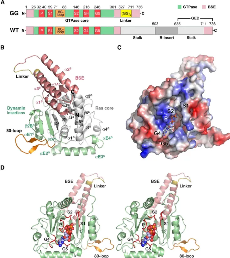
element (BSE, B) of human DNM1L (the so-called GTPase domain - GED (GG) construct [13]). The stalk (S), which participates in higher-order oligomerization, and the B-insert were replaced by a GSGSGSGS linker, which continued with the C-terminal helix of the BSE (Fig. 1A) [38]. This construct was expressed inE. coliand purified and crystallized in the absence of nucleotides. To obtain structural insights into nucleotide binding, nucleotide-free crystals were soaked with the non-hydrolyzable GTP analogue GMP-PNP [38]. Both crystals diffracted to 2.3 A˚ resolution, and the structures were solved by molecular replace-ment using the GG construct of human dynamin-1 as a search model (Tables 1, 2). Both structures exhibited a monomeric GG construct in the asymmetric unit. All molecular contacts generated by symmetry operations of the space group P21212 appear to be typical crystal contacts, as indicated by the PISA server (http:// www.ebi.ac.uk/msd-srv/prot_int/cgi-bin/piserver). However, a polar interface, generated by the 2-fold axis, might form a functional DNM1L assembly, which is discussed in detail further below.
The GTPase domain of human DNM1L consists of a central eight-strandedb-sheet surrounded by sevena-helices and two one-turn helices resembling the GTPase core of mammalian dynamin-1 [53]. The b-sheet is composed of six parallel and two anti-parallel (ap) strands in the spatial orderb6G,b5G,b4G,b1G,b3G,
b2G(ap),b2AG(ap), andb2BG. The latter twob-strands represent an insertion including the helix aE1G with respect to the prototypic, canonical GTPase domain of h-Ras (Fig. 1B) [54]. Helices a1Gtoa5Gare present in h-Ras and dynamin-1, while DNM1L shares helicesaE2GtoaE4Gthat are inserted between
b6Ganda5Gonly with dynamin-1, comprising residues 248 to 281 (Fig. 1B). Although the GTPase domain of DNM1L is similar to that of dynamin-1 at the sequence and structural level, it possesses a unique 16-residue insertion between Ser71 and Glu88, which shall be designated as ‘‘80-loop’’, according to its central residue (Fig. 1).
The BSE is formed by the N-terminal helicesa1B(residues 4– 16) and a1AB (20–24), helix a2B (303–323) and the C-terminal helix a3B (708–729, with residues S708GS710 from the artificial linker), arranged in a three-helix bundle with an additional one-turn helix (Fig. 1B). Both our structures represent the closed conformation of the BSE in relation to the GTPase domain, which has been called the ‘‘post-fission state’’ [37]. Similar closed conformations were found in nucleotide-free and transition state complexes of mammalian dynamin-1 or the GDP bound form of
A. thalianaDrp1A [3,8,16,17,37].
Conformational Changes upon GTP-binding in the Active Site
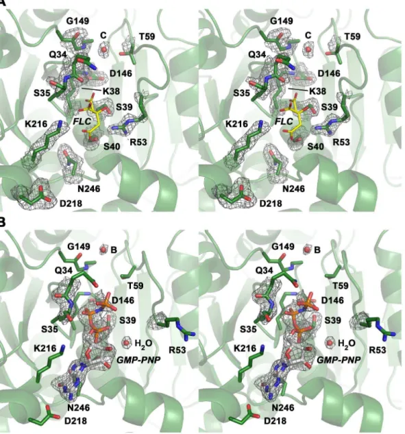
DNM1L contains the five canonical guanine nucleotide binding motifs: G1 or P-loop (phosphate binding loop) with the highly conserved sequence G32SQSSGKSS, switch I with the central T59 as G2 element, G3 around switch II (D146LPG) and G4 (T215KLD), which are conserved among all GTPases, such as h-Ras. In addition, DNM1L also possesses the dynamin-specific G5 or G-cap motif consisting of G243VVNRSQ (Figs. 1C, D) [3]. These motifs surround the active site cleft and exhibit a distinct charge distribution: The positively charged P-loop attracts the anionic triphosphate of GTP, while a less-charged area is complementary to the ribose moiety. The polar part of the guanine base is directed towards the negatively charged G4/G5 region (Fig. 1C). Eventually, the G4 and G5 elements bind guanine, while the P-loop, G4 and G5 bind the ribose moiety and theatoc-phosphates, and the switch I and II regions mediatec -phosphate stabilization and hydrolysis. In the nucleotide-free form,
a citrate from the crystallization buffer is bound to the P-loop, mimicking the phosphate moiety of GTP (Fig. 2A). Apparently, the citrate compensates charges in the active site of DNM1L, while the GMP-PNP occupied active site exhibits a distinct conforma-tion in the P-loop and switch I (Fig. 2B).
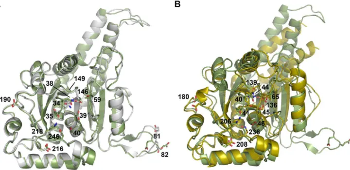
The overall root mean square deviation (RMSD) between the nucleotide-free and –bound DNM1L GG construct is only 0.69 A˚ for the Caatoms, indicating almost identical structures (Fig. 3A).
The nucleotide-bound form and the corresponding dynamin GDP-AlF42 complex (2X2F) exhibit an RMSD value of 2.10 A˚ (Fig. 3B). However, both DNM1L forms differ significantly in their active site (Figs. 2A, B, and 3A), which shall be discussed in the following.
The Phosphate Binding Loop (G1)
The P-loop fixes the triphosphate analogue moiety of GMP-PNP with an intricate hydrogen bonded network (Figs. 4A–C). The a-phosphate is bound by Ser40 via main and side chain interactions, and by backbone interactions of Ser39 and Lys38. Additionally, a single water molecule is coordinated by the a -phosphate (Fig. 4A). Theb-phosphate is coordinated by several main chain interactions of Ser35, Ser36, Gly37 and Lys38, which form a tight loop around theb-phosphate. The hydroxyl group of Ser39 rotates about 180uwith respect to the nucleotide-free active site. In human dynamin-1 with 59 -Guanylylmethylenedipho-sphonate (GMP-PCP) (3ZYC) and GDP-AlF42 (2X2E), or plant Drp1A with GDP-AlF42 (3T34), the side chains of Lys44/47 (corresponding to Lys38 in DNM1L) and Ser45/48 (Ser39 in DNM1L) are involved inbandc-phosphate binding. In contrast, the side chain of Lys38 of DNM1L is in a similar position in the nucleotide-free and GMP-PNP bound forms and does not bind to theb-phosphates (Fig. 4B). Also, Ser39 in the DNM1L structure does not make significant interactions with theb-phosphate.
Compared to the GMP-PCP-bound crystal structure of dynamin-1 (3ZYC), thec-phosphate of GMP-PNP in DNM1L is shifted by about 2.5 A˚ towards the Gln34 side chain. This shift is supported by hydrogen bonds of the Gln34 side chain and the Ser35 main chain with thec-phosphate. Remarkably, the peptide bond between Gln34-Ser35 flips by 180ufrom its apo-conforma-tion upon GMP-PNP binding, accompanied by a Ser35 side chain rotation of nearly 180u (Fig. 4C). The same peptide bond flip is observed when the apo and GDP-bound structures ofD. discoideum
dynamin-A are compared (1JX2 and 1JWY), as well as the Ser35 and Ser39 side chain rotations, strongly supporting the idea that DNM1L and dynamin use a similar mechanism for nucleotide binding [3].
Ser41 in dynamin, corresponding to Ser35 in DNM1L, is required for binding a catalytic cation, such as Na+
, in the presence of a transition state analogue of the GTPase reaction (2X2E) [8]. In our DNM1L structure, we did not find a cation at this position. Also, in the GMP-PCP bound structure of a dynamin-1 GG construct (3ZYC), no cation has been found at this position, since it might only be recruited during GTP hydrolysis. It is possible that the bridging nitrogen atom in GMP-PNP favors an unusual conformation of thec-phosphate, which shifts about 2.5 A˚ away from the catalytic water with respect to the transition state analogue GDP-AlF42 in dynamin-1. This shift might interfere with cation binding of Ser35 in our structure.
The Catalytic Switches I and II Interestingly, no Mg2+
the side chains of Ser45, Asp146, Thr59, and two water molecules. However, in DNM1L, the Caatom of the central residue from
switch I, Thr59, is shifted 2–3 A˚ away from the canonical Mg2+
binding site with respect to corresponding dynamin-GTP analogue complexes (Fig. 5A).
Although the switch I region is not very well defined in the electron density, a large movement of Arg53 is observed (Fig. 5A). In GMP-PNP-DNM1L, the side chain of Arg53 extends to the bulk solvent above the GTP-binding groove, whereas in the nucleotide-free form, this side chain is hydrogen-bonded to Ser40
and Glu43, in a position similar to that of the nucleotide-loaded dynamin complex. This translocation in the nucleotide-free state may be an artificial charge compensation for the negative charge of the P-loop bound citrate.
Based on the crystal structure of the AlF4–bound GTPase domain dimer of dynamin-1, two water molecules were suggested to directly contribute to catalysis: A catalytic (C) water molecule, positioned near Thr65 in switch I and Gln139 in switch II, is thought to mediate the nucleophile attack on the GTP. A second water molecule orients the catalytic water and additionally bridges
Fig. 1B, except for the whole GTPase domain shown in green. Atom colors are carbon in grey, oxygen in red, nitrogen in blue and phosphorus in orange. This nucleotide-free structure corresponds to the closed or post-fission state.
doi:10.1371/journal.pone.0071835.g001
Figure 2. Close-up views of the active site cleft in the nucleotide-free and bound structures of the DNM1L GG construct in stereo. (A) The nucleotide-free form with the most relevant residue side chains of the five GTP binding stretches and citrate (FLC, yellow) displayed as stick models. Electron density of a 2Fo-Fcmap is shown in grey and contoured at 1s. The red sphere designates the catalytic water (C). (B) GMP-PNP complex of the DNM1L GG construct. The nucleotide is shown as stick model, while the red spheres represent water molecules, such as the bridging water (B) and one, which binds to thea-phosphate. The electron density of a 2Fo-Fcmap is shown in grey and contoured at 1s, surrounding the nucleotide and relevant parts of the structure with significant conformational changes with respect to the nucleotide-free form.
Gln40 in the P-loop with Gly139 in switch II (the bridging or B water) [8].
Interestingly, the water molecule in our nucleotide-free DNM1L GG construct is in the same position as the catalytic water in
dynamin-1. This catalytic water connects switch I with switch II via hydrogen bonds to the Thr59 carbonyl oxygen and the Gly149 NH, respectively (Figs. 2A and 5A). In contrast, in the nucleotide-loaded DNM1L complex, the carbonyl oxygen of Gly149 binds to
Figure 3. Superposition of the two DNM1L GG structures and dynamin-1 GG.(A) Overlay of the nucleotide-free DNM1L GG structure in white with the GMP-PNP-bound structure in green (shown without ligands). Side chains that were mutated in our study are shown as stick models with sequence number labels. (B) Overlay of dynamin-1 (PDB code 2X2F) in yellow with the structure of GMP-PNP-bound DNM1L in green. Mutated residues of DNM1L that are equivalent to those of dynamin (see Fig. 3A) are displayed as side chain stick models with dynamin sequence numbers (depicted without ligands).
doi:10.1371/journal.pone.0071835.g003
Figure 4. Detailed views of GTP-binding in the P-loop of DNM1L.(A) Four hydrogen bonds fix thea-phosphate to the P-loop: O1ato Ser40 Oc(distance 2.48 A˚ ) and NH (3.45 A˚), O3ato NHs of Ser39 (3.34 A˚ ) and Lys38 (3.30 A˚). Additionally, the O2abinds an H2O (3.39 A˚ ). Lys38 is stabilized by a hydrogen bond to Asp146 from switch II. The overlaid apo-structure in grey shows that the Ser39 side chain rotates about 180uupon GTP binding, to a conformation (in green) that is suitable to stabilize a GTP transition state as seen in other structures of dynamins, e.g. in complex with GDP-AlF42. (B) Interactions that fix theb-phosphate: O1bforms hydrogen bonds to the NHs of Ser36 (3.31 A˚ ) and of Ser35 (2.94 A˚), while the O2b only binds the Lys38 NH (3.49 A˚). The Nfof Lys38 is more than 4 A˚ away from the O2b, but has the capacity to stabilize together with the Ser39 side chain the phosphate portion of the GTP, as seen in other dynamin-1 nucleotide analogue complexes. (C) Thec-phosphate forms hydrogen bonds via its O2cto the Ser35 NH (3.48 A˚) and to the Ne2 of Gln34 (3.14 A˚ ), which also binds the O1c(2.72 A˚ ). The Gln34 side chain rotates significantly from the apo-conformation (light grey) to the nucleotide-conformation (green). The 180upeptide flip between Gln34-Ser35 brings Ser35 NH in a position suitable for O1band O2cbinding, accompanied by a 180uside chain rotation of Ser35. Similar peptide flips occur in apo-nucleotide pairs of mammalian andD. discoideumdynamins.
a single water molecule, which corresponds to the bridging water molecule in dynamin-1 (Figs. 2B and 5A). Asp146 in switch II is crucial for Mg2+binding in dynamin-1 andAtDrp1A [8,37]. It has
a similar position in the apo- and GMP-PNP structures but interacts only with the Lys38 side chain (Figs. 2B and 4A).
Guanosyl Moiety Binding Elements (G4 and G5)
Overall, the rather rigid guanosyl binding pocket of DNM1L with the G4 and G5 elements exhibits more characteristics of a lock-and-key enzyme whereas structural changes in the more flexible P-loop, and the catalytic residues in switch I and II resemble the induced-fit principle. Thus, the P-loop, switch I and switch II in DNM1L undergo major conformational changes upon nucleotide binding, while no conformational changes were observed in G4 and G5. This phenomenon is found in all related dynamin structures, in which G4 and G5 exhibit strikingly similar conformations independent of the nature of the bound nucleotide. The ribose O4 atom is bound by Lys216 in G4, while the 29OH group of the ribose is stabilized by water-mediated contacts with the Arg247 main chain in G5 and the Ser40 side chain of the P-loop (Fig. 5B). The guanine moiety of GMP-PNP is located between the G4 and G5 stretches, whereby the Lys216 side chain covers one flat side of the aromatic ring system. Nucleotide specificity is mediated by Asp218, which coordinates the guanine base via two hydrogen bonds (Fig. 5C). Another interaction with the G5 stretch completes the substrate binding elements of DNM1L: Asn246 in G5 forms part of the guanine-binding pocket, and its side chain could interact with N7 of the aromatic ring system via a water molecule, as in dynamin-1 (Fig. 5C).
The BSE Conformation
Our nucleotide-free and GMP-PNP DNM1L structures exhibit virtually the same closed or post-fission conformation of the BSE
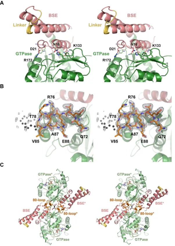
relative to the GTPase domain, with the little defined linker replacing the stalk and B-insert domains (Fig. 1A, B). The interface between the BSE and the GTPase domain is formed by charged, polar, and hydrophobic residues (Fig. 6A). As in the GDP?AlF42 dynamin-1 and theAtDrp1A-GDP complex, the connected helices
a5Ganda2Bare kinked at the Leu301-Pro302 bond by about 60u. In dynamin-1, these helices stretch into a single straight helix upon GTP binding, accompanied by a rotation of the complete BSE around the Leu293-Pro294 kink, corresponding to Leu301-Pro302 in DNM1L.
A comparison of the mammalian dynamin-1 apo (pdb code: 3SNH), GDP?AlF4
_
(2X2E) and GMP-PCP (3ZYC) forms and the corresponding AtDrp1 structures (3T34, 3T35) suggests that the mechanochemical energy conversion is accompanied by a twist of the central b-sheet in the GTPase domain, involving a shift of strands b2G and b3G by about 5 A˚ [8,12,16,17,37]. The GTP analogue complex does not exhibit the twist of the centralb-sheet which might be initiated by GTP-Mg2+
binding to the switch I Thr59 and switch II Asp146 together with Gly149. Since our GG construct is tightly packed in the crystals, the GTP hydrolysis transition state and the pre-fission conformation of the BSE are most likely not accessible. Furthermore, conformational changes induced by GTPase domain dimerization might also contribute to the open conformation of the BSE.
The Unique DNM1L 80-loop
Between the two strandsb2Gandb2AGof the GTPase domain, DNM1L possesses a unique insertion (residues 72–87), which has only a counterpart inS. cerevisiaeDNM1 (see section Conclusion). This insertion forms a flexible loop, which is clamped by a short antiparallelb-sheet between Arg76 to Thr78 and Val85 to Ala87 exhibiting an excess of negatively charged side chains (Fig. 6B). Given its location at the rim of the centralb-sheet of the GTPase
Figure 5. Detailed views of the GTP-binding elements switch I, switch II, G4 and G5 of DNM1L.(A) The canonical Mg2+
site between O1b
and the O2cis not occupied in the GMP-PNP-DNM1L structure (green). Also, no significant positional shift of Thr59 from the switch I loop takes place between apo- (grey) and nucleotide form (green). The unnatural N3batom may favour thec-phosphate conformation rotated by about 60uwith respect to the transition state of GTP, shifting it about 2.5 A˚ away from the catalytic water. Only the nucleotide-free form exhibits the catalytic water molecule (C, grey) bound at the Thr59 carbonyl O (3.15 A˚) and connected to switch II via the NH of Gly149 (2.87 A˚ ). The bridging H2O (B, red) is only present in the nucleotide complex, bound to the Gly149 carbonyl O (3.21 A˚). Upon GMP-PNP binding, the Arg53 side chain moves out of the active site, making room for an H2O, which binds O2 of thea-phosphate. (B) The ribose of GMP-PNP forms bonds with the ether oxygen O4 to the Nfof Lys216 (3.16 A˚ ), and with the hydroxyl group of O2 to an H2O (3.25 A˚ ), which is bonded to the carbonyl O of Arg247 (2.45 A˚) and the Ser40 Oc (3.01 A˚ ). Another bond is formed by the ribose O2 to Gln249 NH (2.79 A˚). (C) The Lys216 side chain, depicted as thin stick model for clarity, covers the aromatic rings of the guanine part, while the Asp218 carboxylate binds the amino N2 (2.86 A˚) and the N1 (3.12 A˚ ). A further interaction from the Asn246 Od1 to the N7 (3.54 A˚ ) might be mediated by an unresolved H2O, which could be bound to the carbonyl O of Gly37, as seen in other dynamin-nucleotide complexes.
Figure 6. BSE-GTPase domain interface, 80-loop, and their interface at the 2-fold axis.(A) The BSE-GTPase interface in the GMP-PNP complex. Both the nucleotide-free and –bound structures represent the closed or post-fission state of dynamin superfamily proteins and exhibit virtually no conformational differences. The interface of GTPase and BSE domain is characterized by mixed charged, polar, and hydrophobic interactions. (B) 80-loop insertion in the GTPase domain of DNM1L from Gln72 to Glu87. The insertion exhibits a short antiparallelb-sheet between Arg76 and Ala87. From Thr79 up to Gly84 the electron density of the 2Fo-Fcmap, shown in grey and contoured at 1s, is not well defined. (C) Polar interface between two symmetry-related DNM1L monomers A and A*. The 2-fold crystallographic axis (black oval) generates an interface that involves 14 hydrogen bonds and salt bridges of polar and charged side chains from the BSE and the 80-loop, together with roughly 20 water molecules (shown as sticks and red spheres, respectively). Both the GTPase domain and the stalks could form higher oligomers, while this dimer remains intact.
domain, the 80-loop might have a role in the BSE domain movement upon GTP hydrolysis or in oligomerization. Due to its distant location from the active site, the 80-loop is unlikely to participate in the GTPase domain dimer interface. In our crystal structures, the 2-fold axis of space group P21212 relates two DNM1L molecules via a polar interface of about 839 A˚2, which resembles a typical crystal contact (Fig. 6C). Various residues of the BSE (amino acids 2, 9, 10) and the 80-loop (amino acids 72, 73, 75, 76, 86) form altogether 14 hydrogen bonds and salt bridges, involving up to 20 water molecules. Nevertheless, we cannot exclude that this novel interface in the dynamin superfamily serves as additional element for the assembly of higher DNM1L oligomers, since its architecture would allow further GTPase-GTPase domain interaction, while the two BSEs could be connected to opposing stalk filaments. Furthermore, small interfaces mediating low affinity interactions are often found in membrane-bound oligomeric complexes [55].
While the overall negatively charged 80-loop may interact with positively charged regions of other protein molecules, some of the seven glutamates and aspartates could serve as cation binding ligands for Mg2+
or even Ca2+
, which sometimes reaches micromolar levels at mitochondrial membranes [56,57]. Intrigu-ingly, the 80-loop sequence resembles those of Ca2+-binding
proteins, such as serine proteases and phospholipid binding proteins [58,59].
GTPase Activity Determinants
Based on the DNM1L GG fusion structure, a systematic analysis of critical residues for GTP binding and turnover was undertaken using full-length DNM1L mutants. The enzyme-coupled assay with increasing GTP concentration resulted in characteristic saturation curves for the GTPase reaction of 1.2mM DNM1L, resembling Michaelis-Menten kinetics (Fig. 7A, Table 3). The apparent maximal GTPase rate was about 6 min-1, with a GTP concentration for half-activation of about 460mM. However,
Michaelis-Menten kinetics assumes a simple enzyme-substrate reaction and does not take into account the underlying multi-step GTPase reaction mechanism of dynamin superfamily proteins [8]. Thus, increasing nucleotide or protein concentrations might induce GTP-dependent interactions of the GTPase domains resulting in higher apparent reaction rates.
To determine whether DNM1L shows liposome-stimulated GTPase activity, GTPase reactions were measured at saturating GTP concentrations (1.5 mM) and higher protein concentrations (10mM) allowing cooperative membrane binding. DNM1L binds
equally well to liposomes constituted of a typical mitochondrial membrane composition or phosphatidylserine (PS) [33,39,52]. Since the liposomes employed in these assays interfered with the continuous coupled reaction, an HPLC-based GTPase assay was used. Furthermore, since divalent cations induce clustering of negatively-charged liposomes, lower concentrations of Mg2+
(0.5 mM) were employed compared to the continuous coupled assay. Previous experiments indicated that this Mg2+
concentra-tion is optimal to observe the DNM1L stimulated GTPase activity with PS liposomes [33]. Under these conditions, DNM1L showed a similar maximal GTPase rate of 7 min21that could be 2.5-fold stimulated by liposomes (Fig. 7B). This extent of GTPase stimulation is, however, much lower than the 200-fold stimulation observed in dynamin-1 under similar conditions [14].
At a concentration of 1.2mM, the DNM1L GG fusion protein showed a reduced maximal GTPase activity of 12%, most likely due to its inability to oligomerize via the stalks [38]. At higher protein concentrations of 10mM, the GG construct exhibited 70% of the GTPase activity of DNM1L pointing to a protein-concentration dependent increase of the GTPase (Figs. 7A, B). Addition of liposomes did not stimulate the GTPase rates, which can be attributed to the missing stalks and/or B-inserts mediating lipid binding.
Mutagenesis of single active site residues within the GTPase binding motifs G1–G5 of full-length DNM1L (Figs. 2, 4, and 5) Table 3.Kinetic parameters of DNM1L basal GTPase activities.
DNM1L kobs(min21)a K (mM)a kcat/Km(M21min21) relative kobsb Motif Function
WT 5.8460.18 462630 126306420 100
GG fusion 0.6960.05 120626 57406160 12
Q34A 0.0 – – 0 G1 (P-loop) c-phosphate binding, G-dimerization
S35A 10.8960.64 298644 3654061730 187 G1 (P-loop) b,c-phosphate binding, G-dimerization
S35Acooperative model 7.7760.14 13865 Hill coefficient2.260.1 133
K38A 0.0 – – 0 G1 (P-loop) a,b-phosphate binding
S39A 0.0 – – 0 G1 (P-loop) a-phosphate binding
S40Ac .1.0 .1000 1670
6640 .20 G1 (P-loop) a-phosphate binding, ribose via H20
T59A 0.0 – – 0 G2 (switch I) coordination of catalytic H2O and Mg2+
E81A 3.5060.06 10265 343106180 60 80-loop unknown
E81A/E82A 3.7960.06 6964 549706210 65 80-loop unknown
D146A 0.0 – – 0 G3 (switch II) coordination of K38
G149A 0.0 – – 0 G3 (switch II) coordination of catalytic and bridging H2O
D190A 1.6460.05 1662 1021906210 28 trans-stabilizing loop G-dimerization
K216A 0.0 – – 0 G4 ribose binding and guanine orientation
D218A 0.0 – – 0 G4 guanine binding
N246Ac .1.0 .1000 1800
6790 .20 G5 guanine binding
aIn all cases except for the cooperative model with the mutant S35A, k
obsand K correspond to kcatand Kmof the applied Michaelis-Menten model; bWT = 100;ck
obsand K could not be determined in a reliable manner, since the substrate did not reach the range of saturating levels.
resulted in most cases in a complete loss of GTPase activities (Fig. 7A, B, and Table 3). Thus, the P-loop mutants K38A and S39A, which are involved in phosphate binding, exhibited essentially no GTP turnover independent of the protein concen-tration or the presence of liposomes. A similar lack of activity has been reported before for mutant K38A [60]. Switch I residue Thr59 is involved in the positioning of the catalytic water molecule and Mg2+
coordination in dynamin-1 [8]. The corresponding T59A mutant in DNM1L as well as the switch II mutants D146A and G149A were also inactive (Fig. 7A, B). The D146A mutation is expected to destabilize the conformation of P-loop residue Lys38, whereas G149A might prevent binding of the catalytic water molecules. Moreover, the G4 motif mutant K216A was
inactive, most likely due to a reduced coordination of the GTP ribonucleotide moiety. The P-loop mutation S40A and the G5 motif mutation N246A showed some residual GTPase activity, which could be stimulated by liposomes (Fig. 7B). However, these mutants could not be saturated with GTP pointing to a reduced affinity for this nucleotide (Fig. 7A).
To explore the function of the 80-loop, we mutated some of the negatively-charged residues and characterized its effect. The two loop mutants E81A and E81A/E82A exhibited slightly different GTP saturation curves compared to wild-type (WT) DNM1L in the continuous-coupled activity assay, such as lower Vmax (kcat) and increased affinity for GTP at half-maximal turnover (Km) (Fig. 7C, Table 3). Also, at higher protein concentrations kobs Figure 7. GTPase activity of DNM1L and the mutants.(A) Basal GTPase activities of wild-type DNM1L, DNM1L GG fusion protein and full-length mutants. Steady-state GTPase activities of full-length wild-type DNM1L, GG fusion protein, active site mutants and predicted GTPase domain dimerization mutants (Q34A, S35A, D190A) were measured as described in the Methods section. Amino acid substitutions Q34A, K38A, S39A, T59A, D146A, G149A, K216A and D218A completely abolished GTP hydrolysis. The Q34A mutant is shown as one representative example for the inactive mutants. Among all these mutants, only S35A, S40A, D190A and N246A exhibited significant GTPase turnover. For S35A, both the simple Michaelis-Menten equation fit (label MM, orange dots) and the curve using a cooperative model (continuous orange line) with a Hill coefficient of 2.2 are depicted. Data are means of at least three independent experiments6standard deviation (displayed as error bars) evaluated by nonlinear regression analysis. (B) Liposome-stimulated GTP hydrolysis of DNM1L and its mutants determined by multiple-turnover assays. Reactions were performed for 12 min at 37uC in the absence (grey bars) or presence (black bars) of PS liposomes. Initial hydrolysis rates kobswere determined by applying a linear fit to the data, with bars representing mean value6standard deviation of three independent experiments. For mutants Q34A, S39A, T59A, D146A, G149A, and K216A less then 4% of the GTP was hydrolyzed within 12 minutes. (C) Basal GTP activity of full length DNM1L and the two loop mutants E81A and E81A/E82A. Although the three variants exhibit similar Michaelis-Menten curves, both mutants displayed lower Vmax(kobs) and faster saturation with GTP compared to WT.
values of both mutants were significantly reduced, reaching about 62% (E81A) and 52% (E81A/E82A) of the WT DNM1L turnover (Fig. 7C). Interestingly, the GTPase activity of these mutants was enhanced by PS liposomes to only 46% of the WT activity in case of the E81A mutant and only 40% for E81A/E82A (Fig. 7B). These data might indicate a function of the 80-loop in higher-order oligomerization, or in the efficient mechanochemical energy conversion of DNM1L, perhaps mediated by the 2-fold BSE/80-loop interface.
Determinants of GTPase Domain Dimerization Linked to Activity
GTPase domain dimerization of human dynamin-1 and
Arabidopsis thaliana dynamin-related protein A (AtDrp1A) was shown to be mediated via a conserved interface across the nucleotide-binding site. A prominent feature of this G-dimer is a hydrogen bond network formed between an aspartic acid side chain of the trans-stabilizing loop (2X2E: Asp180, 3T35: Asp186) with a glutamine and a serine of the P-loop in the opposing molecule (2X2E: Gln40, Ser41, 3T35: Gln43, Ser44). Addition-ally, a serine and a glycine from switch I (2X2E: Ser61 and Gly62, 3T35: Thr64 and Gly65) form hydrogen bonds with Asp180. Overall, these interactions occur twice per G-dimer in a symmetrical manner, involving a rotational 2-fold axis [8]. All of these residues are conserved in DNM1L. Thus, we postulated that DNM1L employs a similar dimerization-dependent GTPase mechanism. Using the GDP-AlF4–bound AtDrp1A dimer as a template, we created a model for the DNM1L dimeric GTPase
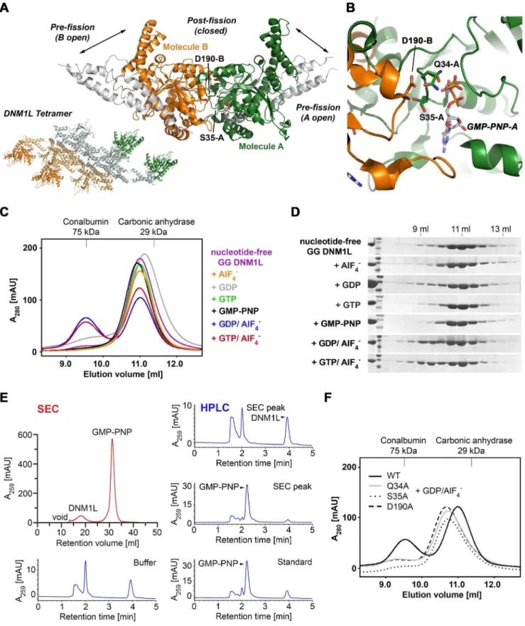
domain (Fig. 8A). In this model, Asp190 in DNM1L, which corresponds to Asp180 in dynamin-1, mediates GTPase domain dimerization by binding to Gln34 and Ser35 in the P loop of the opposing catalytic site. Concomitantly, Asp190 binds to Thr55 and Gly56 from switch I (Fig. 8B).
To confirm this model, size-exclusion chromatography (SEC) experiments in the absence and presence of guanine nucleotides were performed. These measurements revealed that the DNM1L GG fusion protein is predominantly monomeric in the absence of nucleotides and upon incubation with GDP, GTP or GMP-PNP (Fig. 8C, D). In fact, GMP-PNP was efficiently separated from DNM1L over the gel filtration run pointing to a low affinity interaction of nucleotides with DNM1L (Fig. 8E). However, in the presence of GDP/AlF42, a transition state analogue of the GTPase reaction, a significant shift to a dimeric species was observed (Fig. 8C).
In order to investigate if Gln34, Ser35 and Asp190 are involved in GTPase domain dimerization, as predicted by the dimer model, these residues were replaced by alanine residues in the DNM1L GG fusion protein. Indeed, all three mutants dimerized with reduced efficiency in comparison to the wild-type DNM1L GG fusion protein in the presence of the transition state mimic (Fig. 8F). In both GTPase assays, the Q34A mutant did not show any GTPase activity. Since Gln34 is not only involved in dimerization, but also in nucleotide binding, this mutation might interfere with GTP binding. However, both D190A and the Ser35A mutant showed efficient GTP hydrolysis. The D190A mutant displayed even higher GTPase rates at lower nucleotide concentrations, which leveled off at high GTP concentrations. The S35A substitution led to a roughly 2-fold higher basal turnover rate at low and at high GTP concentrations. Remarkably, this mutant exhibited a distinct sigmoidal curve for the turnover of GTP, which agrees best with a cooperative model, resulting in a Hill coefficient of 2.2 (Table 3, Fig. 7A). In contrast, no significant positive cooperativity was observed for the other investigated DNM1L variants with respect to increasing substrate concentra-tions. Both S35A and D190A completely lost their liposome-stimulated GTPase activity (Fig. 7B). These data suggest that Ser35 and Asp190 in DNM1L are indeed involved in GTPase domain dimerization and the subsequent stimulation of the GTPase reaction.
Conclusions
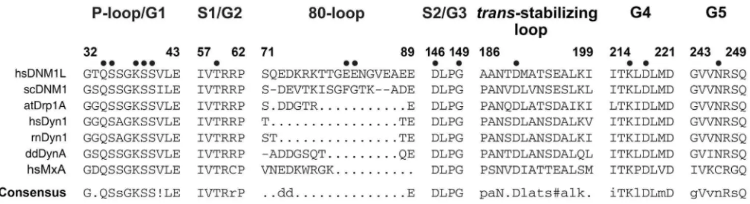
Our study provides a mutational and kinetic analysis of GTP recognizing and hydrolyzing residues in DNM1L, which were identified in the nucleotide-free and GMP-PNP crystal structures. These findings are summarized in a structure-function map of the DNM1L active site (Fig. 9). A sequence comparison of DNM1L with dynamin-1-like proteins from S. cerevisiae and A. thaliana, dynamins-1 of human, rat, and D. discoideum, as well as human Myxovirus resistance protein A (MxA, Mx1), highlights the significance of our investigation for understanding the common mechanisms of these mechanochemical GTPases (Fig. 10). The importance of a functional GTPase in DNM1L is emphasized by studies on cultured mammalian cells, which formed unnaturally large mitochondrial clusters or networks upon transfection with P-loop and switch I mutants of DNM1L, such as K38A and T59A
indicated. The dimeric protein eluted at a retention volume of 9.5 ml and monomeric protein at 11 ml. (D) SDS PAGE analysis of the SEC runs. Lane 1 shows purified GG fusion protein (41 kDa) followed by a molecular weight protein ladder (from top to bottom: 55 kDa, 43 kDa, 34 kDa). Elution volumes are indicated above. (E) Analysis of the DNM1L GMP-PNP complex stability under SEC conditions as in Fig. 8C. SEC elution (red) and further analysis of the peaks by HPLC (blue), with the indicated controls. (F) SEC of GG fusion protein mutants Q34A, S35A and D190A under conditions as in Fig. 8C in the presence of GDP?AlF42. Retention volumes of molecular weight standards are shown above.
doi:10.1371/journal.pone.0071835.g008
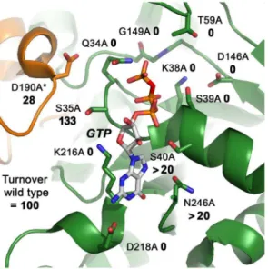
Figure 9. Structure-function map of the modelled DNM1L active site dimer.All active site and dimerization residues that have been mutated to alanine are represented as stick models, as well as the GTP. The turnover numbers of the respective mutants as determined by the GTPase assay for basal activity are shown, whereby the WT was defined as 100%. Molecule A of the dimer is depicted in green, while the second molecule B is shown in orange, with the corresponding D190A*.
[29]. An analysis of rat DNM1L mutants corresponding to K38A and D218N showed similar effects, leading to cellular lethality [61]. Similarly, the K38A DNM1L mutation affects the segrega-tion of peroxisomes [62]. Thus, it can be expected that other GTPase defective DNM1L mutations will result in failure of mitochondria and peroxisome division. Moreover, mutations at the DNM1L G-dimer interface, such as Q34A, S35A, and D190A, might have similar effects on mitochondrial remodeling. Certainly, the formation of higher-ordered oligomers of DNM1L is crucial for its physiological function, as corroborated by the oligmeriza-tion-deficient point mutation in the stalk of human DNM1L, A395D, which is lethal for infants [63].
Future studies have to explore the fine details of mechano-chemical energy conversion by full-length DNM1L with structural methods, and link the underlying mechanisms to the dynamic reality of mitochondrial and peroxisomal segregation. In partic-ular, the roughly 100-fold lower stimulatory effect of lipids on the GTPase activity of DNM1L compared to dynamin-1 has to be elucidated. The large difference of these GTPases in the extent of their stimulated activity is not evident on the basis of the basic architecture of their GTPase domains. However, DNM1L oligomerization at the mitochondrial surface and stronger GTPase stimulation might depend on specific membrane receptors such as
MFF, which was not present in our assays [34]. Moreover, the lower GTPase activation of DNM1L might be based on the different oligomerization modes via the stalks, which have been proposed for dynamin and DNM1L [32,33]. DNM1L is implicated in several neuronal diseases, such as Alzheimer’s, Parkinson’s, Huntington’s, and amyotrophic lateral sclerosis [64]. Eventually, the structure-based knowledge of regulating the function of DNM1L by pharmacological means may help to target these neurodegenerative diseases successfully.
Acknowledgments
Data collection was performed at the Helmholtz-Zentrum Berlin fu¨r Materialien und Energie, Elektronenspeicherring BESSY II, Berlin, Germany. We are thankful to Matthew Locke, Katja Faelber, and Hans Brandstetter for critical reading of the manuscript.
Author Contributions
Conceived and designed the experiments: JW EK OD PG. Performed the experiments: JW EK CF CE AG MH SP. Analyzed the data: JW EK CF CE AG MH SP OD PG. Contributed reagents/materials/analysis tools: JW EK CF OD PG. Wrote the paper: JW EK CF OD PG. Critical reading of the manuscript and corrections: JW EK CF CE AG MH SP OD PG.
References
1. Heymann Jr AW, Hinshaw JE (2009) Dynamins at a glance. Journal of Cell Science 122: 3427–3431.
2. Praefcke GJK, McMahon HT (2004) The dynamin superfamily: universal membrane tubulation and fission molecules? Nat Rev Mol Cell Biol 5: 133–147. 3. Niemann HH, Knetsch MLW, Scherer A, Manstein DJ, Kull FJ (2001) Crystal structure of a dynamin GTPase domain in both nucleotide-free and GDP-bound forms. EMBO J 20: 5813–5821.
4. Saraste M, Sibbald PR, Wittinghofer A (1990) The P-loop – a common motif in ATP- and GTP-binding proteins. Trends in Biochemical Sciences 15: 430–434. 5. Vetter IR, Wittinghofer A (2001) The Guanine Nucleotide-Binding Switch in
Three Dimensions. Science 294: 1299–1304.
6. Tuma PL, Stachniak MC, Collins CA (1993) Activation of dynamin GTPase by acidic phospholipids and endogenous rat brain vesicles. Journal of Biological Chemistry 268: 17240–17246.
7. Sever S, Muhlberg AB, Schmid SL (1999) Impairment of dynamin’s GAP domain stimulates receptor-mediated endocytosis. Nature 398: 481–486. 8. Chappie JS, Acharya S, Leonard M, Schmid SL, Dyda F (2010) G domain
dimerization controls dynamin’s assembly-stimulated GTPase activity. Nature 465: 435–440.
9. Song BD, Leonard M, Schmid SL (2004) Dynamin GTPase Domain Mutants That Differentially Affect GTP Binding, GTP Hydrolysis, and Clathrin-mediated Endocytosis. Journal of Biological Chemistry 279: 40431–40436.
10. Ghosh A, Praefcke GJK, Renault L, Wittinghofer A, Herrmann C (2006) How guanylate-binding proteins achieve assembly-stimulated processive cleavage of GTP to GMP. Nature 440: 101–104.
11. Marks B, Stowell MHB, Vallis Y, Mills IG, Gibson A, et al. (2001) GTPase activity of dynamin and resulting conformation change are essential for endocytosis. Nature 410: 231–235.
12. Chappie JS, Mears JA, Fang S, Leonard M, Schmid SL, et al. (2011) A Pseudoatomic Model of the Dynamin Polymer Identifies a Hydrolysis-Dependent Powerstroke. Cell 147: 209–222.
13. Chappie JS, Acharya S, Liu Y-W, Leonard M, Pucadyil TJ, et al. (2009) An Intramolecular Signaling Element that Modulates Dynamin Function In Vitro and In Vivo. Molecular Biology of the Cell 20: 3561–3571.
14. Faelber K, Held M, Gao S, Posor Y, Haucke V, et al. (2012) Structural Insights into Dynamin-Mediated Membrane Fission. Structure 20: 1621–1628. 15. Gao S, von der Malsburg A, Paeschke S, Behlke J, Haller O, et al. (2010)
Structural basis of oligomerization in the stalk region of dynamin-like MxA. Nature 465: 502–506.
16. Ford MGJ, Jenni S, Nunnari J (2011) The crystal structure of dynamin. Nature 477: 561–566.
17. Faelber K, Posor Y, Gao S, Held M, Roske Y, et al. (2011) Crystal structure of nucleotide-free dynamin. Nature 477: 556–560.
18. Hinshaw JE, Schmid SL (1995) Dynamin self-assembles into rings suggesting a mechanism for coated vesicle budding. Nature 374: 190–192.
Figure 10. Conserved sequence motifs of the dynamin superfamily.Sequence alignments of representative members of the dynamin superfamily comparing conserved GTP binding motifs and thetrans-stabilizing loops important for G dimerization. Conserved key residues that were mutated to alanine in our study are marked with a dot. Species abbreviations are hs,Homo sapiens; rn,Rattus norvegicus; sc,Saccharomyces cerevisiae; dd,Dictyostelium discoideum; at,Arabidopsis thaliana; MxA, interferon-induced GTP-binding protein A; ! represents one of the amino acids of IV;
19. Ramachandran R, Surka M, Chappie JS, Fowler DM, Foss TR, et al. (2007) The dynamin middle domain is critical for tetramerization and higher-order self-assembly. EMBO J 26: 559–566.
20. von der Malsburg A, Abutbul-Ionita I, Haller O, Kochs G, Danino D (2011) Stalk Domain of the Dynamin-like MxA GTPase Protein Mediates Membrane Binding and Liposome Tubulation via the Unstructured L4 Loop. Journal of Biological Chemistry 286: 37858–37865.
21. Otera H, Ishihara N, Mihara K (2013) New insights into the function and regulation of mitochondrial fission. Biochimica et Biophysica Acta (BBA) -Molecular Cell Research 1833: 1256–1268.
22. Koch A, Thiemann M, Grabenbauer M, Yoon Y, McNiven MA, et al. (2003) Dynamin-like Protein 1 Is Involved in Peroxisomal Fission. Journal of Biological Chemistry 278: 8597–8605.
23. Strack S, Cribbs JT (2012) Allosteric Modulation of Drp1 Mechanoenzyme Assembly and Mitochondrial Fission by the Variable Domain. Journal of Biological Chemistry 287: 10990–11001.
24. Elgass K, Pakay J, Ryan MT, Palmer CS (2013) Recent advances into the understanding of mitochondrial fission. Biochimica et Biophysica Acta (BBA) -Molecular Cell Research 1833: 150–161.
25. Grohm J, Kim SW, Mamrak U, Tobaben S, Cassidy-Stone A, et al. (2012) Inhibition of Drp1 provides neuroprotection in vitro and in vivo. Cell Death Differ 19: 1446–1458.
26. Reddy PH, Reddy TP, Manczak M, Calkins MJ, Shirendeb U, et al. (2011) Dynamin-related protein 1 and mitochondrial fragmentation in neurodegener-ative diseases. Brain Research Reviews 67: 103–118.
27. Chang C-R, Manlandro CM, Arnoult D, Stadler J, Posey AE, et al. (2010) A Lethal de Novo Mutation in the Middle Domain of the Dynamin-related GTPase Drp1 Impairs Higher Order Assembly and Mitochondrial Division. Journal of Biological Chemistry 285: 32494–32503.
28. Binns D, Barylko B, Grichine N, Atkinson ML, Helms M, et al. (1999) Correlation Between Self-Association Modes and GTPase Activation of Dynamin. Journal of Protein Chemistry 18: 277–290.
29. Smirnova E, Griparic L, Shurland D-L, van der Bliek AM (2001) Dynamin-related Protein Drp1 Is Required for Mitochondrial Division in Mammalian Cells. Molecular Biology of the Cell 12: 2245–2256.
30. Friedman JR, Lackner LL, West M, DiBenedetto JR, Nunnari J, et al. (2011) ER Tubules Mark Sites of Mitochondrial Division. Science 334: 358–362. 31. Montessuit S, Somasekharan SP, Terrones O, Lucken-Ardjomande S, Herzig S,
et al. (2010) Membrane Remodeling Induced by the Dynamin-Related Protein Drp1 Stimulates Bax Oligomerization. Cell 142: 889–901.
32. Mears JA, Lackner LL, Fang S, Ingerman E, Nunnari J, et al. (2011) Conformational changes in Dnm1 support a contractile mechanism for mitochondrial fission. Nat Struct Mol Biol 18: 20–26.
33. Fro¨hlich C, Grabiger S, Schwefel D, Faelber K, Rosenbaum E, et al. (2013) Structural insights into oligomerization and mitochondrial remodelling of dynamin 1-like protein. EMBO J 32: 1280–1292.
34. Kageyama Y, Zhang Z, Sesaki H (2011) Mitochondrial division: molecular machinery and physiological functions. Current Opinion in Cell Biology 23: 427–434.
35. Otera H, Mihara K (2011) Discovery of the membrane receptor for mitochondrial fission GTPase Drp1. Small GTPases 2: 167–172.
36. Ingerman E, Perkins EM, Marino M, Mears JA, McCaffery JM, et al. (2005) Dnm1 forms spirals that are structurally tailored to fit mitochondria. The Journal of Cell Biology 170: 1021–1027.
37. Yan L, Ma Y, Sun Y, Gao J, Chen X, et al. (2011) Structural basis for mechanochemical role of Arabidopsis thaliana dynamin-related protein in membrane fission. Journal of Molecular Cell Biology 3: 378–381.
38. Klinglmayr E, Wenger J, Mayr S, Bossy-Wetzel E, Puehringer S (2012) Purification, crystallization and X-ray diffraction analysis of human dynamin-related protein 1 GTPase-GED fusion protein. Acta Crystallographica Section F 68: 1217–1221.
39. Song W, Chen J, Petrilli A, Liot G, Klinglmayr E, et al. (2011) Mutant huntingtin binds the mitochondrial fission GTPase dynamin-related protein-1 and increases its enzymatic activity. Nat Med 17: 377–382.
40. Mueller U, Darowski N, Fuchs MR, Forster R, Hellmig M, et al. (2012) Facilities for macromolecular crystallography at the Helmholtz-Zentrum Berlin. Journal of Synchrotron Radiation 19: 442–449.
41. Leslie AG (2006) The integration of macromolecular diffraction data. Acta Crystallogr D Biol Crystallogr 62: 48–57.
42. Evans P (2006) Scaling and assessment of data quality. Acta Crystallogr D Biol Crystallogr 62: 72–82.
43. McCoy AJ, Grosse-Kunstleve RW, Adams PD, Winn MD, Storoni LC, et al. (2007) Phaser crystallographic software. J Appl Crystallogr 40: 658–674. 44. Winn MD, Ballard CC, Cowtan KD, Dodson EJ, Emsley P, et al. (2011)
Overview of the CCP4 suite and current developments. Acta Crystallographica Section D 67: 235–242.
45. Adams PD, Afonine PV, Bunkoczi G, Chen VB, Davis IW, et al. (2010) PHENIX: a comprehensive Python-based system for macromolecular structure solution. Acta Crystallogr D Biol Crystallogr 66: 213–221.
46. Emsley P, Cowtan K (2004) Coot: model-building tools for molecular graphics. Acta Crystallogr D Biol Crystallogr 60: 2126–2132.
47. Collaborative Computational Project N (1994) The CCP4 suite: programs for protein crystallography. Acta Crystallographica Section D 50: 760–763. 48. Chen VB, Arendall WB III, Headd JJ, Keedy DA, Immormino RM, et al. (2010)
MolProbity: all-atom structure validation for macromolecular crystallography. Acta Crystallographica Section D 66: 12–21.
49. Krissinel E, Henrick K (2004) Secondary-structure matching (SSM), a new tool for fast protein structure alignment in three dimensions. Acta Crystallographica Section D 60: 2256–2268.
50. DeLano WL (2002) The PyMOL Molecular Graphics System. DeLano Scientific, San Carlos, CA, USA.
51. Fenner G (1931) Das Genauigkeitsmass von Summen, Differenzen, Produkten und Quotienten der Beobachtungsreihen. Die Naturwissenschaften 19: 310– 310.
52. Bossy B, Petrilli A, Klinglmayr E, Chen J, Lu¨tz-Meindl U, et al. (2010) S-Nitrosylation of DRP1 does not affect enzymatic activity and is not specific to Alzheimer’s disease. J Alzheimers Dis 20: S513–526.
53. Reubold TF, Eschenburg S, Becker A, Leonard M, Schmid SL, et al. (2005) Crystal structure of the GTPase domain of rat dynamin 1. Proceedings of the National Academy of Sciences of the United States of America 102: 13093– 13098.
54. Pai EF, Krengel U, Petsko GA, Goody RS, Kabsch W, et al. (1990) Refined crystal structure of the triphosphate conformation of H-ras p21 at 1.35 A resolution: implications for the mechanism of GTP hydrolysis. EMBO J 9: 2351– 2359.
55. Schmid EM, McMahon HT (2007) Integrating molecular and network biology to decode endocytosis. Nature 448: 883–888.
56. Dudev M, Lim C (2007) Discovering structural motifs using a structural alphabet: Application to magnesium-binding sites. BMC Bioinformatics 8: 106. 57. Rapizzi E, Pinton P, Szabadkai G, Wieckowski MR, Vandecasteele G, et al. (2002) Recombinant expression of the voltage-dependent anion channel enhances the transfer of Ca2+microdomains to mitochondria. The Journal of Cell Biology 159: 613–624.
58. Bode W, Schwager P (1975) The single calcium-binding site of crystallin bovin beta-trypsin. FEBS Lett 56: 139–143.
59. Nelsestuen GL, Ostrowski BG (1999) Membrane association with multiple calcium ions: vitamin-K-dependent proteins, annexins and pentraxins. Current Opinion in Structural Biology 9: 433–437.
60. Zhu PP, Patterson A, Stadler J, Seeburg DP, Sheng M, et al. (2004) Intra- and intermolecular domain interactions of the C-terminal GTPase effector domain of the multimeric dynamin-like GTPase Drp1. J Biol Chem 279: 35967–35974. 61. Pitts KR, Yoon Y, Krueger EW, McNiven MA (1999) The dynamin-like protein DLP1 is essential for normal distribution and morphology of the endoplasmic reticulum and mitochondria in mammalian cells. Mol Biol Cell 10: 4403–4417. 62. Koch A, Thiemann M, Grabenbauer M, Yoon Y, McNiven MA, et al. (2003) Dynamin-like protein 1 is involved in peroxisomal fission. J Biol Chem 278: 8597–8605.
63. Waterham HR, Koster J, van Roermund CWT, Mooyer PAW, Wanders RJA, et al. (2007) A Lethal Defect of Mitochondrial and Peroxisomal Fission. New England Journal of Medicine 356: 1736–1741.