ISSN: 2067-533X
INTERNATIONAL JOURNAL
OF
CONSERVATION SCIENCE
Volume 4, Issue 2, April-June 2013: 145-152 www.ijcs.uaic.ro
SCIENTIFIC INVESTIGATION OF THE MATERIALS AND
TECHNIQUES USED IN A 19
THCENTURY EGYPTIAN CEMETERY
WALL PAINTING (HAWSH AL-BASHA)
Sawsan Sayed DARWISH *
Conservation Department, Faculty of Archaeology, Cairo University, Giza, Egypt
Abstract
The present research was carried out to obtain more information on materials and painting techniques used in Egyptian wall paintings during the 19th century. The Hawsh al-Basha courtyard, dating back to Mohammed Ali's family period (1805-1952,) was studied for this purpose. The obtained results will be used to set up a scientific plan for restoration and preservation. Pigments, including white zincite, earth green, blue synthetic ultramarine, yellow massicot, black a mixture of magnetite & graphite, brownish red lead and brass were identified. The binding medium in the painting was identified as animal glue. Two preparation layers were identified: the inner coarse ground layer, composed of gypsum as a major component, with calcite and small amounts of quartz and the outer, fine ground layer, composed of calcite only. Optical Microscopy (OM), Scanning Electron Microscopy coupled with Energy Dispersive X-ray Microanalysis (SEM-EDX), X-ray Diffraction (XRD) and Fourier Transform Infrared Spectroscopy with Attenuation Total Reflection (FTIR-ATR) were used in our study.
Keywords: Wall painting; Hosh Al-Basha, OM; SEM-EDX; XRD; FTIR-ATR.
Introduction
Hawsh al-Basha is an Arabic name meaning, “courtyard of the Pasha”, which was given to the Royal family graveyards and, more specifically, to the descendents of Mohammed Ali's family. The graveyards were built on a rectangular area located behind the tomb of Al-Imam al-Shafei, dating back to the 12th century, south of the Cairo citadel. The graveyard is dating back to 1815, but it was reported that Mohammed Ali bought this courtyard in 1805 [1-2]. At first he built a simple graveyard, consisting of two domes, that was completed later to be a six-domed complex. The tomb complex was built for the burial of his family members and his distinguished statesmen. The interiors of the chamber of Khediew Tawfik's mother, Nour Shafaq, were richly decorated with inscriptions, precious marbles and amazingly detailed, colorful paintings. The analysis of pigments used in wall paintings provides useful information in defining the gamut of pigments available at a local, regional or even wider scale and in understanding the techniques of color preparation and application. In addition, through pigment study, it is possible to discover lines of communication and trade exchanges [3].
This paper aims to characterize the materials and techniques used in the wall paintings of Hawsh al-Basha. The results obtained will provide the conservators with the essential
*
information needed to choose the suitable materials and methods that can be used in conservation and restoration works.
Experimental
Painted layers samples
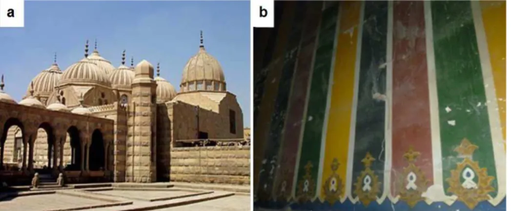
Some representative samples were collected from the detached parts of painted layers with different colors on the chamber wall of Khediew Tawfik's mother, Nour Shafaq, in the Hawsh al-Basha graveyard (Fig. 1). Preliminary morphological observations of the samples were carried out by using digital optical light microscopy coupled with a computer to investigate their stratigraphic sequence. Then the samples were prepared for characterization studies.
Fig. 1. The tomb complex of Hawsh al-Basha in Al-Imam Al-Shafei: a – outside view, b - interiors of the chamber of Khediew Tawfik's mother, Nour Shafaq
Investigation methodologies
Different analytical techniques were performed to characterize the pigments, grounds and binding media employed in a number of pigment fragments. Coupling of the used techniques, most of which being non-destructive, suitable for the determination of anionic groups, crystalline phases, structure and elemental composition, allowed us to obtain a complete enough characterization of the used pigments, materials and techniques.
Scanning electron microscopy
The microstructure of the painted samples was observed by scanning electron microscope (SEM), type Philips XL30, equipped with an EDX micro-analytical system. This examination was performed to detect the elements contents qualitatively and quantitatively in the samples. Images were acquired in backscattered mode (BSE). It was useful for semi quantitative elemental analysis, to make up for the deficiencies of XRD [4]. In some cases, doubt arose about specific minerals that could not be readily identified by XRD [5].
X-ray diffraction
The mineralogical composition of the painting specimens was obtained by XRD analysis. Fine powders of the pigments and subsurface layer were analyzed with a diffractometer (PW 3071 Philips - CuKα 40kV, 30mA). The scanned 2θ range was 5 to 60 degrees.
Fourier transformed infrared spectroscopy
MATERIALS AND TECHNIQUES USED IN A 19 CENTURY EGYPTIAN CEMETERY WALL PAINTING
Results and Discussions
The structure of the wall paintings and the pigments used
Ground layers
The preliminary observation of the collected specimens revealed the presence of three main layers: a thin paint layer, a white finely grained preparation layer underneath and the bottom – the coarse ground layer. The preparation technique used seems to apply the first layer of a coarse ground layer directly on the lime stone wall followed by another smooth layer prepared for the painting process. This three-layer structure was observed on all samples and could also be visualized in the SEM results. Based on our experimental results obtained, XRD analysis revealed that the inner coarse ground layer was composed of gypsum CaSO4⋅2H2O as a
major component, with calcite CaCO3 and small amounts of quartz (SiO2), while the fine
ground layer was composed of calcite only (Fig. 2).
Fig. 2. Microscopic images (350 X) and spectrums:
a – OM image, b – SEM image, 1- thin paint layer; 2- finely grained preparation layer, 3- coarse ground layer, c - XRD pattern of coarse ground layer, d - XRD pattern of fine preparation layer.
Binding media
The spectroscopic study was essentially addressed to characterize the coloring medium used in the pigment samples. In all the analyzed samples, the stretching vibrations of calcium carbonate, CaCO3, peaked at 1396, 873 and the 712cm-1 assignable to CO32- group were
identified since the substrate was just a calcarenite [6]. The use of animal glue was revealed in this FTIR- ATR spectrum (Fig. 3c) by the presence of a band at ≈1650cm-1
, assignable to C=O stretching (amide I) and a band at 1530cm-1 associated with C–N stretching and the deformation vibration of the N–H (amide II) [7].
Pigments identification
Green pigments
Green Earth K[(Al,FeIII),(FeII,Mg)](AlSi3,Si4)O10(OH)2 could be recognized from the
the area 1100-900cm-1 of Si-O stretching and O–H stretching in the region 3300-3600cm-1 lead to characterize the green earth as celadonite. XRD results confirm this conclusion (Fig. 3d).
Fig. 3. The analysis of green pigment:
a – EDX spectrum, b – SEM image (1000X), c – FTIR Spectrum, d – XRD pattern.
Blue pigment
EDX analysis (Fig. 4a) reveals the presence of Na, Al, Si, S and Zn, which leads to characterize the blue colour as Ultramarine. XRD analysis (Fig. 4c) shows the presence of Ultramarine, zincite and calcite, indicating that the blue colour was composed of Ultramarine pigment (Na8-10Al6Si6O24S2-4) and zincite ZnO. Zincite was probably added to various colored
pigments by manufacturers as a lighting agent. The presence of calcite is related to preparation layer. The FTIR spectrum shows the presence of Si-O stretching bands in the area 1100-900cm
-1
confirming the characterization of the blue pigment as Ultramarine. The SEM image (Fig. 4b) shows spherical particles in the microscope, suggesting the blue pigment to be synthetic Ultramarine. Although the chemical composition of both natural and synthetic Ultramarine is almost identical, we can recognize the difference among the two pigments, especially well under artificial illumination. Synthetic Ultramarine pigments shows, contrary to the Lapis lazuli spherical equal sized particles in the microscope [12]. Synthetic Ultramarine was synthesized by both the French Jean Baptiste Guimet and the German chemist Christian Gottlob Gmelin in the late 1820s and early 1830s. Schmauderer [13] reported that a short time after 1830 synthetic Ultramarine was used for painting.
Fig. 4. The analysis of blue pigment:
MATERIALS AND TECHNIQUES USED IN A 19 CENTURY EGYPTIAN CEMETERY WALL PAINTING
White pigment
The mineralogical compositions of the studied white samples (Fig. 5c) show that zincite (ZnO) is responsible for the white color. It was called red, orange, yellow and white zinc, the oxide mineral that was mined and crushed to produce various colours of zinc paints during 1830 to 1850 in Europe and America. Welsh [14] reported that the New Jersey Zinc Company not only mined the ore but also manufactured the zinc oxide pigment and the paints at their plant in New-York starting in the late 1840s. In addition, they successfully marketed paints made with the crushed ore, franklinite and zincite in a variety of colors. The presence of zinc in the EDX data confirms this result. The presence of chloride and sodium is related to halite (NaCl) whose presence is due to the materials used in the ground layer or/and salty water leaked through the wall (Fig. 5a). The SEM image (Fig. 5b) shows the distinguishing characteristic of the acicular form of zinc white pigment i.e., many of the smallest particles in the paint have a unique crystal form of two, three, or four needles combined, like arms radiating from a central join.
Fig. 5. The analysis of white pigment:
a - EDX spectrum, b - SEM image (148 X), c - XRD pattern.
Yellow pigment
EDX analysis (Fig. 6a) revealed the presence of lead, zinc, calcium, sodium and chlorine. XRD analysis (Fig. 6c) confirmed these results and showed that the presence of massicot mixed with small amount of zincite was responsible for the yellow color. The SEM image (Fig. 6b) shows the orthorhombic- dipyramidal crystal form of massicot, the morphology of the crystals look like fish scales. Massicot has been used as a yellow pigment since antiquity [15] and was identified as a yellow pigment by other studies [16-17]. It was also used in Abdeen palace (one of the official residences of the president of Egypt) dating to the Mohamed Ali pasha period, the 19th century [18].
Black pigment
The results of EDX analysis of the black paint samples (Fig. 7a) reveal the presence of iron (Fe) and carbon (C). XRD analysis (Fig. 7c) shows the presence of crystalline phases of magnetite Fe3O4 and graphite, indicating that in this instance the black paint is a mixture. The
presence of silicon, calcium and sulphur is related to the preparation layers below. The SEM image (Fig. 7b) shows that the crystals have a small size and occur in massive granular form, both coarse and fine.
Brownish red
could be prepared by the use of red lead (minium, Pb3O4) [19]. The SEM image (Fig. 8b) shows
indistinguishable crystal form of the brownish red pigment. Browning of red lead occurs under
the effect of environmental parameters mainly humidity, light and salts [20-21]. Browning more notably occurred when red lead was applied in water color or tempera media,
resulting in the formation of brown lead oxide. Recent investigations show that both composition and micro-structural features of red lead grains, which result from the
manufacturing process, are determining factors in the pigment alteration process [21].
Fig. 6. The analysis of yellow pigment:
a - EDX spectrum, b - SEM image (266 X), c - XRD pattern.
Fig. 7. The analysis of black pigment:
a - EDX spectrum, b - SEM image (148 X), c - XRD pattern
MATERIALS AND TECHNIQUES USED IN A 19 CENTURY EGYPTIAN CEMETERY WALL PAINTING
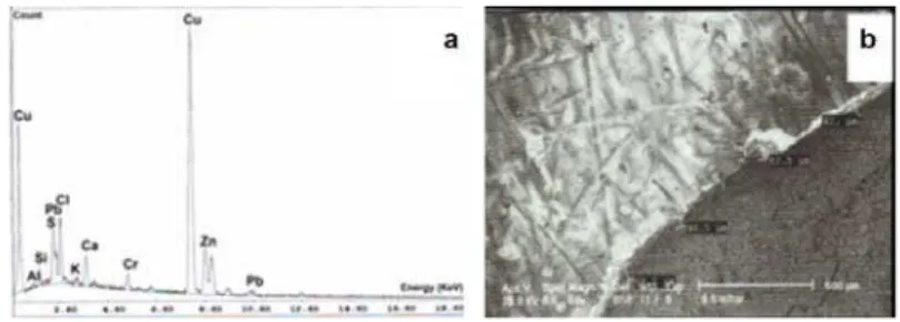
Golden pigment
SEM-EDX results show that the golden color was composed mainly from copper and zinc [Fig. 9], indicating that the golden color is brass (Cu-Zn alloy). The same results were obtained in Abdeen palace, dating to the Mohamed Ali pasha period, the 19th century [18]. The shape and composition of the brass thin layer of the painting was identified by using SEM (Fig. 9b).
Fig. 9. The analysis golden colour: a - EDX spectrum, b – SEM image
Conclusions
In our study, the painting technique, pigments and binding media used in wall paintings of Hawsh al-Basha courtyard were characterized. The painting was done in three layers: the first one is the ground layer, made of gypsum as a major component, with calcite and small amounts of quartz, followed by the fine preparation layer, made of calcite only and at the top the paint layer. This is a very typical Egyptian wall painting technique. White zincite, earth green, blue ultramarine, yellow massicot, brownish red lead, black a mixture of (magnetite and graphite) and golden brass were among the common pigments used in Egyptian wall paintings of the 19th century. The browning of red lead is assumed to have occurred through the effect of environmental parameters, mainly humidity, light and salts. The binding medium in the painting was identified as animal glue, also found in other Egyptian wall paintings. The results obtained will provide the conservators with the essential information needed to choose the suitable materials and methods to be used in conservation and restoration works.
Acknowledgements
The author would like to thank Dr. Amany Abd el Hafez and Mr. Mourd Fawzy at The Conservation Department, Faculty of Archaeology, Cairo University, Egypt for their useful help especially in XRD interpretation.
References
[1] C. Williams, Islamic Monuments in Cairo: The Practical Guide, American University of Cairo Pre-ss, Cairo,2002.
[2] F.A. Mostafa, Cairo cemetery architectures in 19th century, archeological and
architectural study, MA Thesis, Faculty of Archaeology, Cairo University, 2003.
[3] G.A. Mazzochin, F. Agnoli, S. Mazzochin, I. Colpo, Analysis of pigments from Roman
[4] J. Hanlan, The Scanning Electron Microscope and Microprobe: application to
conservation and historical research. 4th Triennial Meeting ICOM Committee for
Conservation, Venice, 13‐18 October, ICOM Paris, 1975, p.6.
[5] V. Perdikatsis, V. Kilikoglou, S. Sotiropoulou, E. Chryssikopoulou, Physiochemical
characterization of pigments of Theran wall paintings, The Wall Paintings of Thera
(Editor: S. Sherratt), Proceedings of the First International Symposium, Petros M. Nomikos Conference Centre, Thera, Hellas, 30 August–4 September 1997, Petros M. Nomikos and the Thera Foundation, Athens, 2000, pp.103–118.
[6] W. Griffith, Advances in the Raman and infrared spectroscopy of minerals.
Spectroscopy of inorganic based materials, Spectroscopy of Inorganic‐Based
Materials (Editors: R.J.H. Clark and R.E. Hester), Chapter 2, Wiley, Chichester, 1987, pp. 315-325.
[7] M.R. Derrick, D. Stulik, J.M. Landry, Infrared Spectroscopy in Conservation Science, The Getty Conservation Institute, Los Angeles, 1999, pp. 97-98.
[8] R.D. Harley, Artists’ Pigmentsc. 1600-1835, Archetype Publications, London, 2001. [9] R.J.H. Clark, Pigment Identification by Spectroscopic Means: an Arts/SciencPigments
in the History of Painting, Applied Clay Science, 22, 2002, pp. 223-236
[10]D. Hradil, T. Grygar, J. Hradilová, P. Bezdićka, Clay and Iron Oxide Interface,
Academie des Sciences, 5(1), 2003, pp. 7-20.
[11]I. Kakoulli, Roman wall paintings in Cyprus: a scientific investigation of their
technology, Roman Wall Painting: Materials, Techniques, Analyses and
Conservation, (Editors: H. Bearat, M. Fuchs, M. Maggetti and D. Paunier),
Proceedings of the International Workshop, Fribourg, 7-9 March 1996, Institute of Mineralogy and Petrology, Fribourg University, 1997, pp. 131-142.
[12]H. Becker, Blau als Pigment - oder blau ist nicht gleich blau, Blau: Farbe der Ferne
(Editor: H. Gehrcke), Heidelberg, Wunderhorn, 1990, pp. 36-52
[13]E. Schmauderer, Die Entwicklung der Ultramarin-Fabrikation im 19. Jahrhundert, Tradition, Bruckmann, München, vol.14, 1969, pp. 127-152
[14]S.F. Welsh, Identification of 1850s Brown Zinc Paint Made with Franklinite and
Zincite at the U.S. Capitol, APT Bulletin, 39, 2008, pp. 17-30.
[15]K. Wehlte, The Materials and Techniques of Painting, Van Nostrand Reinhold, London, 1975.
[16]S.I. Augusti, Colori Pompeiani, De Luca, Roma, 1967.
[17]E. Doorylée, M. Anne, I. Bardies, J.L. Hodeau, P. Martinetto, S. Rondot, J. Saloman, G.B.M. Vaughan, P. Walter, Non-destructive synchroton X-ray diffraction mapping of
a Roman painting,Journal of Applied Physics, A81, 2005, pp. 663- 667.
[18]M. F. Ali, S. S. Darwish, Comparative Analytical Study of the materials used in Wall
Painting of Historical Palaces, EgyptianJournal of Archaeological & Restoration
Studies, 1(1), 2011, pp. 91-100.
[19]I.M. Bell, R.J.H. Clark, P.J. Gibbs, Raman Spectroscopic Library of Natural and
Synthetic Pigments (Pre-~1850 AD), Spectrochimica Acta, Part A, 53, 1997, pp.
2159-2179.
[20]S. Aze, J. Vallet, M. Pomey, A. Baronnet, O. Grauby, Red lead darkening in wall paintings: natural ageing of experimental wall paintings versus artificial ageing tests,
European Journal of Mineralogy, 19(6), 2007, pp. 883-890.
[21]S. Aze, J.M. Vallet, V. Detalle, O. Grauby, A. Baronnet, Chromatic alterations of red
lead pigments in art works. A review,Phase Transition, 81(2-3), 2008, pp.145-154.