Drug Transporter Genetic Variants Are Not
Associated with TDF-Related Renal
Dysfunction in Patients with HIV-1 Infection:
A Pharmacogenetic Study
Takeshi Nishijima1,2, Tsunefusa Hayashida1, Takuma Kurosawa3,4, Noriko Tanaka4, Shinichi Oka1,2, Hiroyuki Gatanaga1,2*
1AIDS Clinical Center, National Center for Global Health and Medicine, Tokyo, Japan,2Center for AIDS Research, Kumamoto University, Kumamoto, Japan,3Department of Mathematical Science for Information Sciences, Graduate School of Science, Tokyo University of Science, Tokyo, Japan,4Biostatistics Section, Department of Clinical Research and Informatics, Clinical Science Center, National Center for Global Health and Medicine, Tokyo, Japan
Abstract
Objective
To investigate whether single nucleotide polymorphisms (SNP) of drug transporter proteins for TDF is a risk factor for TDF-related renal function decrement.
Methods
This study investigated the association between 3 SNPs (ABCC2–24, 1249, andABCB1 2677), which are shown to be associated with TDF-induced tubulopathy, and clinically important renal outcomes (>10ml/min/1.73m2decrement in eGFR relative to baseline, >25% decrement in eGFR, and eGFR<60ml/min/1.73m2) in 703 HIV-1-infected Japanese
patients who initiated TDF-containing antiretroviral therapy (ART). Genotyping was per-formed by allelic discrimination using TaqMan 5’-nuclease assays.
Results
95% of the study patients were males and 66% were treatment-naïve, with median CD4 count of 249/μl, median baseline eGFR of 96ml/min/1.73m2(IQR 84.6–109.2), and median
exposure to TDF of 3.66 years (IQR 1.93–5.59). The frequencies of genotypes at -24, 1249
ofABCC2, and 2677 ofABCB1were neither different between patients with decrement in
eGFR of>10ml/min/1.73m2and those without such decrement (ABCC2: -24, p = 0.53,
1249, p = 0.68;ABCB1: 2677, p = 0.74), nor between those without and with the other two renal outcomes (>25% decrement:ABCC2: -24, p = 0.83, 1249, p = 0.97,ABCB1: 2677, p = 0.40; eGFR<60ml/min/1.73m2:ABCC2: -24, p = 0.51, 1249, p = 0.81,ABCB1: 2677, p =
0.94). Logistic regression analysis showed that the risk genotype of the three SNPs were
a11111
OPEN ACCESS
Citation:Nishijima T, Hayashida T, Kurosawa T, Tanaka N, Oka S, Gatanaga H (2015) Drug Transporter Genetic Variants Are Not Associated with TDF-Related Renal Dysfunction in Patients with HIV-1 Infection: A Pharmacogenetic Study. PLoS ONE 10(11): e0141931. doi:10.1371/journal.pone.0141931
Editor:Roger Le Grand, Commissariat a l'Energie Atomique(cea), FRANCE
Received:July 29, 2015
Accepted:October 14, 2015
Published:November 4, 2015
Copyright:© 2015 Nishijima et al. This is an open access article distributed under the terms of the Creative Commons Attribution License, which permits unrestricted use, distribution, and reproduction in any medium, provided the original author and source are credited.
Data Availability Statement:All relevant data are within the paper and its Supporting Information files.
not associated with any of the three renal outcomes, respectively. Logistic regression model that applied either dominant, recessive, or additive model yielded the same results.
Conclusions
SNPs of the drug transporters for TDF are not associated with clinically important renal out-comes in patients who initiated TDF-containing ART.
Introduction
Tenofovir disoproxil fumarate (TDF), a prodrug of tenofovir, is one of the most widely used nucleotide reverse transcriptase inhibitors (NRTI) for the treatment of HIV-1 infection in both resource-rich and resource-limited settings [1,2], and also for the treatment of hepatitis B infec-tion [3]. Furthermore, the use of TDF, either as a fixed dose with emtricitabine (FTC) or alone, has been recently recommended by the WHO and American CDC guidelines, as pre-exposure prophylaxis for prevention of transmission of HIV-1 in high-risk uninfected adults [4,5].
Tenofovir is predominantly excreted by the kidney through the combination of glomerular filtration and active tubular secretion [6]. TDF is known to cause renal proximal tubular dys-function, such as Fanconi’s syndrome [7], and also reduces the estimated glomerular filtration rate (eGFR) more than other NRTIs [8,9]. Although the exact mechanism of tenofovir-induced nephrotoxicity is not fully understood, mitochondria toxicity in proximal renal tubular cells has been suspected as the main cause of TDF-related renal function decrement [10].
Because the severity of tenofovir nephrotoxicity varies widely among individuals, the role of host genetics has drawn a particular attention. Many single nucleotide polymorphisms (SNPs) of the genes encoding transporter proteins in renal tubular cells, such as organic anion trans-porter (OAT) 1 and 3, multidrug resistance protein (MRP) 2, 4, and 7, and P-glycoprotein, have been investigated to elucidate their roles in tenofovir-induced tubulopathy [11–15]. As a result, genotype C/C at -24 (rs717620) and genotype A/A at 1249 (rs2273697) on theABCC2
gene, which encode MRP2, consistently shown the association with tenofovir-induced tubulo-pathy [11,13,16]. However, whether individuals with such SNPs are more susceptible to TDF-related renal function decrement than those without such genetic variants remains to be eluci-dated. This issue is important because HIV-1 infection requires life-long antiretroviral therapy (ART) and renal dysfunction and chronic kidney diseases are important comorbidities that can influence mortality [17,18].
Based on the above background, the present study was designed to elucidate the association between polymorphisms in genes encoding drug transporters in renal tubular cells and tenofo-vir-related renal function decrement among HIV-1-infected patients who initiated TDF-con-taining ART.
Methods
Ethics Statement
This study was approved by the Human Genetics Research Ethics Committee of the National Center for Global Health and Medicine, Tokyo, Japan. Each patient included in this study pro-vided a written informed consent for genetic testing and publication of clinical data for research purposes. The study was also conducted according to the principles expressed in the Declaration of Helsinki.
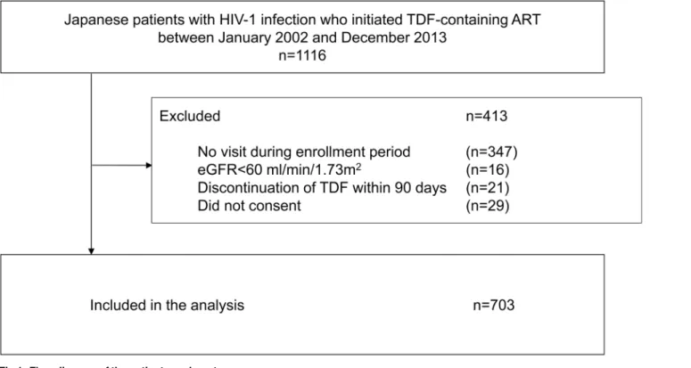
Study Design and subjects
We performed a single-center cohort study to investigate the association between TDF-related renal function decrement and SNPs in genes encoding renal tubular transporters in Japanese HIV-1-infected patients who initiated TDF-containing ART. The inclusion criteria for the study patients were: 1) Japanese patients with HIV infection who initiated TDF-containing ART at our clinic between January 2002 and December 2013, 2) patients who continued TDF for90 days, and 3) patients who provided a written informed consent for the study. Patients with eGFR<60 ml/min/1.73m2at initiation of TDF were excluded. The written informed
con-sent was obtained from the candidate patients between June 2014 and October 2014.
Measurements
The eGFR was calculated using the equation developed by the Japanese Society of Nephrology (JSN): eGFR = 194 × [serum creatinine]-1.094× [age]-0.287× [0.739 if female] [19]. This equation is used because this is more suitable for patients with small body stature, such as Japanese, than The Chronic Kidney Disease Epidemiology Collaboration (CKD-EPI) equation [20,21]. The 2013 practice guidelines for patients with CKD published by JSN also recommend the use of this equation for the Japanese, rather than the CKD-EPI equation [21]. The baseline eGFR was estimated for each patient from age, sex, and serum creatinine measurements made closest to and preceding the initiation of ART by no more than 90 days. Patients visited our clinic at least every three months for monitoring CD4 cell count, HIV-1 viral load, and eGFR, since the pre-scription period under the Japanese health care system is limited to three months [22]. Thus, for calculation of follow-up eGFR values, we collected serum creatinine values measured closest to every 90 day within a range of 45 days from initiation of ART. Follow up eGFR data were collected from the baseline measurement until discontinuation of TDF or at the end of the fol-low-up period (August 2014).
The potential risk factors for renal dysfunction were determined according to previous stud-ies and collected together with the basic demographics from the medical records [23–26]. They included age, sex, body weight, body mass index (BMI) = {body weight (kg) / [(height (m)]2}, history of AIDS, route of HIV-1 transmission, HIV-1 treatment status (either treatment-naïve or experienced), combination of ART, baseline laboratory data (CD4 cell count, HIV viral load, and serum creatinine), and presence or absence of other medical conditions [diabetes mellitus defined by using anti-diabetic agents or fasting plasma glucose>126 mg/dl or plasma
glucose>200 mg/dl on two different days, hypertension defined by current treatment with
antihypertensive agents or two successive measurements of systolic blood pressure>140
mmHg or diastolic blood pressure>90 mmHg at the clinic, dyslipidemia defined by current
treatment with lipid-lowering agents, co-infection with hepatitis B defined by positive hepatitis B surface antigen, co-infection with hepatitis C defined by positive HCV viral load, and current smoking] [22], concurrent use of ritonavir-boosted PIs (PI/r), concurrent use of nephrotoxic drugs, such as ganciclovir and sulfamethoxazole/trimethoprim. At our clinic, body weight and blood pressure were measured on every visit whereas other variables were measured in the first visit and at least once annually [22]. We used the data on or closest to and preceding the day of starting ART by no more than 180 days.
Genetic polymorphisms
P-glycoprotein, was also selected, because this triallelic SNP is functionally significant and appears to influence the absorption of TDF at the intestine and affect exposure of tenofovir [6,27,28].
Pharmacogenetic analyses
Genomic DNA was extracted from peripheral blood leukocytes using the QIAamp DNA Mini-Kit and the protocol provided by the manufacturer (Qiagen, Valencia, CA). All genotyping was performed by allelic discrimination using TaqMan 5’-nuclease assays with standard protocols (TaqMan SNP Genotyping Assays; Applied Biosystems, Foster City, CA). The primer and probe sequences are available on request.
Statistical analysis
Three renal endpoints were applied in this study; we focused primarily on 1) decrement in eGFR of>10 ml/min/1.73 m2relative to the baseline, because this endpoint is considered
appropriate for patients with well maintained renal function [22,29], such as the study popula-tion. We also set two commonly used renal endpoints; 2)>25% decrement in eGFR relative to
the baseline [30,31], and 3) eGFR<60 ml/min/1.73 m2[32].
The baseline characteristics were compared between patients with decrement in eGFR of
>10 ml/min/1.73 m2and those without such decrement by the Student’s t-test or the Wilcoxon
signed-rank test for continuous variables and by either theχ2test or Fisher’s exact test for
cate-gorical variables. Theχ2test was used to test for deviation of allele frequency from the
Hardy-Weinberg equilibrium. Statistical comparisons for genotype frequencies between the two groups were tested by the Fisher’s exact test or theχ2test where appropriate. The logistic
regression model was used to estimate the association of risk genotype/allele of each SNP with the occurrence of these renal endpoints. We applied the following four genetic models for the analysis: genotype model (a model that postulates no mode of inheritance), dominant model, recessive model, and additive model. Each genetic effect in logistic regression models was esti-mated with the adjustment for the variables which were determined a priori; they included baseline eGFR, age, baseline body weight, nephrotoxic drug use, PI/r use, CD4 count, hyperten-sion, and dyslipidemia, which are established risk factors for TDF nephrotoxicity [9,23,24,26]. Sex and diabetes mellitus were not added to the models due to their low frequency. The above statistical analyses were repeated using eGFR calculated by the CKD-EPI equation adjusted with the Japanese coefficient [33].
Statistical significance was defined at two-sidedpvalues<0.05. We used the odds ratio
(OR) with 95% confidence intervals (95% CI) as a measure of the effect of risk allele/genotype on each renal endpoint. All statistical analyses were performed with SAS software, version 9.3 (SAS Institute, Cary, NC).
Results
Of the 703 study patients,>10 ml/min/1.73 m2decrement in eGFR relative to the baseline
occurred in 624 (89%),>25% decrement in 119 (17%), and eGFR<60 ml/min/1.73 m2in 126
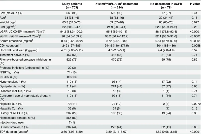
(18%). Patients with>10 ml/min/1.73 m2decrement in eGFR had higher baseline eGFR
(p<0.0001), lower CD4 count (p = 0.0058), had more frequent HBV co-infection (p = 0.0070),
and had longer exposure to TDF (p<0.0001), compared to those without decrement in eGFR
(Table 1).
Table 2summarizes the distribution of genotypes at -24 and 1249 ofABCC2gene and at
2677 ofABCB1gene, for patients with each renal endpoint and those free of decrement in eGFR. All polymorphisms were in Hardy-Weinberg equilibrium. The frequencies of genotypes at -24, 1249 ofABCC2gene and at 2677 ofABCB1gene were not different between patients with>10 ml/min/1.73 m2decrement in eGFR and those without decrement in eGFR (-24 of
ABCC2, p = 0.53, 1249 ofABCC2, p = 0.68, 2677 ofABCB1, p = 0.74), between patients with
>25% decrement in eGFR and those without (-24 ofABCC2, p = 0.83, 1249 ofABCC2,
p = 0.97, 2677 ofABCB1, p = 0.40), and between patients with decrement in eGFR to<60 ml/
min/1.73 m2and those without (-24 ofABCC2, p = 0.51, 1249 ofABCC2, p = 0.81, 2677 of
ABCB1, p = 0.94).
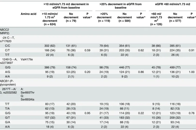
The results of additional analyses using eGFR calculated by the CKD-EPI equation are shown inTable 3. Similarly, the frequencies of genotypes at -24, 1249 ofABCC2gene and at 2677 ofABCB1gene were not different between patients with each renal endpoint and those who reached no such endpoint (>10 ml/min/1.73 m2decrement in eGFR: -24 ofABCC2,
p = 0.59, 1249 ofABCC2, p = 0.20, 2677 ofABCB1, p = 0.95) (>25% decrement in eGFR: -24 of
ABCC2, p = 0.62, 1249 ofABCC2, p = 0.86, 2677 ofABCB1, p = 0.22) (eGFR<60 ml/min/1.73
m2: -24 ofABCC2, p = 0.91, 1249 ofABCC2, p = 1.00, 2677 ofABCB1, p = 0.76).
The logistic regression model which evaluated genotypic effect of theABCC2gene showed that the risk genotype (i.e., genotype CC at -24) was not associated with any of the three renal Fig 1. Flow diagram of the patient enrolment process.
outcomes (Table 4) (>10 ml/min/1.73 m2decrement in eGFR: genotype C/C versus T/T,
adjusted OR 0.5, 95%CI 0.06–3.91, p = 0.62; genotype C/T versus T/T, adjusted OR 0.4, 95%CI 0.05–3.33, p = 0.34) (>25% decrement in eGFR: genotype C/C versus T/T, adjusted OR 1.2,
95%CI 0.42–3.20, p = 0.62; genotype C/T versus T/T, adjusted OR 1.0, 95%CI 0.35–2.83, p = 0.81) (eGFR<60 ml/min/1.73 m2: genotype C/C versus T/T, adjusted OR 0.6, 95%CI 0.20–
2.08, p = 0.61; genotype C/T versus T/T, adjusted OR 0.6, 95%CI 0.17–1.93, p = 0.36). Simi-larly, the risk genotype (genotype A/A) at 1249 ofABCC2or a genotype at 2677 ofABCB1was not associated with either of the three renal outcomes (S1andS2Tables). Furthermore, logistic regression analysis, which applied the dominant model, recessive model, and additive model, showed no association between each allele/genotype at the SNPs and any of the three renal endpoints (S3 Table). Logistic regression analysis using eGFR calculated by the CKD-EPI Table 1. Baseline characteristics of the study patients.
Study patients (n = 703)
>10 ml/min/1.73 m2decrement (n = 624)
No decrement in eGFR (n = 79)
P value
Sex (male), n (%) 669 (95) 592 (95) 77 (97) 0.41
Age†
38 (33–46) 38 (33–46) 39 (34–47) 0.18
Weight (kg)† 63.3 (57.3–70) 63 (57–70) 66 (60–73) 0.077
BMI (kg/m2)†
22 (20.2–24.1) 21.9 (20–24.1) 22.8 (20.9–24.2) 0.26
eGFR: JCKD-EPI (ml/min/1.73m2)†
94.2 (88.3–100.3) 95.4 (89–101.1) 88.4 (76.8–92.4) <0.0001 eGFR: JeGFR (ml/min/1.73m2)†
96 (84.6–109.2) 98.2 (86.7–112.2) 82.1 (68.3–91.6) <0.0001 Serum creatinine (mg/dl)† 0.74 (0.65–0.82) 0.72 (0.65–0.80) 0.84 (0.79–0.96) <0.0001
CD4 count (/μl)† 249 (127–385) 244.5 (110–377.5) 304 (188–436) 0.0058
HIV RNA viral load (log10/ml)† 4.51 (2.66–5.11) 4.5 (2.6–5.1) 4.4 (2.8–4.9) 0.52
Treatment naive, n (%) 467 (66) 416 (67) 51 (64) 0.71
Ritonavir-boosted protease inhibitors, n (%)
529 (75) 470 (75) 59 (75) 0.89
Protease inhibitors (unboosted), n (%) 22 (3)
NNRTIs, n (%) 71 (10)
INSTIs, n (%) 89 (13)
Hypertension, n (%) 110 (16) 93 (14) 17 (22) 0.14
Dyslipidemia, n (%) 311 (44) 274 (44) 37 (47) 0.63
Diabetes mellitus, n (%) 19 (3) 18 (3) 1 (1) 0.71
Concurrent use of nephrotoxic drugs, n (%)
110 (16) 99 (16) 11 (14) 0.74
Hepatitis B, n (%) 79 (11) 77 (12) 2 (3) 0.0070
Hepatitis C, n (%) 35 (5) 34 (5) 1 (1) 0.16
History of AIDS, n (%) 207 (29) 188 (30) 19 (24) 0.30
Homosexual contact, n (%) 565 (80)
Injection drug user 7 (1)
Current smoker, n (%) 307 (44) 275 (44) 32 (41) 0.63
TDF duration (years)† 3.66 (1.93–5.59) 3.89 (2.14–5.67) 1.52 (0.96–3.15) <0.0001
†
median (interquartile range)
Nine patients were taking both PI/r and NNRTI, 1 patient with NNRIT and INSTI. 1 patient was treated with 2 NRTIs and 1 with 3 NRTIs. Other patients were treated with 2 NRTIs together with either PI, NNRTI, or INSTI.
Differences between the two groups were compared by the Student’s t-test for continuous variables and by Fisher’s exact test for categorical variables, except for CD4 count, HIV RNA viral load, and TDF duration, which were compared by the Wilcoxon signed-rank test.
BMI: body mass index, TDF: tenofovir disoproxil fumarate, eGFR: estimated glomerularfiltration rate, NNRTI: non- nucleoside reverse transcriptase inhibitor, INSTI: integrase strand transfer inhibitor.
equation yielded the same results. Post-hoc analysis with the logistic models after further adjustment for duration of TDF therapy also yielded the same results.
Discussion
This pharmacogenetics study investigated the association between drug transporter genetic variants and TDF-related renal function decrement in Japanese HIV-1-infected patients who initiated TDF-containing ART. The results showed that none of the three examined SNPs was associated with any of the three selected renal outcomes:>10 ml/min/1.73 m2decrement in
eGFR relative to the baseline,>25% decrement in eGFR, and eGFR<60 ml/min/1.73 m2. The
results were reproduced using the dominant, recessive, and additive models, in addition to the genotype model for the estimation of association between genetic variants and renal outcomes.
Two main aspects of our study are important. First, this study showed that genetic factors do not need to be taken into account as predisposing factors for TDF–related renal dysfunc-tion, using three clinically important renal outcomes (>10 ml/min/1.73 m2decrement in eGFR
relative to the baseline [22,29],>25% decrement [30,31], and eGFR<60 ml/min/1.73 m2[32],
which are known to be associated with morbidity and mortality in HIV-1-infected patients Table 2. Genotype frequencies at three SNPs ofABCC2andABCB1in patients with and without three renal outcomes.
>10 ml/min/1.73 m2decrement in eGFR from baseline
>25% decrement in eGFR from baseline
eGFR<60 ml/min/1.73 m2
Amino acid >10 ml/min/ 1.73 m2 decrement
(n = 624)
No decrement
(n = 79) P value*
>25% decrement
(n = 119)
No decrement
(n = 584) P value*
<60 ml/ min/1.73
m2
(n = 126)
No decrement
(n = 577) P value*
ABCC2 (MRP2)
-24 C!T, rs717620
C/C 382 (61) 51 (65) 76 (64) 357 (61) 83 (66) 350 (61)
C/T 215 (35) 27 (34) 0.53 38 (32) 204 (35) 0.83 38 (30) 204 (35) 0.51
T/T 27 (4) 1 (1) 5 (4) 23 (4) 5 (4) 23 (4)
1249 G!A, rs2273697
Val417Ile
G/G 483 (78) 61 (77) 93 (78) 451 (77) 100 (79) 444 (77)
A/G 132 (21) 16 (20) 0.68 24 (20) 124 (21) 0.97 24 (19) 124 (21) 0.81
A/A 9 (1) 2 (3) 2 (2) 9 (2) 2 (2) 9 (2)
ABCB1 (P-glycoprotein)
2677T!A/ G, rs2032582
A: Ser893Thr G: Ser893Ala
T/T 112 (18) 13 (16) 19 (16) 106 (18) 21 (17) 104 (18)
T/A 77 (12) 13 (16) 22 (18) 68 (11) 18 (14) 72 (12)
G/G 122 (20) 13 (16) 0.74 20 (17) 115 (20) 0.40 21 (17) 114 (20) 0.94
G/T 195 (31) 29 (37) 39 (33) 185 (32) 41 (32) 183 (32)
G/A 96 (15) 9 (12) 17 (14) 88 (15) 20 (16) 85 (15)
A/A 22 (4) 2 (3) 2 (2) 22 (4) 5 (4) 19 (3)
*By use of Fisher’s exact test for 2×3 table (2×6 table for rs2032582).
[17,18]. In this regard, one study from Thailand reported the association between -24 C/C genotype ofABCC2gene and lower eGFR in treatment-naïve patients who initiated TDF-con-taining non-NRTI based regimen [34]. However, the relatively small sample size of 117 patients and, more importantly, the cross-sectional analysis used for the assessment of the Table 3. Genotype frequencies of three SNPs ofABCC2andABCB1in patients with and without three renal outcomes calculated by the CKD-EPI equation.
>10 ml/min/1.73 m2 decrement in eGFR from baseline
>25% decrement in eGFR from baseline
eGFR<60 ml/min/1.73 m2
Amino acid >10 ml/min/ 1.73 m2
decrement (n = 624)
No decrement
(n = 79) P value*
>25% decrement
(n = 119)
No decrement
(n = 584) P value*
<60 ml/ min/1.73
m2
(n = 126)
No decrement
(n = 577) P value*
ABCC2 (MRP2)
-24 C!T, rs717620
C/C 302 (62) 131 (61) 79 (64) 354 (61) 38 (66) 395 (61)
C/T 166 (34) 76 (36) 0.59 39 (31) 203 (35) 0.62 18 (31) 224 (35) 0.91
T/T 22 (4) 6 (3) 6 (5) 22 (4) 2 (3) 26 (4)
1249 G!A, rs2273697
Val417Ile
G/G 386 (79) 158 (74) 98 (79) 446 (77) 45 (78) 499 (77)
A/G 95 (19) 53 (25) 0.20 24 (19) 124 (21) 0.86 12 (21) 136 (21) 1.00
A/A 9 (2) 2 (1) 2 (2) 9 (2) 1 (1) 10 (2)
ABCB1 (P-glycoprotein)
2677T!A/ G, rs2032582
A: Ser893Thr G: Ser893Ala
T/T 83 (17) 42 (20) 19 (15) 106 (18) 9 (15) 116 (18)
T/A 62 (13) 28 (13) 24 (19) 66 (11) 8 (14) 82 (13)
G/G 95 (19) 40 (19) 0.95 21 (17) 114 (20) 0.22 12 (21) 123 (19) 0.76
G/T 157 (32) 67 (31) 41 (33) 183 (32) 15 (26) 209 (32)
G/A 75 (15) 30 (14) 17 (14) 88 (15) 12 (21) 93 (14)
A/A 18 (4) 6 (3) 2 (2) 22 (4) 2 (3) 22 (4)
*By use of Fisher’s exact test for 2×3 table (2×6 table for rs2032582).
doi:10.1371/journal.pone.0141931.t003
Table 4. Effects of SNP at -24 ofABCC2on three renal outcomes in patients who initiated TDF-containing antiretroviral therapy: Multivariate logis-tic regression with genotype model.
>10 ml/min/1.73 m2decrement in eGFR
>25% decrement in eGFR eGFR<60 ml/min/1.73 m2
OR 95%CI P value OR 95%CI P value OR 95%CI P value
Genotype C/C versus T/T 0.5 0.06–3.91 0.62 1.2 0.42–3.20 0.62 0.6 0.20–2.08 0.61
Genotype C/T versus T/T 0.4 0.05–3.33 0.34 1.0 0.35–2.83 0.81 0.6 0.17–1.93 0.36
Odds ratios for each genotype were adjusted for baseline eGFR, age, CD4 count, body weight, nephrotoxic drug use, hypertension, dyslipidemia, and use of PI/r. OR: odds ratio, CI: confidence interval, eGFR: estimated glomerularfiltration rate, PI/r: ritonavir-boosted protease inhibitor.
association between eGFR and genotype at 48 and 96 weeks undermine the reliability of their findings, because in using such design, the value of eGFR at 48 or 96 weeks is inevitably affected by the baseline eGFR. Furthermore, another recent Thai study of 238 patients showed that SNPs of drug transporters, including -24 and 1249 ofABCC2gene, were not associated with a change in creatinine clearance from the baseline to 1 and 3 years of TDF exposure [35]. Never-theless, our sample size is the largest (n = 703) among the studies investigating the effect of genetic variants of drug transporters on TDF-related renal dysfunction, and by using clinically relevant renal outcomes, our study showed that SNPs were not associated with TDF
nephrotoxicity.
Second, to our knowledge, this is the first study to apply not only genotype model (a model that postulates no mode of inheritance), but also dominant, recessive, and additive models, to investigate the association between genetic variants and TDF nephrotoxicity [16,34,35]. It is noteworthy that none of the four genetic models showed any association between genetic vari-ants of transporter proteins and TDF-associated renal dysfunction, especially considering that it is unknown which genetic model is appropriate for the evaluation of the effect of SNPs on TDF nephrotoxicity. Another strength of this study is that the results were reproduced with eGFR calculated by the CKD-EPI equation, in addition to the results based on eGFR calculated by the JSN equation.
It is noteworthy that although the association between the SNPs ofABCC2investigated in this study and TDF-induced tubulopathy has been well established [11,13,16], the exact mecha-nism by which these SNPs pose a risk for TDF tubulopathy remains unknown [13,16]. In this regard, MRP2 encoded byABCC2is not likely to take part in the transportation of TDF at the luminal membrane of kidney tubular cells [16,36]. At this point, because the results of this study showed that genetic variants of the drug transporters for TDF are not associated with clinically important renal outcomes, we think that these SNPs do not count as a risk factor for TDF-related renal dysfunction, at least in the clinical setting, and efforts should rather be focused on the management of traditional risk factors for renal dysfunction, such as diabetes mellitus and hypertension [37], as well as the management of PI/r, antiretroviral agents that are reported to increase TDF exposure [38] and thus are a risk factor for TDF-related renal dys-function [23]. It is also notable that among PI/r, ritonavir-boosted atazanavir and lopinavir/ ritonavir are reported to be associated with CKD [39].
Several limitations need to be acknowledged. First, the patients who discontinued TDF within 90 days from the initiation of this therapy were excluded from the study. It is difficult to completely exclude the possibility that inclusion of such patients would have resulted in mis-leading results, because the subjects would have included some who experienced substantial decrement in renal function due to causes other than TDF, such as death shortly after initiation of ART or immune reconstitution inflammatory syndrome of opportunistic infections, consid-ering that two-third of the study patients were treatment-naive. Second, although we selected the target SNPs that have been identified to associate with TDF-induced tubulopathy reported in previous studies, there might be other unknown transporter proteins for tenofovir excretion or transportation that contribute to susceptibility to tenofovir nephrotoxicity. Third, this study did not measure TDF plasma concentration, which could correlate with TDF-induced renal dysfunction [40]. Fourth, our cohort was characterized by the high prevalence of PI/r use, which can affect plasma concentration of TDF [41], and it is difficult to completely exclude the impact of concurrent PI/r in this study.
Supporting Information
S1 Table. Effects of SNP at 1249 ofABCC2on three renal outcomes in patients who initi-ated TDF-containing antiretroviral therapy: Multivariate logistic regression with genotype model.
(DOCX)
S2 Table. Effects of SNP at 2677 of ABCB1 on three renal outcomes in patients who initi-ated TDF-containing antiretroviral therapy: Multivariate logistic regression with genotype model.
(DOCX)
S3 Table. Effects of SNPs on three renal endpoints using dominant, recessive, and additive genetic models for multivariate logistic analysis.
(DOCX)
Acknowledgments
The authors thank the study patients. The authors also thank Yoshimi Kikuchi, Katsuji Teruya, Kunihisa Tsukada, Junko Tanuma, Haruhito Honda, Ei Kinai, Koji Watanabe, Takahiro Aoki, Kiyoto Tsuchiya, Daisuke Mizushima, Yasuaki Yanagawa, and all other clinical staff at the AIDS Clinical Center for their help in completion of this study.
Author Contributions
Conceived and designed the experiments: TN SO HG. Performed the experiments: TN TH TK NT. Analyzed the data: TN TH TK NT. Contributed reagents/materials/analysis tools: NT SO HG. Wrote the paper: TN NT SO HG.
References
1. Panel on Antiretroviral Guidelines for Adults and Adolescents. Guidelines for the use of antiretroviral agents in HIV-1-infected adults and adolescents. Department of Health and Human Services.http:// www.aidsinfo.nih.gov/ContentFiles/AdultandAdolescentGL.pdf(18 December 2013, date last accessed).
2. World Health Organization. Consolicated Guidelines on the Use of Antiretroviral Drugs for Treating and Preventing HIV Invection: Recommendations for a Public Health Approach.http://apps.who.int/iris/ bitstream/10665/85321/1/9789241505727_eng.pdf. (19 January 2014, date last accessed).
3. Fung S, Kwan P, Fabri M, Horban A, Pelemis M, Hann HW, et al. (2013) Randomized Comparison of Tenofovir Disoproxil Fumarate vs Emtricitabine and Tenofovir Disoproxil Fumarate in Patients with Lamivudine-resistant Chronic Hepatitis B. Gastroenterology.
4. Consolidated guidelines on HIV prevention, diagnosis, treatment and care for key populations. World Health Organization. July 2014 Available athttp://www.who.int/hiv/pub/guidelines/keypopulations/en/ Accessed 19 February 2015.
5. Preexposure Prophylaxis for the Prevention of HIV Infection in the United States 2014 Clinical Practice Guideline. American Center for Disease Prevention and Control. Available at:http://www.cdc.gov/hiv/ pdf/PrEPguidelines2014.pdf. Accessed 19 February 2015.
6. Moss DM, Neary M, Owen A (2014) The role of drug transporters in the kidney: lessons from tenofovir. Front Pharmacol 5: 248. doi:10.3389/fphar.2014.00248PMID:25426075
7. Verhelst D, Monge M, Meynard JL, Fouqueray B, Mougenot B, Girard PM, et al. (2002) Fanconi syn-drome and renal failure induced by tenofovir: a first case report. Am J Kidney Dis 40: 1331–1333. PMID:12460055
8. Gallant JE, Winston JA, DeJesus E, Pozniak AL, Chen SS, Cheng AK, et al. (2008) The 3-year renal safety of a tenofovir disoproxil fumarate vs. a thymidine analogue-containing regimen in antiretroviral-naive patients. AIDS 22: 2155–2163. PMID:18832879
treatment-naive patients with HIV infection. PLoS One 7: e29977. doi:10.1371/journal.pone.0029977PMID: 22242194
10. Kohler JJ, Hosseini SH, Hoying-Brandt A, Green E, Johnson DM, Russ R, et al. (2009) Tenofovir renal toxicity targets mitochondria of renal proximal tubules. Lab Invest 89: 513–519. doi:10.1038/labinvest. 2009.14PMID:19274046
11. Izzedine H, Hulot JS, Villard E, Goyenvalle C, Dominguez S, Ghosn J, et al. (2006) Association between ABCC2 gene haplotypes and tenofovir-induced proximal tubulopathy. J Infect Dis 194: 1481–
1491. PMID:17083032
12. Kiser JJ, Aquilante CL, Anderson PL, King TM, Carten ML, Fletcher CV (2008) Clinical and genetic determinants of intracellular tenofovir diphosphate concentrations in HIV-infected patients. J Acquir Immune Defic Syndr 47: 298–303. PMID:18398970
13. Rodriguez-Novoa S, Labarga P, Soriano V, Egan D, Albalater M, Morello J, et al. (2009) Predictors of kidney tubular dysfunction in HIV-infected patients treated with tenofovir: a pharmacogenetic study. Clin Infect Dis 48: e108–116. doi:10.1086/598507PMID:19400747
14. Pushpakom SP, Liptrott NJ, Rodriguez-Novoa S, Labarga P, Soriano V, Albalater M, et al. (2011) Genetic variants of ABCC10, a novel tenofovir transporter, are associated with kidney tubular dysfunc-tion. J Infect Dis 204: 145–153. doi:10.1093/infdis/jir215PMID:21628669
15. Rodriguez-Novoa S, Labarga P, Soriano V (2009) Pharmacogenetics of tenofovir treatment. Pharma-cogenomics 10: 1675–1685. doi:10.2217/pgs.09.115PMID:19842939
16. Nishijima T, Komatsu H, Higasa K, Takano M, Tsuchiya K, Hayashida T, et al. (2012) Single nucleotide polymorphisms in ABCC2 associate with tenofovir-induced kidney tubular dysfunction in Japanese patients with HIV-1 infection: a pharmacogenetic study. Clin Infect Dis 55: 1558–1567. doi:10.1093/ cid/cis772PMID:22955427
17. Choi AI, Rodriguez RA, Bacchetti P, Bertenthal D, Volberding PA, O'Hare AM (2007) The impact of HIV on chronic kidney disease outcomes. Kidney Int 72: 1380–1387. PMID:17805235
18. Brennan A, Evans D, Maskew M, Naicker S, Ive P, Sanne I, et al. (2011) Relationship between renal dysfunction, nephrotoxicity and death among HIV adults on tenofovir. AIDS 25: 1603–1609. PMID: 21646902
19. Matsuo S, Imai E, Horio M, Yasuda Y, Tomita K, Nitta K, et al. (2009) Revised equations for estimated GFR from serum creatinine in Japan. Am J Kidney Dis 53: 982–992. doi:10.1053/j.ajkd.2008.12.034 PMID:19339088
20. Levey AS, Stevens LA, Schmid CH, Zhang YL, Castro AF 3rd, Feldman HI, et al. (2009) A new equation to estimate glomerular filtration rate. Ann Intern Med 150: 604–612. PMID:19414839
21. Horio M, Imai E, Yasuda Y, Watanabe T, Matsuo S (2013) Performance of GFR equations in Japanese subjects. Clin Exp Nephrol 17: 352–358. doi:10.1007/s10157-012-0704-5PMID:23080048
22. Nishijima T, Kawasaki Y, Tanaka N, Mizushima D, Aoki T, Watanabe K, et al. (2014) Long-term expo-sure to tenofovir continuously decrease renal function in HIV-1-infected patients with low body weight. AIDS 28: 1903–1910. PMID:25259702
23. Goicoechea M, Liu S, Best B, Sun S, Jain S, Kemper C, et al. (2008) Greater tenofovir-associated renal function decline with protease based versus nonnucleoside reverse-transcriptase inhibitor-based therapy. J Infect Dis 197: 102–108. doi:10.1086/524061PMID:18171292
24. Nelson MR, Katlama C, Montaner JS, Cooper DA, Gazzard B, Clotet B, et al. (2007) The safety of teno-fovir disoproxil fumarate for the treatment of HIV infection in adults: the first 4 years. AIDS 21: 1273–
1281. PMID:17545703
25. Gatanaga H, Tachikawa N, Kikuchi Y, Teruya K, Genka I, Honda M, et al. (2006) Urinary beta2-micro-globulin as a possible sensitive marker for renal injury caused by tenofovir disoproxil fumarate. AIDS Res Hum Retroviruses 22: 744–748. PMID:16910829
26. Lucas GM, Ross MJ, Stock PG, Shlipak MG, Wyatt CM, Gupta SK, et al. (2014) Clinical practice guide-line for the management of chronic kidney disease in patients infected with HIV: 2014 update by the HIV Medicine Association of the Infectious Diseases Society of America. Clin Infect Dis 59: e96–138. doi:10.1093/cid/ciu617PMID:25234519
27. Kurata Y, Ieiri I, Kimura M, Morita T, Irie S, Urae A, et al. (2002) Role of human MDR1 gene polymor-phism in bioavailability and interaction of digoxin, a substrate of P-glycoprotein. Clin Pharmacol Ther 72: 209–219. PMID:12189368
28. Neumanova Z, Cerveny L, Ceckova M, Staud F (2014) Interactions of tenofovir and tenofovir disoproxil fumarate with drug efflux transporters ABCB1, ABCG2, and ABCC2; role in transport across the pla-centa. AIDS 28: 9–17. PMID:24413260
thromboembolism and bleeding). Am J Cardiol 111: 1159–1164. doi:10.1016/j.amjcard.2012.12.045 PMID:23337836
30. Chaisiri K, Bowonwatanuwong C, Kasettratat N, Kiertiburanakul S (2010) Incidence and risk factors for tenofovir-associated renal function decline among Thai HIV-infected patients with low-body weight. Curr HIV Res 8: 504–509. PMID:21073439
31. Nishijima T, Komatsu H, Gatanaga H, Aoki T, Watanabe K, Kinai E, et al. (2011) Impact of small body weight on tenofovir-associated renal dysfunction in HIV-infected patients: a retrospective cohort study of Japanese patients. PLoS One 6: e22661. doi:10.1371/journal.pone.0022661PMID:21799928
32. Mocroft A, Kirk O, Reiss P, De Wit S, Sedlacek D, Beniowski M, et al. (2010) Estimated glomerular filtra-tion rate, chronic kidney disease and antiretroviral drug use in HIV-positive patients. AIDS 24: 1667–
1678. PMID:20523203
33. Horio M, Imai E, Yasuda Y, Watanabe T, Matsuo S (2010) Modification of the CKD epidemiology collab-oration (CKD-EPI) equation for Japanese: accuracy and use for population estimates. Am J Kidney Dis 56: 32–38. doi:10.1053/j.ajkd.2010.02.344PMID:20416999
34. Manosuthi W, Sukasem C, Thongyen S, Nilkamhang S, Sungkanuparph S (2014) ABCC2*1C and plasma tenofovir concentration are correlated to decreased glomerular filtration rate in patients receiv-ing a tenofovir-containreceiv-ing antiretroviral regimen. J Antimicrob Chemother 69: 2195–2201. doi:10. 1093/jac/dku129PMID:24788661
35. Sirirungsi W, Urien S, Harrison L, Kamkon J, Tawon Y, Luekamlung N, et al. (2015) No Relationship Between Drug Transporter Genetic Variants and Tenofovir Plasma Concentrations or Changes in Glo-merular Filtration Rate in HIV-Infected Adults. J Acquir Immune Defic Syndr 68: e56–59. PMID: 25559596
36. Ray AS, Cihlar T, Robinson KL, Tong L, Vela JE, Fuller MD, et al. (2006) Mechanism of active renal tubular efflux of tenofovir. Antimicrob Agents Chemother 50: 3297–3304. PMID:17005808
37. Lucas GM, Ross MJ, Stock PG, Shlipak MG, Wyatt CM, Gupta SK, et al. (2014) Clinical Practice Guide-line for the Management of Chronic Kidney Disease in Patients Infected With HIV: 2014 Update by the HIV Medicine Association of the Infectious Diseases Society of America. Clinical Infectious Diseases.
38. Kearney BP, Mathias A, Mittan A, Sayre J, Ebrahimi R, Cheng AK (2006) Pharmacokinetics and safety of tenofovir disoproxil fumarate on coadministration with lopinavir/ritonavir. J Acquir Immune Defic Syndr 43: 278–283. PMID:17079992
39. Ryom L, Mocroft A, Kirk O, Worm SW, Kamara DA, Reiss P, et al. (2013) Association between antire-troviral exposure and renal impairment among HIV-positive persons with normal baseline renal func-tion: the D:A:D study. J Infect Dis 207: 1359–1369. doi:10.1093/infdis/jit043PMID:23382571
40. Poizot-Martin I, Solas C, Allemand J, Obry-Roguet V, Pradel V, Bregigeon S, et al. (2013) Renal impairment in patients receiving a tenofovir-cART regimen: impact of tenofovir trough concentration. J Acquir Immune Defic Syndr 62: 375–380. PMID:23196828