ORIGINAL ARTICLE
Clinical improvement in patients with borderline
personality disorder after treatment with repetitive
transcranial magnetic stimulation: preliminary results
Julian Reyes-Lo´pez,
1Josefina Ricardo-Garcell,
2Gabriela Armas-Castan˜eda,
3Marı´a Garcı´a-Anaya,
4Iva´n Arango-De Montis,
4Jorge J. Gonza´lez-Olvera,
4Francisco Pellicer
41Clı´nica del Sistema Nervioso, Departamento de Investigacio´n Biome´dica, Facultad de Medicina, Universidad Auto´noma de Quere´taro,
Quere´taro, Mexico.2Instituto de Neurobiologı´a, Universidad Nacional Auto´noma de Me´xico, Juriquilla, Quere´taro, Mexico.3Departamento de Psiquiatrı´a y Salud Mental, Facultad de Medicina, Universidad Nacional Auto´noma de Me´xico, Quere´taro, Mexico.4Instituto Nacional de Psiquiatrı´a Ramo´n de la Fuente Mun˜iz, Ciudad de Me´xico, Mexico.
Objective:Current treatment of borderline personality disorder (BPD) consists of psychotherapy and pharmacological interventions. However, the use of repetitive transcranial magnetic stimulation (rTMS) could be beneficial to improve some BPD symptoms. The objective of this study was to evaluate clinical improvement in patients with BPD after application of rTMS over the right or left dorsolateral prefrontal cortex (DLPFC).
Method: Twenty-nine patients with BPD from the National Institute of Psychiatry, Mexico, were randomized in two groups to receive 15 sessions of rTMS applied over the right (1 Hz, n=15) or left (5 Hz, n=14) DLPFC. Improvement was measured by the Clinical Global Impression Scale for BPD (CGI-BPD), Borderline Evaluation of Severity Over Time (BEST), Beck Depression Inventory (BDI), Hamilton Anxiety Rating Scale (HAM-A), and Barratt Impulsiveness Scale (BIS).
Results:Intragroup comparison showed significant (po0.05) reductions in every psychopathologic domain of the CGI-BPD and in the total scores of all scales in both groups.
Conclusions: Both protocols produced global improvement in severity and symptoms of BPD, particularly in impulsiveness, affective instability, and anger. Further studies are warranted to explore the therapeutic effect of rTMS in BPD.
Clinical trial registration:NCT02273674.
Keywords: Borderline personality disorder; neurophysiology; neurosciences; psychosocial factors
Introduction
Borderline personality disorder (BPD) is one of the most common personality disorders in clinical practice. It affects 11 to 5.9%2 of the general population and accounts for 10% of outpatient psychiatry visits and more than 20% of the psychiatric inpatient population,1,2 generating a huge demand for health services.2BPD prevalence is similar in both genders,2 although diagnosis is more common in women. The disorder is characterized by persistent pat-terns of affective instability, problematic relationships, and marked impulsiveness,2 which manifests as self-injurious behavior, substance abuse, suicidality,2-4 and other
high-risk behaviors. Comorbidity with disorders such as depres-sion, anxiety, and posttraumatic stress disorder (PTSD) is common.2
Neuroimaging and neuropsychological studies1,5have shown that the clinical manifestations of BPD are related
to changes in the frontolimbic network,2,3including amy-gdala hyperactivity and hypofunctionality in prefrontal structures6 such as the orbitofrontal cortex (OFC), the ventromedial prefrontal cortex (VMPFC), and the dorso-lateral (DLPFC) cortex.7-9 Particularly, the DLPFC plays a key role in regulating top-down emotional control and impulsiveness.9,10These findings become relevant when considering that the current lines of treatment are psy-chotherapy (maintenance treatment) and pharmacologi-cal interventions (which are used during exacerbations of symptoms).2
Nevertheless, the use of neuromodulation, such as repet-itive transcranial magnetic stimulation (rTMS),11,12 could be beneficial to improve some symptoms of BPD and to normalize the cortical dysfunction associated with these manifestations.13 This technique uses electromagnetic induction14 to stimulate the cerebral cortex focally and
noninvasively, with few side effects,15,16and is relatively pain free. The neuromodulatory action of rTMS involves excitatory and inhibitory neuronal processes and plastic changes.16,17
At present, rTMS has been approved for the treatment of depression in several countries; it is accepted as an evidence-based treatment option by the American
Correspondence: Josefina Ricardo-Garcell, Instituto de Neurobiolo-gı´a, Universidad Nacional Auto´noma de Me´xico, Campus Juriquilla, Boulevard Juriquilla, 3001, 76230, Quere´taro, Mexico.
E-mail: oojrg@yahoo.com
Submitted Sep 13 2016, accepted Feb 06 2017, Epub Jun 12 2017.
Brazilian Journal of Psychiatry Brazilian Psychiatric Association
Psychiatric Association (APA), the Canadian Network for Mood and Anxiety Treatments (CANMAT), and the World Federation of Societies of Biological Psychiatry (WFSBP).11,12,18 The most frequent protocols are those using high frequencies (4 1 Hz to a maximum of 20 Hz) over the left DLPFC16,18-21or low frequencies (p1 Hz) over the right one.16,18Effects have also been demonstrated in psychiatric disorders that share features with BPD, such as impulse control deficit22,23and anxiety symptoms.18,22
Studies have explored the therapeutic potential of rTMS in BPD using high-frequency protocols (10 Hz) on the right13 and left9 DLPFCs, although evidence shows that
use of frequencies in the inhibitory (p1 Hz) or 5-Hz ranges can provide clinical benefits with greater tolerability and reduced risk of adverse events.11,21,24Thus, the aim of this study was to evaluate clinical improvement in patients with BPD after treatment with high-frequency (5 Hz) or low-frequency (1 Hz) rTMS of the left or right DLPFC, respectively.
Material and methods
Participants
Twenty-nine patients with BPD, of both genders (27 women), all right-handed, with an age range of 18-45 years (mean 30.2 years, standard deviation [SD] = 7.6), participated in a randomized clinical trial that was conducted over 12 months. Outpatients from the BPD Clinic of the Ramon de la Fuente Mun˜iz National Institute of Psychiatry (INPRF) in Mexico City, with a DSM-IV-TR25diagnosis of BPD and a score48 on the Spanish version of the Borderline Diagnostic Interview Revised (DIB-R),26-28were included.
Subjects with intracranial metallic objects and medical devices contraindicated in transcranial magnetic stimula-tion (TMS) were excluded, as were subjects with epilepsy, history of seizures, substance dependence, suicidal idea-tion, psychotic symptoms, bipolar affective disorder, current major depressive episode, and other comorbid psychiatric disorders, except generalized anxiety disorder. To reduce the risk of inducing seizures by rTMS, subjects with epilep-tiform activity on an electroencephalogram were also exclu-ded. A safety questionnaire was applied in accordance with international guidelines.11,24
All participants received a complete description of the study and provided informed consent. This study was conducted in compliance with the Declaration of Helsinki, was approved by the INPRF Research Ethics Committee, and was registered in the U.S. National Institutes of Health ClinicalTrials.gov platform (www.clinicaltrials.gov) with accession number NCT02273674.
Clinical evaluation of participants
Six clinical tests were administered to assess BPD, anxiety and depressive symptoms, and impulsiveness. To determine the severity of BPD symptoms and their changes over time, the Clinical Global Impression Scale for BPD (CGI-BPD)29 and the Spanish version of the Borderline Evaluation of Severity Over Time (BEST)30 were applied. The CGI-BPD is an adaptation of the Clinical
Global Impression scale (CGI) that was designed with the objective of evaluating both the severity and the subse-quent change in response to an intervention in patients diagnosed with BPD. The CGI consists of 10 Likert-type items scored on a scale of 1 to 7, which evaluate nine psychopathological domains of BPD, and an additional overall score.
CGI-BPD consists of two formats to assess current severity and change over time. The instrument has dem-onstrated adequate validity, reliability, and sensitivity to change.29BEST, in turn, is a self-administered instrument designed to evaluate the severity and change over time of typical thoughts, emotions, and behaviors in BPD. This scale has also demonstrated adequate sensitivity to change, high internal consistency, and discriminant validity.30
The Barratt Impulsiveness Scale (BIS) was used to assess impulsiveness. It is self-administered and was validated in Spanish by Oquendo et al.31The BIS consists of 30 items grouped into three impulsiveness subscales: cognitive, motor, and unplanned. This test has a high inter-nal consistency.31
The presence, severity, and change over time of anxiety and depressive symptoms were evaluated by the Hamilton Anxiety Rating Scale (HAM-A) and a 21-item version of the Beck Depression Inventory (BDI), respectively. Clinimetric tests were applied by an experienced psychiatrist, before and after 15 rTMS sessions, in order to evaluate changes in BPD, anxiety and depressive symptoms, and impul-siveness.
Repetitive transcranial magnetic stimulation procedure
Participants were randomly assigned to receive one of two different rTMS protocols (5 Hz or 1 Hz), which gen-erated two treatment groups. In both protocols, rTMS pulses were administered at an intensity equal to 100% of each patient’s motor threshold using a Dantec MagPro rapid magnetic stimulator and a 50 mm Dantec MC-B70 butterfly (figure-eight) coil with 150o
angulation.
The resting motor threshold (RMT) was determined at the start of each session, using the visual inspection method as described by Fitzgerald, in which the abductor pollicis brevis muscle (APBM) motor response is eval-uated. Stimulation site was defined as 5 cm above the maximum stimulation point at the APBM region, accord-ing to descriptions in previous clinical guidelines for locating the DLPFC.11,19
In the 1 Hz group (n=15, 14 women), rTMS was applied to the right DLPFC (one 15-minute train, 1 pulse per second continuously, for a total of 900 pulses per session). In the 5 Hz group (n=14, 13 women), rTMS was applied to the left DLPFC (30 trains of 10 seconds each, with a 10-second interval between each train, for a total of 1,500 pulses per session). Both rTMS protocols consisted of one daily session from Monday through Friday for 3 weeks (15 sessions total).
Statistical analysis
using the nonparametric Mann-Whitney U test, while gender distributions were compared by Fisher’s exact test. To analyze changes in clinimetric test scores, the Mann-WhitneyUwas used to compare differences between groups, while the Wilcoxon test was used to evaluate the effect of rTMS within each group. Cohen’s d was calculated in Microsoft Excel to analyze the effect size of rTMS on BPD symptoms.
Results
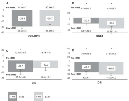
Both treatment groups were relatively homogeneous in terms of age, sex, and baseline symptoms, since there were no statistically significant differences in these var-iables (Table 1). After application of the rTMS protocols, both groups showed significant reductions in total scores of all instruments (Table 2, Figures 1 and 2).
Borderline personality disorder symptoms and repetitive transcranial magnetic stimulation
The change in the patients’ symptoms, evaluated through the CGI-BPD, was obtained considering the score of each of the first nine BPD psychopathological domains, which assess current severity. The total score was also obtained by adding the scores of each of these nine domains. The Wilcoxon test was used for statistical analysis in both groups, and showed a significant reduction in CGI-BPD
total score from baseline after rTMS (z = 3.3 in both groups, p = 0.001 for both groups), with a percent change of 29.4 and 28.7% for groups 1 and 5 Hz, respectively (Figure 1A), and an effect size of d= 2.58 for the 1 Hz group andd= 2.02 for the 5 Hz group.
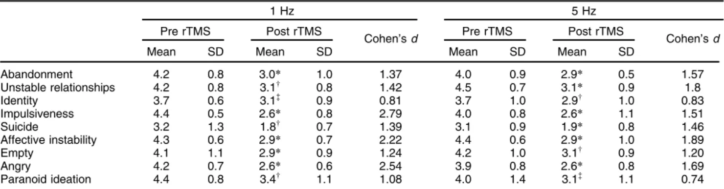
Table 2 shows significant differences before and after rTMS, with effect sizes (obtained by the Wilcoxon test and Cohen’sdrespectively), for the nine CGI-BPD domains in each treatment group. It can be noted that both groups showed significant score reductions in all domains, parti-cularly abandonment, impulsiveness, emotional instabil-ity, and anger, in which highly significant reductions (po
0.005) were observed in both the 1 Hz and 5 Hz groups. In all CGI-BPD domains, Cohen’sdeffect size was40.7 (Table 2). However, no significant differences (Mann-Whit-ney U) in total score at baseline (U = 91.0, p4 0.05) or after rTMS (U= 93.5, p40.05) were found between the two groups (Figure 1), nor were there significant between-group differences in individual CGI-BPD domain scores (1 Hz vs. 5 Hz groups Pre rTMS or 1 Hz vs. 5 Hz groups Post rTMS, Table 2).
Total BEST scale scores in both the 1 Hz and 5 Hz groups also reduced significantly after rTMS (41.0614.5 vs. 30.7612.0, z = 2.3, p = 0.001; 42.869.7 vs. 26.8611.8, z = -2.94, p = 0.003 for 1 and 5 Hz, respectively). This represented 20.4% and 36.9% reductions from baseline for the 1 Hz and 5 Hz groups, with effect sizes of 0.8 and 1.54, respectively. No significant between-group
Table 1 Statistical analysis of sociodemographic variables (age, sex) and baseline clinical test scores in the two treatment groups
Groups
1 Hz (n=15) 5 Hz (n=14) Statistic p-value
Age 29.667.8 30.967.6 U= 76.5 0.32
Sex, male/female (% female) 1/14 (93) 1/13 (92) Fisher’s exact test 0.9
BDI 30.9615.6 31.9614.6 U= 97.5 0.982
HAM-A 20.266.8 15.566.4 U= 33 0.120
CGI-BPD 41.164.7 40.266.0 U= 91.0 0.747
BEST 41.0614.5 42.869.7 U= 65.0 0.510
Barratt Impulsiveness Scale 70.2612.0 73.1614.2 U= 94.0 0.631
Data presented as mean6standard deviation, unless otherwise specified.
BDI = Beck Depression Inventory; BEST = Borderline Evaluation of Severity Over Time; CGI-BPD = Clinical Global Impression Scale for Borderline Personality Disorder; HAM-A = Hamilton Anxiety Rating Scale.
Table 2 Significant differences and effect sizes obtained by comparing values of the nine CGI-BPD domains, in each treatment group, before and after rTMS (Wilcoxon test and Cohen’sd)
1 Hz 5 Hz
Pre rTMS Post rTMS
Cohen’sd Pre rTMS Post rTMS Cohen’sd
Mean SD Mean SD Mean SD Mean SD
Abandonment 4.2 0.8 3.0* 1.0 1.37 4.0 0.9 2.9* 0.5 1.57
Unstable relationships 4.2 0.8 3.1w 0.8 1.42 4.5 0.7 3.1
* 0.9 1.8
Identity 3.7 0.6 3.1= 0.9 0.81 3.7 1.0 2.9w 1.0 0.83
Impulsiveness 4.4 0.5 2.6* 0.8 2.79 4.0 0.8 2.6* 1.1 1.51
Suicide 3.2 1.3 1.8w 0.7 1.39 3.1 0.9 1.9
* 0.8 1.46
Affective instability 4.3 0.6 2.9* 0.7 2.22 4.4 0.6 2.9* 1.0 1.89
Empty 4.1 1.1 2.9* 0.9 1.24 4.2 1.0 3.1w 0.9 1.20
Angry 4.2 0.7 2.6* 0.6 2.54 3.9 0.8 2.6* 0.8 1.69
Paranoid ideation 4.4 0.8 3.4w 1.1 1.08 4.0 1.4 3.1=
1.1 0.74
CGI-BPD = Clinical Global Impression Scale for Borderline Personality Disorder; rTMS = repetitive transcranial magnetic stimulation; SD = standard deviation.
*po0.005;wp
differences in BEST total score were found, whether at baseline (U= 65.0, p 4 0.05) or after rTMS (U = 56.0, p40.05) (Figure 1B).
Regarding individual BEST scale dimensions, signifi-cant reductions were observed in both groups for Thoughts and Feelings (1 Hz = 27.168.2 vs. 19.968.4,
Figure 1 Percent change in total Clinical Global Impression Scale for Borderline Personality Disorder (CGI-BPD), Borderline Evaluation of Severity Over Time (BEST), Barratt Impulsiveness Scale (BIS), and Beck Depression Inventory (BDI) scores after repetitive transcranial magnetic stimulation (rTMS). Both groups showed reductions in total score of all instruments (Wilcoxon test). Central squares show percent change. Data presented as mean 6 standard deviation. * po0.005;w
po0.05.
Figure 2 Changes in Borderline Evaluation of Severity Over Time (BEST) dimensions. Data presented as mean6standard deviation. * po0.005;w
z = 2.3, p = 0.021, Cohen’sd= 0.90; 5 Hz = 27.266.4 vs. 17.068.2, z = 2.9, p = 0.003, Cohen’sd= 1.44), repre-senting a percent change of 23.5% for the 1 Hz group and 38.6% for the 5 Hz group, and in Negative Behaviors, with a 17% reduction for the 1 Hz group (9.664.4 vs. 7.063.5, z = 1.3, p40.05, Cohen’sd= 0.68) and a 33.9% reduc-tion for the 5 Hz group (10.963.8 vs. 6.563.2, z = 2.4, p = 0.014, Cohen’sd= 1.3). In the Positive Behaviors dimen-sion, no significant changes were observed in either 1 Hz group (10.663.0 vs. 11.262.2, z = -0.8, p = 0.39) or the 5 Hz groups (10.362.3 vs. 11.761.5, z = 1.4, p = 0.14). There were no significant between-group differences in individual BEST dimension scores at baseline or after rTMS (Figure 2A).
Impulsiveness
Comparison of baseline and post-treatment BIS scores showed significant reductions in total scores in the 1 Hz group (70.2612.0 vs. 57.3614.8, z = 3.2, p = 0.001, Cohen’s d = 0.99) and the 5 Hz group (73.1614 vs. 63.3612.1, z = 2.3, p = 0.017, Cohen’sd = 0.78), with change percentages of 18.96% and 11.83% respectively. No between-groups differences in impulsiveness scores were observed at baseline (U= 94.0, p40.05) or after magnetic stimulation sessions (U = 87.0, p 4 0.05) (Figure 1C).
Analysis of changes in BIS dimension scores showed significant reductions for both groups in motor impulsive-ness (1 Hz, 24.465.0 vs. 16.868, z = 2.7 p = 0.007, Cohen’sd= 1.18, 29% change; 5 Hz, 25.567.5 vs. 18.96
7.3, z = 2.8 p = 0.004, Cohen’s d= 0.93, 25% change). Additionally, the 1 Hz group showed a significant reduction in cognitive impulsiveness dimension score (20.863.7 vs. 18.064.6, z = 2.0 p = 0.037, Cohen’s d = 0.69, 13%
reduction compared to baseline). There were no significant changes in the Nonplanning impulsiveness dimension. No significant differences were found on between-group com-parison (Figure 3).
Anxiety and depressive symptoms
BDI scores reduced significantly from baseline after rTMS in both the 1 Hz group (30.9615.5 vs. 1369.1, z = -3.1, p = 0.002, Cohen’sd= 1.46, percent change 49%) and the 5 Hz group (31.9614.5 vs. 14.2611.0, z = -3.3, p = 0.001, Cohen’s d = 1.43, percent change 60%). No between-group differences were found at baseline (U= 97.5, p 4 0.05) or after rTMS (U= 96.5, p40.05) (Figure 1D).
Similarly, HAM-A scores reduced after rTMS treatment in both groups (1 Hz, 20.266.8 vs. 864.7, z = -2.8, p = 0.005, Cohen’s d = 2.16, percent change 60.3%; 5 Hz, 15.566.4 vs. 6.463.5, z = -2.9, p = 0.003, Cohen’sd= 1.83, percent change 58.7%). Again, no between-groups differ-ences at baseline (U= 33, p40.05) or after rTMS (U= 43, p40.05) were found.
Discussion
This is the first study to explore the effect of rTMS, using 5 Hz frequencies on the left DLPFC and 1 Hz on the right DLPFC, on clinical improvement in patients with BPD. Previous studies have demonstrated the effectiveness of these protocols in treating depressive symptoms,16,19 besides reducing discomfort and inducing seizure risk.11,21,24
Although imaging studies and the pathophysiology of BPD suggest dysfunction in the frontolimbic network, inclu-ding the anterior cingulate cortex (ACC), the orbitofrontal and dorsolateral prefrontal cortex, the hippocampus, and
Figure 3 Changes in Barratt Impulsiveness (BIS) dimensions. Data presented as mean6standard deviation. * po0.005;
w
the amygdala,2 limitations in access due to the design of TMS coils make stimulation of these structures more dif-ficult. For instance, stimulation of the ACC or amygdala requires different coil designs, such as a double-cone angulated coil, Hesed-coil (H-coil), C-core coil, or circular crown-coil.18,24Furthermore, considering the physical dis-comfort observed during stimulation of other regions (orbito-frontal cortex and (orbito-frontal pole) using a figure-eight coil, through a pilot study carried out by our research group in healthy volunteers, we decided to use the same anatomical targets reported in previous studies in both BPD and depression.9,11,13,18
Unlike in previous reports and treatment guidelines for conditions such as depression, where treatment is sug-gested to last 2 to 6 weeks of treatment,18reports on the application of rTMS in BPD have used only 10-session, 2-week protocols.9,13In this context, we decided to extend the number of sessions by 50% (15 sessions in 3 weeks), within parameters that have been demonstrated to elicit responses in the left19and right16DLPFC.
It is important to mention that, although it can be con-sidered a soft stimulation parameter, the use of 900 pul-ses per pul-session is greater than that reported in previous studies for conditions such as depression and PTSD, where a clinical effect has been reported even with proto-cols administering 120-1,200 pulses per session.18
Our results showed that both stimulation protocols were effective in reducing BPD symptom severity and several symptoms in particular, such as fear of abandon-ment, impulsivity, emotional instability, and anger. This may have a positive impact on reduction of self-harm and suicidal behavior, as well as improve family and inter-personal relationships through better social functioning.
After application of an inhibitory frequency (1 Hz) over the right DLPFC, we observed scores reductions in every clinimetric scale, particularly in BIS, with a significant decrease in the cognitive impulsiveness subscale. This result is similar to that reported by the Cailhol group,13by stimulating the same cortex, but with an excitatory fre-quency (10 Hz) on the right DLPFC.
Furthermore, using a lower excitatory frequency (5 Hz) on the left DLPFC, we obtained results similar to those reported by Arbabi et al. in a case report, where the same region was stimulated at 10 Hz.9In both studies, reductions
in depressive affective symptoms and impulsiveness level were observed.
The effect of rTMS is influenced by variables such as frequency and number of pulses.32Even if the number of pulses in each rTMS session (1,500) was the same in both protocols; our study was performed in 15 sessions (22,500 total pulses) instead of the 10 sessions (15,000 total pulses) applied in Arbabi’s case,9resulting in a larger amount of total pulses.
It is reasonable to assume that the significant improve-ment in every BPD psychopathological domain observed in our results is related to this larger amount of total pulses applied, as Arbabi et al.9 only found changes in identity, impulsiveness, emotional instability and anger domains.
Evidence supports an association between BPD symp-toms (specifically, impulsiveness and affective instability) with a deficit in top-down regulation of emotional processing,
due to lower modulation of cortical structures (particularly the DLPFC) over subcortical structures (such as the amygdala).10It has also been reported that severity of
self-harm in these patients is associated with level of impulsivity, anger, and somatic anxiety.4These data are consistent with
findings of DLPFC functional disturbances in patients with BPD2,7,33and microstructural damage to the uncinate fascic-ulus white matter (WM),3the largest WM tract interconnect-ing the amygdala with prefrontal structures.34
Given this background, one could infer that using inhi-bitory frequencies (p 1 Hz) on frontal structures would have a potentiating effect on BPD symptoms by further reducing DLPFC top-down regulation on the amygdala. However, our results after stimulation of the right DLPFC with 1 Hz suggest otherwise. Although we have no refer-ences to explain this effect on BPD symptoms, the use of inhibitory frequencies in other entities (i.e., attention-deficit hyperactivity disorder,35 Tourette’s syndrome,23 posttraumatic stress18), which share impulse control fai-lure and anxiety symptoms with BPD, suggest that this beneficial effect of rTMS may be attributable to improve-ment in functional deficits in the frontostriatal circuitry that appear to be associated with impulsivity36 and affective instability in BPD.3
Moreover, the effect of excitatory frequencies on the left DLPFC can be interpreted in light of the Valencia Asymmetry Hypothesis,37which proposes that emotions associated with anxiety are processed predominantly by the right hemisphere, while the left hemisphere processes emotions related to approach behaviors and positive mood states.10 Thus, 5 Hz rTMS applied over the left DLPFC could help increase top-down regulation of the amygdala, improving aspects such as impulsivity and affective instability.
Interestingly, both forms of stimulation (1 Hz and 5 Hz) produced global improvement in BPD symptom sever-ity, particularly in impulsiveness, affective instabilsever-ity, and anger. In these sense, the role of laterality and frequency of rTMS have been controversial technical aspects; for example, in previous studies of rTMS in BPD, the authors described improvement of the symptoms with the use of high-frequency protocols, independently of rTMS later-ality. Fitzgerald21reported absence of a differential effect between right or left rTMS at low frequencies over the DLPFC for depression treatment, while Speer et al. reported that high frequencies (20 Hz) and low frequen-cies (1 Hz), when applied to the left DLPFC at 110% of RMT, had the same antidepressant effect.38 Similarly, there are reports of clinical response to right or left rTMS in PTSD.20
However, different guidelines recommend the use of protocols with high frequencies over the left DLPFC for treatment of depression18,24and PTSD.20Speer et al.39,40
hippocampus, parahippocampus, thalamus, and cerebel-lum; with low frequencies (1 Hz), the authors found decreases in blood flow in the right prefrontal cortex, left medial temporal cortex, left basal ganglia, and left amyg-dala.39 In a second study,40 the same authors showed
that improvement with the use of high-frequency rTMS (20 Hz) was associated with hypoperfusion on baseline PET. In this sense, these papers showed differential effects of high vs. low frequencies in metabolic response to rTMS over the left DLPFC, although clinical response was reported with both protocols of rTMS.40
Among the limitations of this preliminary report, we must consider that the sample size was small, there was no sham group, and we did not use neurophysiology or neuroimaging techniques which might have revealed anatomical and functional changes associated with the clinical benefits of our rTMS protocols. Despite published studies on BPD and rTMS, we did not consider inclusion of a sham group essential, because ours is an exploratory study about the potential therapeutic effect of rTMS in the treatment of BPD. A report by Cailhol et al.13reported
comparative results between five patients who received active treatment with rTMS and five sham subjects, and although they did not find significant differences between the two groups in clinical scales, cognitive improvement was reported in the active group. Therefore, differences in active vs. sham treatment have not been demonstrated yet. It is essential that future studies include sham groups, as partial responses to sham treatment have been found in other conditions, such as depression.
Despite the limitations mentioned above, our results sup-port the use of rTMS as a supplemental treatment for BPD. Considering that BPD is the most common personality disorder in clinical practice, further studies are warranted to explore the potential therapeutic effect of rTMS in this condition.
Acknowledgements
The authors wish to thank the National Council of Science and Technology (CONACYT), Mexico, for granting a doctoral scholarship (CVU/Scholar: 228471/210582).
The authors are grateful to the Ramon de la Fuente Mun˜iz National Institute of Psychiatry, to Erik Daniel Morelos, and to all participants included in the study for their support. We also thank Hector Garcı´a Castro for his support in the English translation of the article.
Disclosure
The authors report no conflicts of interest.
References
1 Ruocco AC, Amirthavasagam S, Choi-Kain LW, McMain SF. Neural correlates of negative emotionality in borderline personality disorder: an activation-likelihood-estimation meta-analysis. Biol Psychiatry. 2013;73:153-60.
2 Leichsenring F, Leibing E, Kruse J, New AS, Leweke F. Borderline personality disorder. Lancet. 2011;377:74-84.
3 Lischke A, Domin M, Freyberger HJ, Grabe HJ, Mentel R, Bernheim D, et al. Structural alterations in white-matter tracts connecting (para-)
limbic and prefrontal brain regions in borderline personality disorder. Psychol Med. 2015;45:3171-80.
4 Mendoza Y, Pellicer F. Percepcio´n del dolor en el sı´ndrome de comportamiento autolesivo. Salud Ment. 2002;25:10-6.
5 Ruocco AC, Medaglia JD, Ayaz H, Chute DL. Abnormal prefrontal cortical response during affective processing in borderline personality disorder. Psychiatry Res. 2010;182:117-22.
6 De la Fuente J, Goldman S, Stanus E, Vizuete C, Morla´n I, Bobes J, et al. Brain glucose metabolism in borderline personality disorder. J Psychiatr Res. 1997;31:531-41.
7 Sala M, Caverzasi E, Lazzaretti M, Morandotti N, De Vidovich G, Marraffini E, et al. Dorsolateral prefrontal cortex and hippocampus sustain impulsivity and aggressiveness in borderline personality dis-order. J Affect Disord. 2011;131:417-21.
8 Tebartz van Elst L, Hesslinger B, Thiel T, Geiger E, Haegele K, Lemieux L, et al. Frontolimbic brain abnormalities in patients with borderline personality disorder: a volumetric magnetic resonance imaging study. Biol Psychiatry. 2003;54:163-71.
9 Arbabi M, Hafizi S, Ansari S, Oghabian MA, Hasani N. High frequency TMS for the management of borderline personality disorder: a case report. Asian J Psychiatry. 2013;6:614-7.
10 Zwanzger P, Steinberg C, Rehbein MA, Bro¨ckelmann AK, Dobel C, Zavorotnyy M, et al. Inhibitory repetitive transcranial magnetic sti-mulation (rTMS) of the dorsolateral prefrontal cortex modulates early affective processing. NeuroImage. 2014;101:193-203.
11 Fitzgerald PB, Daskalakis ZJ. A practical guide to the use of repetitive transcranial magnetic stimulation in the treatment of depression. Brain Stimul. 2012;5:287-96.
12 Cook IA, Espinoza R, Leuchter AF. Neuromodulation for depression: invasive and noninvasive (deep brain stimulation, transcranial mag-netic stimulation, trigeminal nerve stimulation). Neurosurg Clin N Am. 2014;25:103-16.
13 Cailhol L, Roussignol B, Klein R, Bousquet B, Simonetta-Moreau M, Schmitt L, et al. Borderline personality disorder and rTMS: a pilot trial. Psychiatry Res. 2014;216:155-7.
14 Sampson SM. Transcranial magnetic stimulation in neuropsychiatry. J Neuropsychiatry Clin Neurosci. 2000;12:512-3.
15 Kobayashi M, Pascual-Leone A. Transcranial magnetic stimulation in neurology. Lancet Neurol. 2003;2:145-56.
16 Garcı´a-Anaya M, Gonza´lez-Olvera J, Ricardo-Garcell J, Armas G, Miranda E, Reyes E, et al. Clinical and electrophysiological effect of right and left repetitive transcranial magnetic stimulation in patients with major depressive disorder. Salud Ment. 2011;34: 291-9.
17 Hallett M. Transcranial magnetic stimulation: a primer. Neuron. 2007;55:187-99.
18 Lefaucheur JP, Andre´ Obadia N, Antal A, Ayache SS, Baeken C, Benninger DH, et al. Evidence-based guidelines on the therapeutic use of repetitive transcranial magnetic stimulation (rTMS). Clin Neuro-physiol. 2014;125:2150-206.
19 Gonza´lez-Olvera JJ, Ricardo-Garcell J, Garcı´a-Anaya ML, Miranda-Terre´s E, Reyes-Zamorano E, Armas-Castan˜eda G. Ana´lisis de fuentes del EEG en pacientes tratados con estimulacio´n magne´tica transcraneal a 5 Hz como tratamiento antidepresivo. Salud Ment. 2013;36:235-40.
20 Lepping P, Scho¨nfeldt-Lecuona C, Sambhi RS, Lanka SV, Lane S, Whittington R, et al. A systematic review of the clinical relevance of repetitive transcranial magnetic stimulation. Acta Psychiatr Scand. 2014;130:326-41.
21 Fitzgerald PB, Hoy K, Gunewardene R, Slack C, Ibrahim S, Bailey M, et al. A randomized trial of unilateral and bilateral prefrontal cortex transcranial magnetic stimulation in treatment-resistant major depression. Psychol Med. 2011;41:1187-96.
22 George MS, Padberg F, Schlaepfer TE, O’Reardon JP, Fitzgerald PB, Nahas ZH, et al. Controversy: repetitive transcranial magnetic stimulation or transcranial direct current stimulation shows efficacy in treating psychiatric diseases (depression, mania, schizophrenia, obsessive-complusive disorder, panic, posttraumatic stress disorder). Brain Stimul. 2009;2:14-21.
23 Kwon HJ, Lim WS, Lim MH, Lee SJ, Hyun JK, Chae JH, et al. 1-Hz low frequency repetitive transcranial magnetic stimulation in children with Tourette’s syndrome. Neurosci Lett. 2011;492:1-4.
transcranial magnetic stimulation (rtms): safety and therapeutic indi-cations. Neurophysiol Clin. 2011;41:221-95.
25 American Psychiatric Association. Diagnostic and Statistical Manual of Mental Disorders, Fourth Edition, Text Revision (DSM-IV-TR) Arlington: American Psychiatric Publishing; 2000.
26 Zanarini MC, Gunderson JG, Frankenburg FR, Chauncey DL. The revised diagnostic interview for borderlines: discriminating BPD from other Axis II disorders. J Pers Disord. 1989;3:10-8.
27 Barrachina J, Soler J, Campins MJ, Tejero A, Pascual JC, Alvarez E, et al. [Validation of a Spanish version of the diagnostic interview for bordelines-revised (DIB-R)]. Actas Esp Psiquiatr. 2004;32:293-8. 28 Zanarini MC, Gunderson JG, Frankenburg FR, Chauncey DL.
Dis-criminating borderline personality disorder from other axis II dis-orders. Am J Psychiatry. 1990;147:161-7.
29 Pe´rez V, Barrachina J, Soler J, Pascual JC, Campins MJ, Puigde-mont D, et al. Impresio´n Clı´nica Global para Pacientes con Trastorno Lı´mite de la Personalidad (ICG-TLP): una escala sensible al cambio. Actas Esp Psiquiatr. 2007;35:229-35.
30 Pfohl B, Blum N, St. John D, McCormick B, Allen J, Black DW. Reliability and validity of the borderline evaluation of severity over time (BEST): a self-rated scale to measure severity and change in persons with borderline personality disorder. J Pers Disord. 2009;23: 281-93.
31 Oquendo MA, Baca-Garcia E, Graver R, Morales M, Montalvan V, Mann JJ. Spanish adaptation of the Barratt Impulsiveness Scale (BIS-11). Eur J Psychiatry. 2001;15:147-55.
32 Rubens MT, Zanto TP. Parameterization of transcranial magnetic stimulation. J Neurophysiol. 2012;107:1257-9.
33 Schulze L, Schmahl C, Niedtfeld I. Neural correlates of disturbed emotion processing in borderline personality disorder: a multimodal meta-analysis. Biol Psychiatry. 2016;79:97-106.
34 Wakana S, Jiang H, Nagae-Poetscher LM, van Zijl PC, Mori S. Fiber tract-based atlas of human white matter anatomy. Radiology. 2004;230: 77-87.
35 Go´mez L, Vidal B, Morales L, Ba´ez M, Maragoto C, Galvizu R, et al. Low frequency repetitive transcranial magnetic stimulation in children with attention deficit/hyperactivity disorder. Preliminary results. Brain Stimul. 2014;7:760-2.
36 Rubio B, Boes AD, Laganiere S, Rotenberg A, Jeurissen D, Pascual-Leone A. Noninvasive brain stimulation in pediatric attention-deficit hyperactivity disorder (ADHD): a review. J Child Neurol. 2016;31: 784-96.
37 Davidson RJ, Irwin W. The functional neuroanatomy of emotion and affective style. Trends Cogn Sci. 1999;3:11-21.
38 Speer AM, Wassermann EM, Benson BE, Herscovitch P, Post RM. Antidepressant efficacy of high and low frequency rTMS at 110% of motor threshold versus sham stimulation over left prefrontal cortex. Brain Stimul. 2014;7:36-41.
39 Speer AM, Benson BE, Kimbrell TK, Wassermann EM, Willis MW, Herscovitch P, et al. Opposite effects of high and low frequency rTMS on mood in depressed patients: relationship to baseline cerebral activity on PET. J Affect Disord. 2009;115:386-94.