16 artigo 582
ORIGINAL ARTICLE
1 – Orthopedist, Hip Surgery Group, Orthopedics and Traumatology Clinic, Hospital de Clínicas de Porto Alegre (HCPA), Universidade Federal do Rio Grande do Sul (UFRGS); Professor of the Graduate Program in Surgery at UFRGS, Porto Alegre, RS, Brazil.
2 – Doctor of Surgery, Universidade Federal do Rio Grande do Sul (UFRGS), Porto Alegre, RS, Brazil.
3 – Orthopedist with residency completed at the Orthopedics and Traumatology Clinic, Hospital de Clínicas de Porto Alegre (HCPA), Universidade Federal do Rio Grande do Sul (UFRGS), Porto Alegre, RS, Brazil.
4 – Master of Surgery, Universidade Federal do Rio Grande do Sul (UFRGS), Porto Alegre, RS, Brazil.
5 – Head, Orthopedics and Traumatology Clinic, Hospital de Clínicas de Porto Alegre (HCPA), Universidade Federal do Rio Grande do Sul (UFRGS), Porto Alegre, RS, Brazil. Study conducted at the Hospital de Clínicas de Porto Alegre, Porto Alegre, RS.
Correspondence: Rua Ramiro Barcelos, 2,350, Sala de Gesso – Andar Térreo, Largo Eduardo Zaccaro Faraco – 90035-903 – Porto Alegre, RS, Brazil. Email: giujr@hotmail. com, [email protected]
Received for publication: 2/12/2012, accepted for publication: 4/12/2012.
boVine lyoPhilized grafT (blg): hisTological analysis
on behaVior in humans afTer 49 monThs
Carlos Roberto Galia1,2, Giuseppe De Luca Júnior3, Luiz Müller Ávila3, Ricardo Rosito1,4, Carlos Alberto Souza Macedo1,2
The authors declare that there was no conflict of interest in conducting this work
This article is available online in Portuguese and English at the websites: www.rbo.org.br and www.scielo.br/rbort
ABSTRACT
Objective: To analyze the histological behavior of bovine lyophi-lized grafts (BLG) produced according to a protocol developed by the first author, in humans over a 49-month period by mea-suring the graft/bone neoformation ratio in relation to the total mineralized area. Methods: This was a case series involving 12 patients: eight females (66%) and four males (34%), totaling 13 biopsies. BLG was used, and surgical reintervention was subse-quently required during the period 2000 to 2011. The slides pro-duced were stained with hematoxylin-eosin (HE), were analyzed
by a pathologist and were digitized for the proposed evaluation. Results: The mean age was 57 years and the mean follow-up was 49 months (range: 6-115). The average proportion of BLG was 42% (range: 13-85) and neoformed bone, 58% (range: 15-87) in relation to the total area mineralized. Conclusions: This stu-dy demonstrated that the BLG used presented osteoconductive characteristics and biocompatibility. BLG is a therapeutic option that can be used in orthopedic surgery in which bone defects need to be filled.
Keywords – Bone Transplantation; Human; Freeze Drying
INTRODUCTION
The use of bone transplants in orthopedic surgery and dentistry has become indispensable in many procedures, including revisions of total hip (RTHA) and knee arthroplasty (RTKA)(1-3). Despite its great
power of repair, bone tissue does not always respond appropriately when affected by extensive osteolysis(4).
In these cases, which are highly prevalent in orthopedics, high quality bone substitutes or other biomaterials that can fill and restore these gaps need to be sought(4). A
key factor related to the quality of a graft as a bone substitute is the process of bone formation triggered in the host(5). The biological events responsible for
osseointegration are osteogenesis, osteoinduction, and osteoconduction(4). A biopsy is the gold standard method
to determine the phenomena occurring in the graft, and
radiographic studies provide only a macroscopic view of what happens microscopically.
Different types of bone grafts are currently available for orthopedic reconstruction: autologous, homologous and heterologous – xenogenic –, and may be sterilized and preserved by freezing or lyophilization, among other means(5-9). From the point of view of integration,
the autograft is considered the ideal and preferred replacement; however, limitations as the quantity available and the risk of complications – local and systemic, ranging from 21% to 49% – are inherent in the intervention needed for its withdrawal, restricting its applicability(1,10,11). The frozen allogenic graft is
widely used in RTHA, but is not always available, and despite strict controls, poses risk of transmission of infectious diseases and tumors(6,12-14). Heterologous
an option that has been used in several medical areas, due to being easily obtainable, highly available and being highly similar to the human bone(6). To decrease
the antigenicity and preserving only the protein-mineral matrix, the LBG is washed, degreased, and decellularized, and is then dehydrated. The bovine bone – raw material for obtaining the proposed graft – has a chemical composition, porosity, size, shape, and biological behavior similar to that of human bone, providing the support structure and osteoconduction, besides providing high content of calcium and phosphorus, which are essential for the neoformation of bone tissue(4). Several studies show this type of
grafting experiment in animals(15-19); however, only
one performed histological analysis in humans(20).
The results of animal studies are hardly applicable in humans because of existing physical and biological differences between them. Thus, studies that will elucidate the behavior of xenografts in humans are of great value.
The fundamental hypothesis of the study, related to the use of the LBG produced according to the protocol developed by the main author, is its applicability as a temporary structural support that allows osteoconduction and integration of the newly formed bone in deficient areas, promoting satisfactory filling. Our study is justified since there is little detailed information on the subject and the available data are controversial(17-22).
This study aims to analyze the histological behavior of this LBG specifically, in humans, during 49 (six to 115) months by measuring the ratio between the graft and bone formation in relation to total mineralized area.
MATERIALS AND METHODS
The study design is a series of cases that analyzed the histological findings of the LBG produced according to the protocol developed by the main author(11-12), in
partnership with Baumer (Mogi Mirim, São Paulo, Brazil) and the Hospital de Clínicas Porto Alegre (HCPA), in human models. The protocol used for this production was based on a modification of that of Kakiuchi et al(23)
of Osaka University, Japan, published in 1996. After the modifications, the LBG maintained the chemical (protein-mineral composition) and structural nature of the raw material virtually unchanged, fostering a product that was suitable for its intended use, therefore, with applicability in humans(13).
The group studied consists of 12 patients, totaling 13 samples, collected from July 2000 to January 2011 (mean of 49 months). The LBG was used alone as a graft in cases with significant bone defects – subjec-tively evaluated by the orthopedic team – resulting from an acute or chronic pre-existing pathology. The inclusion criterion was the need to use LBG in pri-mary surgeries or reoperations that the patients may have undergone. Most of these were in good clinical condition and had no pathology that contraindicated surgery; however, it is worth clarifying: in the patient JARS, due to the large volume of LBG used in the first surgery, it was possible to collect two biopsies from different areas to be counted as two cases; pa-tient SG showed signs of infection in the first surgery, and the RTHA was performed in the same procedure as grafting.
The biopsies were collected at the time of reinter-vention (due to causes unrelated to grafting).
It is important to emphasize that in no way was any procedure performed with the sole purpose of removing bone fragments for analysis of the behavior of the graft. The follow-up period was estimated from the primary intervention until the time of biopsy. Seven of these were collected using a Yamshidi needle, guided by the image intensifier, with cylindrical shape and measuring approximately 6 mm long and 2 mm in diameter, the remaining six, under direct vision with the aid of a biopsy punch or curette. All were immediately immersed in the appropriate fixative during surgery and sent for further analysis according to protocol. In the pathology laboratory, the samples were fixed for 12 to 16 hours in 10% buffered formalin, with 10 times the volume of the sample. Sequentially, they were washed to remove excess fixative and transferred to a softener, which promoted the removal of calcium phosphate from the bone tissue, using 10% nitric acid (155 ml of 65% nitric acid and 845 ml distilled water) for approximately 24 hours. Afterwards, they were washed with distilled water for six hours to remove the acid and embedded in paraffin, melted at 60°C, to form blocks. Slides were then prepared from serial
transverse sections, that were 200-μm on average, with
Figure 1 – Lyophilized bovine graft (LBG) stained blue with hematoxylin-eosin (HE). 10x magnification. HCPA 2011.
Figure2 – Blue areas denote the lyophilized bovine graft (LBG) stained with hematoxylin-eosin (HE). 100x magnification. HCPA 2011.
compound can clearly be seen and measured. A doctor from the Pathology Clinic – blinded to those cases – was responsible for the histological analysis of the slides, discerning – according to the doctor’s own criteria – the following findings: resorption of the graft, presence of signs of inflammation and/or infection (neutrophils), neoformation bone, and fibrosis. After this, a second group of researchers, independent and similarly blinded with respect to cases, digitized the slides and measured the mineralized areas occupied by the LBG and the newly formed bone. Micrographs were taken through a microscope (Axiolab E, Carl Zeiss, Gottingen, Germany) coupled with a high-definition camera (Sony Inc., Japan). The images were inserted into a graphics program (ImageJ 1.40 – National Institutes of Health, USA, http://rsb.info. nih.gov/ij/) for accurate quantification of each area. Histomorphometric parameters were described as pixels by a semi-automatic analysis system; these data were transferred to Excel for Windows, which
defined the proportions of each component within the total mineralized area.
Statistical analysis was performed using SPSS 13 for Windows (SPSS Inc. Chicago, IL, USA). Descrip-tive analyses are presented as mean, maximum, and minimum values for quantitative variables and per-centage for qualitative variables.
All of the risks, benefits, and the purpose of the sur-gery and bone biopsies were made clear to the patients prior to the study, and each was required to sign an informed consent form agreeing with every phase. This study was conducted by the Hip Group of the Orthope-dics and Traumatology Clinic, Department of Surgery, Hospital de Clínicas de Porto Alegre (HCPA), Univer-sidade Federal do Rio Grande do Sul (UFRGS), in col-laboration with the Pathology Clinic of the institution. This study was approved by the research and ethics committee of the graduate group (GPPG) of HCPA.
RESULTS
Global analysis
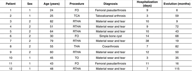
All demographic data are shown in Table 1. The average age in the group was 51 years (24 to 82), consisting of four (34%) male and eight (66%) fe-male patients. The average length of hospitaliza-tion was 10 (3-38) days and mean follow-up was 49 months (6-115).
Histological analysis
Figure3 – Fibrosis represented by whitish light areas in the center of the image. 200x magnification. HCPA 2011.
Table 1 – Demographic data.
Patient Sex Age (years) Procedure Diagnosis Hospitalization (days) Evolution (months)
1 1 24 FO Femoral pseudarthrosis 9 6
2 1 25 TCA Talocalcaneal arthrosis 3 59
3 2 82 RTHA Material wear and tear 10 9
4 2 51 RTHA Material wear and tear 9 72
5 2 64 RTHA Material wear and tear 10 43
6 2 30 FO Simple bone cyst 14 68
7 2 79 RTHA Material wear and tear 38 19
8 2 55 THA Coxarthrosis 7 82
9 2 60 RTHA Material wear and tear 12 50
10 1 45 TO Material wear and tear 3 35
11 1 43 FO Femoral pseudarthrosis 11 16
12 1 48 RTHA Material wear and tear 7 115
1: Male, 2: Female; TO: Tibial osteosynthesis, FO: Femoral osteosynthesis, EBC: Excision of bone callus, TCA: Talocalcaneal arthrodesis, THA: Total hip arthroplasty, RTHA: revision of THA. HCPA 2011
Table 2 – Histological analysis of slides.
Neutrophils Fibrosis absorptionLBG New local bone formation
ALL - + + +
JARS 1 - + + +
JARS 2 - + + +
MTRC - + - +
EG - + + +
AMB - + + +
TGG - + + +
AACS - + + +
MB - + + +
SG + + +
-MH - + + +
HJCS - + + +
MAR - + + +
+: present, -: absent. HCPA 2011
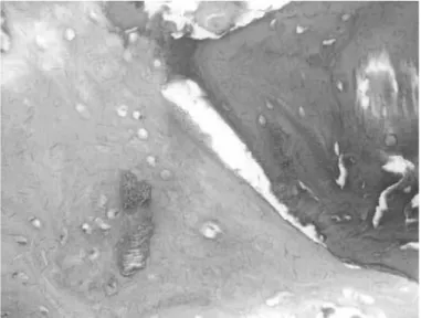
Figure4 – Lyophilized bovine graft (LBG) (blue areas) surrounded by newly formed bone tissue (pink areas). 200x magnification. HCPA 2011.
Figure6 – Well-defined boundaries between the bluish lyophilized bovine graft (LBG) and the pink new bone. 400x magnification. HCPA 2011.
specific analysis
The digitized slides showed a mean proportion of 42% (13-85%) of lyophilized bovine graft and 58% (15-87%) of new bone formation in relation to the total mineralized area (Table 3). Importantly, two pa-tients showed significant differences in relation to the other samples with a ratio of LBG/bone formation of 85/15% and 76/24%, and the slides were reviewed, in order to exclude any measurement errors.
Table 3 –Proportions measured.
LBG Neoformation Follow-up*
ALL 37% 63% 6
JARS 1 34% 66% 59
JARS 2 34% 66% 59
MTRC 23% 77% 9
EG 32% 68% 72
AMB 60% 40% 43
TGG 85% 15% 68
AACA 26% 74% 19
MB 13% 87% 82
SG 76% 24% 50
MH 47% 53% 62
HJCS 38% 62% 16
MAR 39% 61% 115
* months
radiographic analysis
The radiographs of all patients showed satisfactory consolidation in the perimeter of the graft.
DISCUSSION
The possibility of using lyophilized bovine graft (LBG) as an alternative to autologous graft in
orthope-dic surgery has been investigated for some time, par-ticularly in animal models(6). This study contributes
information that is extremely relevant to the use of LBG, produced according to the protocol developed by the main author, in humans, since it demonstrates its osseointegration. Previous studies endorse the use of this LBG both in animals(15,16) and in humans(2,5,18).
Meyer et al(21) brought forth the first data relating
to histological osseointegration in relation to LBG – Tutobone® – in humans. These results were
promis-ing, prompting us to evaluate our biopsies and com-pare the results with those available in the literature. In our study, which has a design similar to Meyer et al(21), we found a proportion of 42% preservation
of the LBG and 58% related to areas of new bone formation compared to the total mineralized area on the slides analyzed in an average period of 49 months. These data are very similar to those presented by this author, who reports having 47% preservation with Tutobone® and 53% of new bone formation in the
course of 11 months in nine samples.
The presence of LBG in the biopsies allows us to infer that the graft proposed presents undeniable osteoconductivity, being only partially reabsorbed in humans, unlike the complete remodeling which occurs in animals. We found no linear correlation between the duration of follow-up and proportions of LBG/bone formation in relation to the total mineralized area, and it was not possible to define an exact pattern of behavior, as the sample with nine months of follow-up had a higher rate of neoformed bone than at 62 months. The fact that the slide that had the highest rate of new bone formation (87%) was one of the longest durations of follow-up (82 months) is also relevant. Thus, we can say that the LBG used promotes satisfactory, efficient, and durable osseointegration, with human bone tissue during the follow-up period measured.
samples showed no difference in the rate of consoli-dation, or worse clinical outcomes.
This leads us to think that the type and quantity of graft applied are not solely responsible for the ca-pacity for new local bone formation, but that every organism, with its different biological characteristics, affects the response of the osseointegration process. Elucidating this is not the purpose of the present study, but it could justify the behavior. The absence of a foreign body reaction in all slides demonstrates the biocompatibility of the lyophilized bovine graft developed by our group in this study.
The presence of satisfactory osseointegration in radiographic examinations shows that the LBG in this study presents a visual pattern similar to other commonly used types of grafts. Previous experi-ments showed full osseointegration in 75.8% after six months and nearly all cases at 12 months(21).
We did not use a control group, since the study
design did not allow for it. The objective of the study is to evaluate the histological behavior of LBG in humans and not to discuss its superiority.
CONCLUSION
This is the first histological study using lyophilized bovine graft (LBG), produced according to the pro-tocol developed by the main author, in humans. The results show that this graft had satisfactory biocom-patibility and did not harm the patients. Additionally, it showed good osteoconduction and integration in an average follow-up period of 49 months, fulfilling the role that was expected of it.
The results obtained are promising and contribute to strengthening the role of the LBG as a treatment option for dental and orthopedic surgeries that require some type of graft to fill bone defects.
REFERENCES
1. Finkemeier CG. Bone-grafting and bone-graft substitutes. J Bone Joint Surg Am. 2002;84(3):454-64.
2. Rosito R, Galia CR, Macedo CA, Moreira LF, Quaresma LM, Palma HM. Acetabular reconstruction with human and bovine freeze-dried bone grafts and a reinforcement device. Clinics (Sao Paulo). 2008;63(4):509-14.
3. Lexer E. Joint transplantations and arthoplasty. Tradução de Frank H’Doubler. Surg Gynecol Obstet. 1925;40:782-809.
4. Oliveira RC, Sicca CM, Silva TL, Cestari TM, Oliveira OT, Buzalaf MAR. Efei-to da temperatura de desproteinização no preparo de osso cortical bovino microgranular. Avaliação microscópica e bioquímica da respota celular em subcutâneo de ratos. Revista FOB. 1999;7(3/4):85-93.
5. Galia CR. Enxertos ósseos liofilizados impactados humanos e bovinos em revisão de artroplastia total de quadril [tese]. Porto Alegre/RS: Universidade Federal do Rio Grande do Sul; 2004.
6. Sugihara S, van Ginkel AD, Jiya TU, van Royen BJ, van Diest PJ, Wuisman PI. Histopathology of retrieved allografts of the femoral head. JBone Joint Surg Br. 1999;81(2):336-41.
7. Godwin L. Tissue banking and allograft transplantation. [periódico online]. 2000. Disponível em: www.iscpubs.com/articles/abl/b0006god.pdf
8. Buck BE, Malinin TI. Human bone and tissue allografts. Preparation and safety. Clin Orthop Relat Res. 1994;(303):8-17.
9. Kreuz FP, Hyatt GW, Turner TC, Bassett AL. The preservation and clinical use of freeze-dried bone. J Bone Joint Surg Am. 1951;33(4):863-73.
10. Autograft, allograft, and xenograft. Disponível em: www.pharmacy.wisc.edu/ courses/718-430/handouts/ tisgraft.pdf
11. Seiler JG 3rd, Johnson J, Hand G, Microsurgery Clinic. Iliac crest autogenous bone grafting: donor site complications. Journal of the Southern Orthopedic Association Disponível em: www.medscape.com/viewarticle/410431.
12. Lind M, Krarup N, Mikkelsen S, Horlyck E. Exchange impaction allografting for femoral revision hip arthroplasty: results in 87 cases after 3.6 years’ follow-up.
J Arthroplasty. 2002;17(2):158-64.
13. Heliotis M, Tsiridis EE. Fresh frozen bone in femoral impaction grafting: can develop-ments in bone regeneration improve on this? Med Hypotheses. 2001;57(6):675-8. 14. Palmer SH, Gibbons CL, Athanasou NA. The pathology of bone allograft. J
Bone Joint Surg Br. 1999; 81(2):333-5.
15. Galia CR, Macedo CAS, Rosito R, Mello TM. Uso de enxerto ósseo homólogo e heterólogo em diáfise femural de ratos: comparação entre enxerto ósseo congelado e liofilizado. Rev Bras Ortop. 2005;40:141-6
16. Galia CR, Macedo CA, Rosito R, Mello TM, Camargo LM, Moreira LF. In vitro and in vivo evaluation of lyophilized bovine bone biocompatibility. Clinics (Sao Paulo). 2008; 63(6):801-6.
17. Williams D. Revisiting the definition of biocompatibility. Med Device Technol. 2003;14(8):10-3.
18. Nuss KM, Auer JA, Boos A, von Rechenberg B. An animal model in sheep for biocompatibility testing of biomaterials in cancellous bones. BMC Musculoskelet Disord. 2006;7:67.
19. Pearce AI, Richards RG, Milz S, Schneider E, Pearce SG. Animal models for implant biomaterial research in bone: a review. Eur Cell Mater. 2007;13:1-10. 20. Conn RA, Peterson LFA, Stauffer RN, Ilstrup D. Management of acetabular deficiency: Long-term results of bone grafting the acetabulum in total hip ar-throplasty. Orthopaedics Trans. 1985;9:451-4
21. Meyer S, Floerkemeier T, Windhagen H. Histological osseointegration of Tu-tobone: first results in human. Arch Orthop Trauma Surg. 2008;128(6):539-44. 22. Galia CR, Macedo CA, Rosito R, Mello TM, Camargo LM, Moreira LF. Femoral and acetabular revision using impacted nondemineralized freeze-dried bone allograft. J Orthop Sci. 2009;14(3):259-65.