15 artigo 438
ORIGINAL ARTICLE
1 – Member of the Brazilian Society of Knee Surgery (Sociedade Brasileira de Cirurgia do Joelho); Member of the Brazilian Society of Orthopedics and Traumatology (Sociedade Brasileira de Ortopedia e Traumatologia); Former Resident of the Knee Group of the Orthopedics and Traumatology Clinic of Hospital São Lucas, PUCRS – Porto Alegre, RS, Brazil.
2 – Member of the Brazilian Society of Orthopedics and Traumatology; Former Resident Physician of the Orthopedics and Traumatology Clinic of Hospital São Lucas, PUCRS – Porto Alegre, RS, Brazil.
3 – Master’s degree in Human Movement Sciences; Doctor’s degree in Medicine; Member of the Knee Group of the Orthopedics Clinic of Hospital São Lucas, PUCRS – Porto Alegre, RS, Brazil.
4 – Master’s degree in Human Movement Sciences; Head of the Orthopedics and Traumatology Clinic of Hospital São Lucas, PUCRS – Porto Alegre, RS, Brazil. Study conducted at the Knee Surgery Group of the Orthopedics and Traumatology Clinic of Hospital São Lucas da Pontifícia Universidade Católica do Rio Grande do Sul. Mailing address: Avenida Ipiranga, 6.690 – 90610-000 – Porto Alegre, RS. Email: [email protected]
Study received for publication: 10/21/2010, accepted for publication: 12/23/2011
PHYSICAL EXAMINATIONS FOR DIAGNOSING MENISCAL INJURIES:
CORRELATION WITH SURGICAL FINDINGS
Ricardo da Rocha Gobbo1, Victor de Oliveira Rangel2, Francisco Consoli Karam3, Luiz Antônio Simões Pires4
7KHDXWKRUVGHFODUHWKDWWKHUHZDVQRFRQIOLFWRILQWHUHVWLQFRQGXFWLQJWKLVZRUN
This article is available online in Portuguese and English at the websites: www.rbo.org.br and www.scielo.br/rbort
Rev Bras Ortop. 2011;46(6):726-29 ABSTRACT
Objective: A set of five maneuvers for meniscal injuries (McMurray, Apley, Childress and Steinmann 1 and 2) was evaluated and their sensitivity, specificity, accuracy and like-lihood were calculated. The same methods were applied to each test individually. Methods: One hundred and fifty-two patients of both sexes who were going to undergo videoar-throscopy on the knee were examined blindly by one of five residents at this hospital, without knowledge of the clinical data and why the patient was going to undergo an opera-tion. This examination was conducted immediately before the videoarthroscopy and its results were recorded in an electronic spreadsheet. The set of maneuvers was considered
positive when one was positive. In the individual analysis, it was enough for the test to be positive. Results: The analysis showed that the set of five meniscal tests presented sensitivity of 89%, specificity of 42%, accuracy of 75%, positive likeli-hood of 1.53 and negative likelilikeli-hood of 0.26. Individually, the tests presented accuracy of between 48% and 53%. Conclu-sion: The set of maneuvers for meniscal injuries presented a good accuracy and significant value, especially for ruling out injury. Individually, the tests had less diagnostic value, although the Apley test had better specificity.
Keywords – Knee; Arthroscopy; Physical Examination; Video-Assisted Surgery; Comparative Study
INTRODUCTION
Meniscal injuries (MI) habitually occur in patients who suffer rotational trauma to the knee under com-pression. They can occur separately or in associa-tion with ligament ruptures and chondral pathologies. They are quite often found in the orthopedic prac-tice(1) and MIs usually occur during sports(1).
In a recent case, the findings of the physical exa-mination may be limited, insofar as the patient may present a painful knee, restricted range of motion
(ROM) and joint effusion(2). When it is a
long-stan-ding injury, the efficacy of the tests for detection of
MI will be compromised(3). The posterior horn of the
medial meniscus is the most common site of meniscal
conditions, while longitudinal ruptures represent the most frequent injuries(4).
su-727
Rev Bras Ortop. 2011;46(6):726-29 PHYSICAL EXAMINATIONS FOR DIAGNOSING MENISCAL INJURIES: CORRELATION WITH SURGICAL FINDINGS
perior value to magnetic resonance imaging.
Kocabey et al(18) evaluated the painful joint line, McMurray, Steinmann and modified Apley tests. This set of tests presented accuracy of 80% for the medial meniscus and 92% for the lateral meniscus(18). The aim of our study is to calculate the sensitivity, speci-ficity, accuracy, positive and negative likelihood of the McMurray, Steinmann, Apley and Childress (duck waddle) tests both separately and jointly.
MATERIAL AND METHODS
A cross-sectional study, with prospective data col-lection performed between January 2008 and June 2009. Two of the five Orthopedics and Traumatology residents of Hospital São Lucas, PUCRS, examined 162 patients (163 knees) with meniscal and/or ACL injury who were going to undergo knee videoarthos-copy afterwards. The examiners, who received speci-fic training to perform meniscal maneuvers, were not familiar with the patients’ clinical data, or the reason for surgery. The physical examination was performed prior to surgery and its results were compared with the surgical findings. The residents selected 117 male patients and 45 female patients over 18 years of age (mean age of 39.03 years) with traumatic or degene-rative knee injuries.
The set of maneuvers performed for the MI
diag-nosis was composed of the McMurray apud Tria(19),
Apley(20), Childress, Steinmann I and II tests(19). The physical examination was conducted by two residents; in the event of a tie, a third resident was re-cruited to examine the patient. The result was written down by this examiner, who marked the test positive or negative. For the set of maneuvers, it was conside-red a positive physical examination when one of them was positive. The maneuvers were also considered positive or negative in separate form.
The arthroscopic assessment of the knee was per-formed at the surgical center, always by one of the two orthopedists specialized in the knee, on patients who presented previous surgical indication, but did not present exams and clinical data reported to the three orthopedic residents who were performing the physical examination proposed in the study. Arthros-copies were performed through the classic parapa-tellar, anterolateral and anteromedial portals. After insertion of the arthroscope through the lateral pa-rapatellar portal, a routine inspection was carried
out on the whole joint in all the cases, analyzing the medial and lateral compartments (condyles, plateaus and menisci), intercondyle (cruciate ligaments), and finally the femoropatellar joint (patellar and synovial cartilages). Investigation through medial and lateral suprapatellar portals was performed as necessary. The injuries were identified and recorded for comparison with the physical examination. After the inspection, the lesions were surgically corrected as necessary. Videoarthroscopies were not performed in patients without indication for surgical treatment (videosur-gery) for their disease.
Any type of meniscal injury found in the tran-soperative period was considered a positive finding, regardless of whether it was radial or longitudinal, simple or complex, traumatic or degenerative.
The study was submitted and approved by the re-search ethics committee of the institution where it was conducted, and all the patients read and signed the informed consent form. The data were filed in a MS Office-Excel 2007 spreadsheet and evaluated by BioEstat 5.0 software. The accuracy, sensitivity and specificity of the tests were evaluated jointly.
RESULTS
Of the 162 patients included in the study, 124 presented meniscal injury, while 82 (66.12%) had an injury involving the medial meniscus (MM), 42 (33.87%) the lateral meniscus (LM) and 10 (8.06%) had both menisci injured (Table 1).
As observed in the Table 2, the analysis showed that the set of the five meniscal tests presented 89% of sensitivity for the MM and 85% of sensitivity for the LM. As regards specificity, the values were 31% for the MM and 24% for the LM, which led us to a calculation of 60% of accuracy for the MM and 40% for the LM. The positive likelihood was 1.29 in the MM and 1.13 in the LM. The negative likelihood was 0.35 for the MM and 0.59 for the LM.
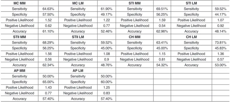
In Table 3 we analyzed each test separately, with their respective values of accuracy, likelihoods, sen-sitivity and specificity. The Apley test presented Table 1 – 5HVXOWVUHODWLQJWRWKHGLVWULEXWLRQRIPHGLDODQGODWHUDO
PHQLVFDOLQMXULHV
MM LM Both M TOTAL
728
Rev Bras Ortop. 2011;46(6):726-29
specificity of 65% for the MM and 60% for the LM; therefore these values are higher than those of the other tests. As regards accuracy, Steinmann I and II tests presented values of around 62% for the detection of MM injury. The accuracy of the isolated tests was greater in the MM examination, with the exception of the Apley test in which it was the same.
DISCUSSION
To verify the value of the physical examination in the detection of meniscal injuries of the knee, we used arthroscopy as the standard method, like in the vast majority of similar studies(18,20-23).
Some provocative tests are described with the in-tention of identifying symptoms involving the me-nisci. The tests used in this study can be divided into two groups. The first group includes the tests that depend on palpation or clicking sensation at the joint line, such as the McMurray and Steinmann II tests. The positive McMurray test for medial meniscus is
demonstrated with external rotation of the tibia and passive movement from flexion to extension. For late-ral meniscus, it is demonstrated with internal rotation of the tibia and passive movement from flexion to extension. The Steinmann II test demonstrates pain at the interline that moves posteriorly when the knee is flexed and anteriorly when the knee is extended.
The second group contains the tests that depend on pain with rotation. The Apley test is carried out through compression and distraction between the ti-biofemoral joint surface in flexion. If the distraction promotes less discomfort than the compression, it in-dicates meniscal pain instead of a joint disorder. The Childress (duck waddle) test provokes compressive force on the posterior horn of the meniscus causing pain. The Steinmann I test is carried out with the knee flexed at 90 degrees and a sudden external ro-tatory force is applied on the tibia to test the medial meniscus. The result is pain along the medial joint line. Internal tibial rotation is used for lateral me-niscal pain(19).
Manzotti et al(23) demonstrated in their study that the McMurray’s maneuver presents greater sensitivity for medial meniscus injuries when compared to the lateral meniscus, a fact that was confirmed in our study. Evans et al(21), taking into account just joint snapping for the positivity of the McMurray test, con-cluded that this has specificity of 98% and sensitivity of 16%. Our study showed a lower specificity, yet a Table 2 – 6HQVLWLYLW\VSHFLILFLW\SRVLWLYHOLNHOLKRRGQHJDWLYHOLNHOL
KRRGRIWKHVHWRIWHVWV
MM Set LM Set
Sensitivity 89.02% Sensitivity 85.71% Specificity 31.25% Specificity 24.17% Positive Likelihood 1.29 Positive Likelihood 1.13 Negative Likelihood 0.35 Negative Likelihood 0.59 Accuracy 60.49% Accuracy 40.00%
MM: medial meniscus; LM: lateral meniscus.
Table 3 – 6HQVLWLYLW\VSHFLILFLW\SRVLWLYHOLNHOLKRRGQHJDWLYHOLNHOLKRRGRIWKHWHVWVVHSDUDWHO\
MC MM MC LM STI MM STI LM
6HQVLWLYLW\ 6HQVLWLYLW\ 6HQVLWLYLW\ 6HQVLWLYLW\
6SHFLILFLW\ 6SHFLILFLW\ 6SHFLILFLW\ 6SHFLILFLW\
3RVLWLYH/LNHOLKRRG 3RVLWLYH/LNHOLKRRG 3RVLWLYH/LNHOLKRRG 3RVLWLYH/LNHOLKRRG
1HJDWLYH/LNHOLKRRG 1HJDWLYH/LNHOLKRRG 1HJDWLYH/LNHOLKRRG 1HJDWLYH/LNHOLKRRG
$FFXUDF\ $FFXUDF\ $FFXUDF\ $FFXUDF\
STII MM STII LM CH MM CH LM
6HQVLWLYLW\ 6HQVLWLYLW\ 6HQVLWLYLW\ 6HQVLWLYLW\
6SHFLILFLW\ 6SHFLILFLW\ 6SHFLILFLW\ 6SHFLILFLW\
3RVLWLYH/LNHOLKRRG 3RVLWLYH/LNHOLKRRG 3RVLWLYH/LNHOLKRRG 3RVLWLYH/LNHOLKRRG
1HJDWLYH/LNHOLKRRG 1HJDWLYH/LNHOLKRRG 1HJDWLYH/LNHOLKRRG 1HJDWLYH/LNHOLKRRG
$FFXUDF\ $FFXUDF\ $FFXUDF\ $FFXUDF\
AP MM AP LM
6HQVLWLYLW\ 6HQVLWLYLW\
6SHFLILFLW\ 6SHFLILFLW\
3RVLWLYH/LNHOLKRRG 3RVLWLYH/LNHOLKRRG
1HJDWLYH/LNHOLKRRG 1HJDWLYH/LNHOLKRRG
$FFXUDF\ $FFXUDF\
729
Rev Bras Ortop. 2011;46(6):726-29 PHYSICAL EXAMINATIONS FOR DIAGNOSING MENISCAL INJURIES: CORRELATION WITH SURGICAL FINDINGS
higher sensitivity in relation to Evans’ study for this test. Meserve et al(24), in their meta-analysis, showed that the Apley test presents superior specificity when compared to McMurray’s maneuvers and painful pal-pation of the joint line. However, as regards sensitivi-ty, the Apley test presented much lower values. These data were similar to those of our study when the Apley test was compared to another four maneuvers.
Steinmann I and II tests are not often used to test the accuracy of the physical examination in studies of greater relevance. Kocabey et al(18) cited Steinmann’s maneuver in their study, yet did not specify whether it was Steinmann I or II, showing the data together with other maneuvers. Through our results it is possible to notice that both Steinmann I and II have superior accuracy over the other tests in relation to the medial meniscus.
Fowler and Lubliner(17) stressed that no menis-cal test is predictive for the diagnosis, and that the set of maneuvers should be used. Kocabey et al(18)
compared the accuracy of the set of maneuvers with that of nuclear magnetic resonance (NMR) in their study. Through their data, they concluded that the physical examination has superior accuracy to that of NMR with sensitivity results that resemble ours (MM 87% – LM 75%). Accordingly, in patients with strong suspicion of injury, the set of maneuvers can be very useful for ruling out this suspicion.
All the isolated tests, except for the Apley test, presented greater sensitivity than specificity. A test with high sensitivity is used mainly to exclude the presence of a pathology.
CONCLUSION
The set of maneuvers for meniscal injuries has good accuracy and significant value, particularly to exclude injuries. The isolated tests have lower diag-nostic value, while the Apley test is that with the best specificity.
REFERENCES
1. 0DMHZVNL0+DOEHW66WHLQEUFN.(SLGHPLRORJ\RIDWKOHWLFNQHHLQMXULHV$ 10-year study. Knee. 2006;1(3):184-8.
2. DeHaven KE. Diagnostic of acute knee injuries with hemarthrosis. Am J Sports Med. 1980;8(1):9-14.
3. Wagemakers HP, Heintjes EM, Boks SS, Berger MY, Verhaar JA, Koes BW et al. Diagnostic value of history-taking and physical examination for as-sessing meniscal tears of the knee in general practice. Clin J Sports Med. 2008;18(1):24-30.
4. Miller RH. Knee injuries. In: Canale ST, editor. Campbell’s orthopaedics surgery. 10th ed. Philadelphia: Mosby,2003. p. 2165-337.
5. Karam FC, Silva JLB, Fridman MW, Abreu A, Arbo RDM, Abreu M et al. A ressonância magnética para lesões condrais, meniscais e dos ligamentos cruzados dos joelhos. Radiol Bras. 2007;40(3):179-82.
6. Schneider I, Schueda MA, Demore AB. Análise comparativa da ressonância nuclear magnética com a artroscopia no diagnóstico das lesões intra-articulares do joelho. Rev Bras Ortop. 1996;31(5):373-6.
7. Oei EH, Nikken JJ, Verstijnen AC, Ginai AZ, Myriam Hunink MG. MR imag-ing of the menisci and cruciate ligaments: a systematic review. Radiology. 2003;226(3):837-48.
8. Vincken PW, ter Braak BP, van Erkell AR, de Rooy TP, Mallens WM, Post W, et al. Effectiveness of MR imaging in selection of patients for arthroscopy of the knee. Radiology. 2002;223(3):739-46.
9. Severino NR, Camargo OPA, Aihara T, Cury RPL, Oliveira VM, Vaz CES et al. Comparação entre a ressonância magnética e a artroscopia no diagnóstico das lesões do joelho Rev Bras Ortop. 1997;32(4):275-8.
10. Yousef JW, Thiele ES, Scuisato DL. Correlação diagnóstica da ressonância magnética com artroscopia nas lesões intra-articulares do joelho. Rev Bras Ortop. 1999;34;(6):375-80.
11. Barretto JM, Couto P. Artroscopia do joelho sob anestesia local e sedação: possibilidades diagnóstica e terapêutica. Rev Bras Ortop. 1997;32(4):289-92.
12. Andrade MA, Vassalo CC, Cunha MG. Artroscopia cirúrgica em pacientes acima de 40 anos. Rev Bras Ortop. 1998;33;(5):401-5.
13. Calmbach WL, Hutchens M. Evaluation of patients presenting with knee pain: Part I. History, physical examination, radiographs, and laboratory tests. Am Fam Physician. 2003;68(5):907-12.
14. Smith BW, Green GA. Acute knee injuries: Part I. History and physical examina-tion. Am Fam Physician. 1995;51(3):615-21.
15. Jackson JL, O’Malley PG, Kroenke K. Evaluation of acute knee pain in primary care. Ann Intern Med. 2003; 139(7):575-88.
16. Heintjes EM, Bierma SM, Bernsen RM. Physical examination of knee injuries. JAMA. 2002;287(1):40; author reply 41.
17. Fowler PJ, Lubliner JA. The predictive value of five clinical signs in the evalu-ation of meniscal pathology. Arthroscopy. 1989;5(3):184-6.
18. Kocabey Y, Tetik O, Isbell WM, Atay A, Johnson DL. The value of clinical ex-amination versus magnetic resonance imaging in the diagnosis of meniscal tears and anterior cruciate ligament rupture. Arthroscopy. 2004;20(7):696-700. 19. Tria AJ Jr. Clinical examination of the knee. In: Insaii JN, Scott WN, editors.
Surgery of the knee. 3rd ed. New York: Churchill Livingstone; 2001. p 161 74. 20. Apley G. The diagnosis of meniscus injuries. Bone Joint Surg Am. 1947;29(1):78-84. 21. Evans PJ, Bell GD, Frank C. Prospective evaluation of the McMurray test. Am
J Sports Med.1993;21(4):604-8.
22. Anderson AF, Lipscomb AB. Clinical diagnosis of meniscal tears: description of a new manipulative test. Am J Sports Med 1986;14(4):291-3.
23. Manzotti A, Baiguini P, Locatelli A. Statistical evaluation of McMurray’s test in the clinical diagnosis of meniscus injuries. J Sports Traumatol Relat Res. 1997;19:83–9.