REVIEW ARTICLE
Neurobiological underpinnings of bipolar disorder
focusing on findings of diffusion tensor imaging:
a systematic review
Juliana A. Duarte,
1,2,3Jaisa Q. de Arau´jo e Silva,
3Andre´ A. Goldani,
1Raffael Massuda,
1,4,5Clarissa S. Gama
11Laborato´rio de Psiquiatria Molecular, Instituto Nacional de Cieˆncia e Tecnologia – Medicina Translacional (INCT-TM), Hospital de Clı´nicas de
Porto Alegre (HCPA), Universidade Federal do Rio Grande do Sul (UFRGS), Porto Alegre, RS, Brazil.2Departamento de Radiologia e Ressonaˆncia Magne´tica, HCPA, Porto Alegre, RS, Brazil.3Tomoclı´nica, Canoas, RS, Brazil.4UFRGS, Porto Alegre, RS, Brazil. 5Departamento de Psiquiatria, Universidade Federal do Parana´ (UFPR), Curitiba, PR, Brazil.
Objective:
To review the available data on diffusion tensor imaging (DTI) of subjects with bipolar
disorder (BD), with a particular focus on fractional anisotropy (FA) in white matter (WM) tracts.
Methods:
The PubMed/MEDLINE database was searched for relevant articles, which were included in
a systematic review of the literature. FA reductions and WM abnormalities were divided anatomically
into three groups: commissural tracts, association tracts, and projection tracts.
Results:
Eighteen studies met the inclusion criteria. The corpus callosum was the main impaired
commissural tract as demonstrated by FA reductions. Five studies reported FA reductions in the
cingulum. Two studies reported decreased FA in the anterior thalamic radiation, and one in the
corticospinal tract. Conversely, three studies found increased FA values in WM tracts involved in BD
pathophysiology.
Conclusion:
Despite considerable heterogeneity, these results indicate a direct link between
executive cognitive functioning and abnormal WM microstructural integrity of fronto-limbic tracts in
patients with remitted BD, providing further evidence of the neuronal disruption that underlies BD
symptomatology.
Keywords:
Bipolar disorder; diffusion tensor imaging; neuroimaging; diffusion tractography
Introduction
Bipolar disorder (BD) is a severe psychiatric disorder that
affects approximately 1.5% of the world population
1,2and
remains one of the leading worldwide causes of disability,
morbidity, and mortality.
3,4The progression of BD is
frequently associated with an increased number of
epi-sodes,
5-8subclinical symptoms in the interepisodic
per-iod,
9,10higher rates of comorbidities,
11increased risk of
suicide,
12a higher number of hospital admissions,
13and
poorer response to treatment.
6Furthermore, several
stu-dies have shown a strong association between number of
mood episodes and unfavorable clinical outcomes,
espe-cially cognitive and functional impairment.
14,15In BD, neural substrate reactivity is changed by
repeated mood episodes, which ultimately promote a
brain rewiring that leads to increased vulnerability to life
stress.
16-18Changes in brain structure have been widely
reported in BD patients.
19Over the past decade,
subs-tantial effort has been made in neuroimaging research to
understand the neural system abnormalities that underlie
BD, and significant progress has been made in identifying
regional brain differences that could contribute to the
symptoms of acute episodes.
20Morphometric studies have demonstrated that patients
with BD exhibit enlargement of the third and lateral
ventricles; a reduction in the gray matter volumes of the
orbital and medial prefrontal cortex, ventral striatum, and
mesotemporal cortex; and enlargement of the amygdala.
Such neuroanatomical changes tend to be more
pro-nounced in patients who have experienced repeated
epi-sodes. With respect to neuropathological findings, recent
data suggest that changes in neuroplasticity, particularly
in cell resilience and connectivity, are the main findings
associated with BD.
21-25Other studies suggest that structural brain changes are
found mainly in the frontal, temporal, and limbic white
matter (WM) regions.
26-31WM abnormalities have been
widely detected in subjects with the pathophysiological
features of BD, especially with diffusion tensor imaging
(DTI) techniques.
29Diffusion imaging principles are based on
measure-ment of the motion of water molecules within tissues.
32Free water usually moves equally in all directions in an
isotropic fashion. When the movement of water
mole-cules is restricted, however, preferential directions are
taken, and movement consequently becomes anisotropic.
Correspondence: Clarissa S. Gama, Hospital de Clı´nicas de Porto Alegre, Rua Ramiro Barcelos, 2350, CEP 90035-903, Porto Alegre, RS, Brazil. E-mail: [email protected]
Submitted Aug 10 2015, accepted Sep 02 2015.
Revista Brasileira de Psiquiatria. 2016;38:167–175 Associac¸a˜o Brasileira de Psiquiatria
Therefore, water mobility in the brain is markedly reduced
in compact tissue, such as WM, is reduced to a lesser
extent in gray matter (GM), and is almost free in the
cerebrospinal fluid (CSF). Pathological processes that
alter the normal brain structure may affect water motion
and thereby affect the resulting diffusion indexes.
33Diffusion images can be acquired from a minimum of
three gradient directions that yield two different types of
sequences: diffusion-weighted imaging (DWI) and DTI.
The use of more than six encoding directions improves
the accuracy of tensor measurement for any arbitrary
orientation (Figure 1).
34WM tracts can be divided anatomically into three
groups: commissural tracts, projection tracts, and
asso-ciation tracts. Commissural tracts are fibers that
inter-connect the hemispheres of the brain, such as the
corpus callosum (CC). Association tracts are groups of
fibers that interconnect cortical areas within the same
hemisphere, and projection tracts are efferent and
af-ferent fibers that interconnect the cortex to subcortical
structures.
35-38Previous investigations have hypothesized that
micro-structural changes in the WM of frontal-subcortical circuits
leads to a disconnection syndrome between the frontal
and subcortical regions.
30These network alterations have
been associated with clinical symptoms of BD, which
suggests that DTI is a promising technique for evaluation
of the underpinnings of neuropathology in BD.
39The aim of this paper is to conduct a systematic review
of studies that have used DTI in patients with BD, with
particular emphasis on fractional anisotropy (FA) findings,
and discuss the relevance and connection of these
find-ings to BD pathophysiology.
Methods
Systematic review
The recorded variables for each article included imaging
technique (magnetic resonance imaging [MRI], DTI),
imag-ing analysis (whole-brain/region of interest [ROI]), field
strength, gender, mean age, exposure to medication, brain
regions analyzed, and principal findings (BD vs. controls).
Selection procedures
The inclusion criteria were: a) English-language original
articles published in peer-reviewed journals, in which
study participants were diagnosed with BD type I (BD-I) or
BD type II (BD-II), and which employed structural or
neurochemical imaging techniques. Studies were
inde-pendently assessed for eligibility by two researchers.
Search strategies
The PubMed/MEDLINE database was searched using
the following queries based on Medical Subject Headings
(MeSH) descriptors: ‘‘imaging, diffusion tensor and bipolar
disorder,’’ ‘‘diffusion tractography and bipolar disorder,’’ and
‘‘tractography, diffusion and bipolar disorder.’’ There were
no limits regarding year of publication, and the search
included papers published through January 2015.
Results
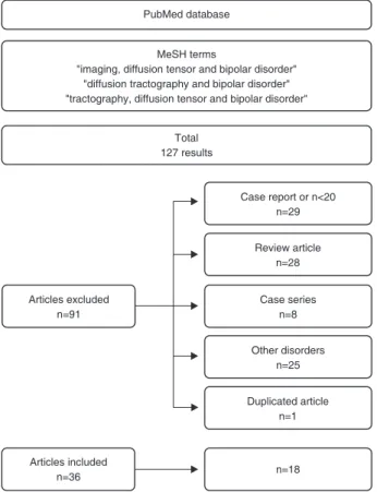
The search yielded 127 articles. Search strategies and
exclusion criteria are summarized in Figure 2. We found 18
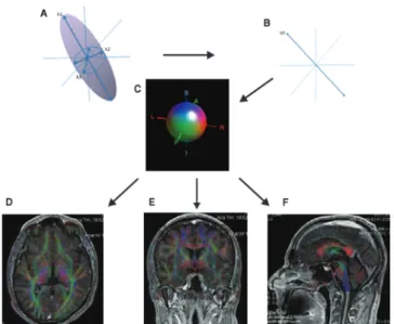
Figure 1
Diffusion measurements along multiple axes. The shape and the orientation of a diffusion ellipsoid is estimated. From
the estimated ellipsoid (A), the orientation of the longest axis can be found (B), which is assumed to represent the local fiber
orientation. This orientation information is converted to a color at each pixel. By combining the intensity of the anisotropy map
and color, a color-coded orientation map is created (C). The diffusion tensor image map is rendered in the axial (D), coronal (E),
and sagittal (F) planes. The color coding depicts the local fiber orientation, i.e., the principal eigenvector of the diffusion tensor,
with red indicating mediolateral, green denoting anteroposterior, and blue representing superoinferior. Color coding is also
indicated by the red-green-blue sphere (C).
Rev Bras Psiquiatr. 2016;38(2)
published
DTI
studies that
identified
WM
changes
in subjects with BD. We assessed FA in three different
anatomical groups: commissural tracts, projection tracts,
and association tracts. Tracts for which FA findings
were reported in the included studies are presented
in Figure 3.
Results are highly heterogeneous, and most
pub-lished papers have reported decreased FA values in
WM tracts (Table 1). Overall, the most common finding
is decreased FA values in commissural and association
tracts,
particularly
in
the
fronto-limbic
tracts
(Table 2).
43,53,54FA and WM tracts
With respect to the commissural tracts, most authors found
decreased FA values in the CC.
29-31,40,43-46,49Regarding association tracts, five studies found
de-creased FA values in the cingulum.
29,43,45,48,49With respect
to the projection tracts, two studies noted decreased FA in
the ATR, and one study found decreased FA in the
cortico-spinal tract (CST) (Table 2).
Conversely, three studies reported increased FA values.
Wessa et al.
55found increased FA values in the medial
frontal, precentral, inferior parietal, and occipital WM. Mahon
et al.
56observed higher FA levels within the right and left
frontal WM, while Versace et al.
42observed increased FA in
the left uncinate fasciculus (UF) (reduced radial diffusivity
distally and increased longitudinal diffusivity centrally), left
optic radiation (increased longitudinal diffusivity), and right
anterior thalamic radiation (ATR).
Discussion
Most studies reported decreased FA values in regions
involved in emotion processing, such as the commissural
tracts,
especially
the
CC,
and
the
association
tracts.
43,53,54,57-60The latter include the UF,
47,61-63the
ATR,
62,63and the cingulum.
45,61Figure 2
Flowchart of identification and selection of studies for a
systematic review of diffusion tensor imaging in bipolar disorder.
Figure 3
White matter tract reconstruction based on reported findings of decreased fractional anisotropy on diffusion tensor
imaging: uncinate fasciculus,
40-42corpus callosum/forceps,
29,31,40,43-48cingulum,
29,43,45,48,49anterior thalamic radiation,
31,42superior longitudinal fasciculus,
29,31,40,43,50inferior longitudinal fasciculus,
40,43,50,51corticospinal tract.
52Table 1
Diffusion tensor imaging studies in bipolar disorder
Study Patients Age I/II/ otherBD type
Disease duration
(years) Moodstate Substanceuse Drugs directionTesla | B-value Software Voxel size (mm3) Measures Results
Maller43 31 BD
31 HC 43.2939.58669.1310.7 16/15/0 N/A N/A N/A 7 drug users (6.288) withBD-I/10 (5.738) with BD-II 1.5 | 12 0/1,000 FMRI Diffusiontoolbox/TBSS 0.9x0.9x3 FA, AD, RD Widespread, significant FAdifferences between controls and all BD subjects, primarily along the CC, cingulum bundles, fornices, SLF, ILF/FOFs, thalami, and UF. Significant differences in FA and all its constituent values between controls and BD-I and BD-II subjects separately. Oertel-Kno¨chel31 21 BD
20 HC
35.67610.6 36.90611.0
21/0/0 7.62 Euthymic N/A Mood stabilizers (n=21), antidepressants (n=9), neuroleptics (n=12), anxiolytics (n=3)
3 | 60 0/1,000 TBSS FSL 4.1 3x3x3 FA, MD, RD, AD
Patients with BD showed significantly higher MD, RD, and AD scores in comparison with HCs in the left superior longitudinal fascicle. FA scores were not significantly different between groups.
Sarrazin29 118 BD 86 HC
36.32610.4 37.26611.2
118/0/0 15.57 Euthymic 25 alcohol Lithium (n=39), other mood stabilizers (n=64), antipsychotics (n=52), antidepressants (n=54)
3 | 41 0 /1,000 Connectomist 2.0 and BrainVisa 4.2
2x2x2 GFA Compared with controls, BD-I patients had significant reductions in mean GFA values along the body and splenium of the CC, the left cingulum, and the anterior part of the left arcuate fasciculus when controlling for age, gender, and acquisition site. Patients with a history of psychotic features had a lower mean GFA value along the body of the CC than those without such a history.
Oertel-Kno¨chel30 30 BD 32 HC
39.22612.3 39.22610.3
30/0/0 10.2 Euthymic N/A 8.30 (7.40) years of medication use
3 | 60 0/1,000 FMRIB/MRIST/ TBSS
1x1x1 AD, RD, FA, MD
Significantly lower FA values in BD patients than in controls. The CC tended to show lower FA and higher RD in BD patients compared with controls. The splenium and truncus showed significantly lower FA and the truncus showed higher RD in BD patients compared with controls. FA values were significantly reduced in BD patients in the right thalamic radiation and showed trend-level significance in the left ATR.
Canales-Rodrı´guez44 40 HC40 BD 40.640.468.9
69.3
40/0/0 15.9 Euthymic Excluded 39 mood stabilizers (n=39) or lithium alone in combination with others (n=30), valproate (n=2), lamotrigine (n=2), others (n=5); antidepressants (n=9). antipsychotics (n=22), combination (n=1)
1.5 | 55 0/1,500 Brain Extraction Tool (FSL)
2x2x3 FA, MD, PTO, GFA
Significant reductions in FA were observed in the splenium of CC and right insula. There was a widespread pattern of increased MD in gray and WM tissues including anterior cingulum, left insula, and subcortical nuclei, without significant decreases in BD patients. Three of the contrasts (FA, mean diffusivity, and GFA) revealed abnormalities in subcortical structures, including the hippocampus, thalamus, and caudate nucleus.
Ambrosi40 20 BD
21 HC 41.9534.616613.110.8 0/20/0 12.6 Euthymic Excluded 19 lithium (n=7),anticonvulsants (n=10), antidepressants (n=8), antipsychotics (n=10)
1.5 | 12 N/A FSL/FMRIB/
TBSS N/A FA Significant, widespread FAreduction in patients with BD-II compared with controls in all major WM tracts studied, including cortico-cortical association tracts, i.e., uncinate, inferior fronto-occipital, inferior longitudinal, and superior longitudinal fasciculi,
interhemispheric tracts, as well as limbic tracts and parahippocampal tract.
Continued on next page
Rev
Bras
P
siquiatr
.
20
16;38(2)
170
JA
Duarte
et
Table 1.(Continued)
Study Patients Age
BD type I/II/ other Disease duration (years) Mood state Substance use Drugs Tesla |
direction B-value Software Voxel size (mm3) Measures Results
Emsell45 35 BD 43 HC
44610 42610
35/0/0 12 Euthymic 7 alcohol users
10 (7) years’ lithium use 1.5 | 64 0/1,300 Explore DTI 2.5x2.5x2.5 FA, MD, AD, RD
Significant differences between patients and control subjects in FA, MD, and RD in the CC. In the fornix, significant differences were found in MD, AD, and RD. In all cases, anisotropy decreased and diffusivity increased in patients compared with controls. Leow46 25 BD
24 HC 41.641.66612.710.6 25/0/0 20.8 Euthymic N/A Valproic acid (n=7),carbamazepine (n=1), lamotrigine (n=3), antipsychotic (n=14), SSRI (n=8), antidepressants (n=5), benzodiazepines (n=3)
3 | 64 0/1,000 DTI Studio 1x1x1 FA, MD Statistically significant group differences in FA, in including the genu, body, and splenium of CC. There was no significant between-group difference in MD for any WM structure, as the genu exhibited higher MD values in the bipolar group.
Torgerson52 27 BD 26 HC
44.2612.9 41.5612.1
27/0/0 23.2 Euthymic 13 alcohol users, 7 drug abusers Antipsychotics (n=16), anticonvulsants (n=15), antidepressants (n=14), benzodiazepines (n=5)
3 | 64 0/1,000 TrackVis 2x2x2 FA No differences in fiber FA between BD subjects and healthy controls, except for reduced FA in one of the corticospinal tracts (CST-R2).
Benedetti49 40 BD 21 HC
47.79613.2 39.86611.0
40/0/0 13.54 Euthymic Excluded Lithium (n=14) 3 | N/A 0/900 TBSS 1.88x1.87x2.3 MD, FA, DI, RD
Compared with control subjects, patients showed lower FA in the genu of the CC and in the anterior and right superior-posterior corona radiata. Higher radial diffusivity values were found in WM tracts of the splenium, genu and body of CC, right mid-dorsal part of the cingulum bundle, left anterior and bilateral superior and posterior corona radiata, bilateral SLF, and right posterior thalamic radiation.
Wessa55 22 BD 21 HC
45.41612.6 42.95613.1
14/08/2 22 Euthymic Excluded Lithium (n=10), anticonvulsants (n=11), atypical antipsychotics (n=5)
1.5 | 41 0/700 BrainVisa 3/ TBSS
1.88x1.87x2.3 MD, FA FA was significantly increased in BD patients relative to healthy controls in medial frontal, precentral, inferior parietal, and occipital WM. No group differences in mean diffusivity were found.
Wang41 33 BD 31 HC
30.4610.8 31.869.6
33/0/0 N/A 10 manic/ hypomanic, depression, 16
euthymic
17 (13 alcohol and/ or nine other substances, four other substances) Lithium (n=9), anticonvulsants (n=16), antipsychotics (n=14), antidepressants (n=12), benzodiazepines (n=7)
3 | 32 0 /1,000 WFU Pick Atlas tool
1.5x1.5x1.5 FA An association was found between pACC-amygdala functional connectivity measurements and the structural integrity of ventro-frontal WM, including the UF, where FA was significantly decreased in the BD group.
Zanetti50 37 BD 26 HC
34.169.1 28.869.5
37/0/0 11.6 16 depressed/21 remission
Excluded Depression: lithium (n=6), valproate (n=6), lamotrigine (n=5), antipsychotics (n=8), antidepressants (n=7), benzodiazepines (n=6). Remission: lithium (n=6), valproate (n=2), carbamazepine (n=3), antipsychotics (n=11), antidepressants (n=9), benzodiazepine (n=4)
3 | 6 0/850 BioImage Suite 2.0
1.6x1.6x3 FA, MD Significantly decreased FA and increased MD in bilateral prefronto-limbic-striatal WM and right inferior fronto-occipital, superior, and inferior longitudinal fasciculi were found in all BD-I patients vs controls and in depressed BD-I patients compared both to controls and to remitted BD-I patients. These findings suggest that depression in BD-I may be associated with acute microstructural WM changes.
Mahon56 30 BD 38 HC
33.468.7 31.968.6
25/2/3 N/A N/A 11 alcohol
or other substances
All treated with antidepressants and/or mood stabilizers
1.5 | 25 0/1,000 DTI Studio N/A AD, RD, FA Voxelwise analysis of WM revealed three regions with higher FA in the right and left frontal WM and one region of lower FA in the left cerebellum in BD patients compared to healthy volunteers.
Continued on next page
Table 1.(Continued)
Study Patients Age
BD type I/II/ other
Disease duration (years)
Mood state
Substance use Drugs
Tesla |
direction B-value Software Voxel size (mm3) Measures Results
These findings suggest that, compared to healthy volunteers, adult patients with BD have higher FA in the bilateral frontal WM, corresponding approximately to fibers of the corticopontine/ corticospinal tract and SLF, as well as superior thalamic radiation fibers. In addition, FA was lower in the left cerebellar WM, thus corresponding approximately to the pontine crossing tract, in patients compared to healthy volunteers.
Versace42 21 BD 25 HC
35.968.9 29.4869.4
31/0/0 12.29 depressed/ 11.82 remitted
14 depressed/17 remitted
10 alcohol or other substances
All medicated 3 | N/A 0/850 FSL/TBSS N/A N/A Subjects with BD had significantly greater FA in the left UF (reduced radial diffusivity distally and increased longitudinal diffusivity centrally), left optic radiation (increased longitudinal diffusivity), and right ATR (no significant diffusivity change), as well as significantly reduced FA in the right UF (greater radial diffusivity), vs. controls. Decreased FA was observed in the left optic radiation and in the right ATR among subjects with BD taking mood stabilizers vs. those with BD not taking mood stabilizers, as well as in the left optic radiation among depressed vs. remitted subjects with BD.
Wang48 BD 42
HC 42 32.628.76610.19.1 42/0/0 N/A hypomanic, 911 manic/ depressed, 22 euthymic
N/A Lithium (n=11), anticonvulsants (n=20), antipsychotics (n=19), antidepressants (n=17), benzodiazepines (n=8), levothyroxine sodium (n=5)
3 | 32 0/1,000 BioImage Suite N/A FA FA was significantly decreased in the anterior cingulum in the BD group compared with healthy controls; however, FA in the posterior cingulum did not differ significantly between groups.
Wang48 BD 33
HC 40 29.2326610.19.2 33/0/0 N/A hypomanic, 77 manic/ depressed, 19 euthymic
11 alcohol, 6 substance abuse, 3
other substances
No medication (n=6), lithium (n=8), anticonvulsants (n=17), atypical antipsychotics (n=17), benzodiazepines (n=8)
3 | 32 0/1,000 BioImage Suite N/A FA Using complementary ROI- and voxel-based DTI methods, the authors found decreased FA values in participants with BD compared to HCs in the anterior and middle CC subregions encompassing the genu, rostral body, and anterior portion of the mid-body.
Bruno51 BD 36 HC 28
39 25/11/0 13.8 N/A N/A Lithium (n=23), sodium valproate (n=3), carbamazepine (n=4), lamotrigine (n=3), neuroleptic (n=9)
1.5 | 7 0/700 SPM2 N/A MD, FA In the patient group, mean diffusivity was increased in the right posterior frontal and bilateral prefrontal WM, while FA was increased in the inferior, middle temporal, and middle occipital regions.
AD = axial diffusivity; ATR = anterior thalamic radiation; BD = bipolar disorder; BD-I = bipolar I disorder; BD-II = bipolar II disorder; CC = corpus callosum; FA = fractional anisotropy; FMRI = functional magnetic resonance imaging; FOFs = fronto-occipital fasciculi; GFA = generalized fractional anisotropy; HC = healthy controls; ILF = inferior longitudinal fasciculi; MD = mean diffusivity; N/A = not mentioned in text; PTO = probability of return to the origin; RD = radial diffusivity; ROI = region of interest; SLF = superior longitudinal fasciculi; SPM = statistical parametric mapping; SSRI = selective serotonin reuptake inhibitors; TBSS = tract-based spatial statistics; UF = uncinate fasciculus; WFU = Wake Forest University School of Medicine PickAtlas; WM = white matter.
Rev
Bras
P
siquiatr
.
20
16;38(2)
172
JA
Duarte
et
The findings of decreased FA values are consistent with
the description of BD as a disconnection syndrome.
64,65The two major symptom domains in BD are mood instability
and poor cognitive control over executive functions.
57Historically, the aforementioned regions have been found
to be involved in emotional processing. In 1937, Papez
66proposed that emotion regulation is enabled through rich
reciprocal connections between parts of the prefrontal
cortex with the amygdala, anterior temporal regions,
sub-genual anterior cingulate cortex, striatum, and thalamus.
Contrary to the predominant findings, Wessa et al.,
55Mahon et al.,
56and Versace et al.
42found increased FA
values in different WM tracts. Despite a lack of support in
the literature, a number of variables may explain these
results. For example, most of these studies were
per-formed before 2009 and used either fewer DTI directions
or older versions of reconstruction software, or involved
patient selection bias.
The main region exhibiting decreased FA values was the
CC, the major interhemispheric WM connection that
integrates emotional, cognitive, motor, and sensory
informa-tion. The anterior CC regions integrate all right and left
pre-frontal cortex, anterior cingulate, and insula regions
impli-cated in emotional deregulation, a core symptom of BD.
With respect to the association tracts, several studies
have reported impairment in WM connection in patients
with BD, with most indicating impairment in the
cingu-lum
29,43-45,48and the UF.
40,42,43The cingulum is a
complex fiber system that forms a central component of
the entire limbic network where the UF carries association
fibers between the medial prefrontal cortex and the
anterior temporal lobe, including the amygdala. These
regions have been extensively related to the
pathophy-siology of BD.
67-69In projection fibers, three studies described decreased
FA values in the ATR. The ATR connects the dorsomedial
and anterior thalamic nuclei with the prefrontal cortex, and
the anterior part of the ATR is connected with the
hippo-campus through the fornix. Alterations in the connections
between the thalamus and limbic areas may be relevant
to cognitive processing and to clinical symptoms
obser-ved in patients with BD.
49,70Alterations in ATR fiber
integrity have been previously reported in BD patients,
consistent with functional magnetic resonance imaging
(fMRI) and structural findings.
19Certain important pathways could also be related to the
pathophysiology of BD. The fornix is a projection tract that
is located underneath the CC and connects the
hippo-campus with the mammillary body as well as with other
cortical and subcortical structures.
30Both structures are
part of the limbic system and known to be involved in
memory processes. The lack of previous reports
regard-ing fornix alterations in BD may be due to the anatomic
characteristics of the fornix and to the spatial resolution of
current MRI methods.
49Previous investigations have hypothesized that
micro-structural changes in the WM of the frontal-subcortical
circuits lead to a disconnection syndrome between the
frontal and subcortical regions.
39These results suggest a
direct link between executive cognitive functioning and
abnormal WM microstructural integrity of the fronto-limbic
tracts in remitted BD patients, and provide further
evi-dence of the neuronal disruption that underlies the
residual symptomatology of BD.
It is not clear whether number of episodes, duration of
illness, and other clinical progression characteristics are
associated with decreased FA values. However, BD has a
poorer long-term outcome than previously thought, with
persistent cognitive impairment and functional decline.
71,72Cognitive impairment has been found to affect executive
functions predominantly, while moderate cognitive deficits
have been observed in other cognitive tests, such as verbal
memory, response inhibition, sustained attention,
psycho-motor speed, abstraction, and set-shifting.
73These
cogni-tive impairment domains seem to have a close correlation
with WM and brain connectivity deterioration.
74-76Recently, a neuroinflammatory component has been
implicated in the pathophysiology of certain psychiatric
disorders,
77and offers a plausible explanation as to why
WM lesions are present in patients with BD.
78Of note, WM
is particularly vulnerable to the inflammatory neurotoxic
Table 2
White matter tracts with decreased fractional
anisotropy values on diffusion tensor imaging studies
White matter
tracts/studies Main results
Commissural tracts
Maller43 Corpus callosum, fornix Sarrazin29 Corpus callosum Oertel-Kno¨chel31 Corpus callosum, fornix Emsell45 Corpus callosum Leow46 Corpus callosum Canales-Rodrı´guez44 Corpus callosum Ambrosi40 Interhemispheric tracts Benedetti49 Corpus callosum Wang48 Corpus callosum
Association tracts
Maller43 Cingulum bundles, superior longitudinal fasciculi, inferior longitudinal fasciculi, fronto-occipital fasciculi, uncinate fasciculi Sarrazin29 Cingulum, arcuate fasciculus
Emsell45 Cingulum
Ambrosi40 Uncinate, inferior fronto-occipital, inferior longitudinal, superior longitudinal fasciculi Canales-Rodrı´guez44 Cingulum bundle, superior
fronto-occipital fasciculus Zanetti50 Prefrontal-limbic-striatal white
matter, inferior fronto-occipital, inferior longitudinal, superior longitudinal fasciculi Versace42 Uncinate fasciculus
Wang48 Cingulum
Bruno51 Inferior longitudinal fasciculus
Projection tracts
Maller43 Thalami (not specified) Canales-Rodrı´guez44 Corona radiata Benedetti49 Corona radiata
Versace42 Optic radiation, anterior thalamic radiation
Oertel-Kno¨chel31 Right thalamic radiation
Other tracts
Torgerson52 Corticospinal tract
effects of BD.
79The cognitive decline that occurs over the
course of the disease seems to be associated, at least in
part, with vulnerability to the toxic effects of inflammation.
79Additionally, immune disturbances have been linked to BD
and symptom severity, mood episodes, staging, effect of
medications, metabolic disturbances, neurotrophin
altera-tions, and increased frequency of comorbid autoimmune
and allergic disorders.
80In this context, DTI findings could
provide a better understanding of the neurobiological
underpinnings of pathophysiology in BD.
Disclosure
The authors report no conflicts of interest.
References
1 Angst J. The emerging epidemiology of hypomania and bipolar II disorder. J Affect Disord. 1998;50:143-51.
2 Narrow WE, Rae DS, Robins LN, Regier DA. Revised prevalence estimates of mental disorders in the United States: using a clinical significance criterion to reconcile 2 surveys’ estimates. Arch Gen Psychiatry. 2002;59:115-23.
3 Goodwin FK, Jamison KR, Ghaemi SN. Manic-depressive illness: bipolar disorders and recurrent depression. New York: Oxford University Press; 2007.
4 Murray CJ, Lopez AD. Evidence-based health policy–lessons from the Global Burden of Disease Study. Science. 1996;274:740-3. 5 Ketter TA, Houston JP, Adams DH, Risser RC, Meyers AL,
Williamson DJ, et al. Differential efficacy of olanzapine and lithium in preventing manic or mixed recurrence in patients with bipolar I dis-order based on number of previous manic or mixed episodes. J Clin Psychiatry. 2006;67:95-101.
6 Swann AC, Bowden CL, Calabrese JR, Dilsaver SC, Morris DD. Differential effect of number of previous episodes of affective disorder on response to lithium or divalproex in acute mania. Am J Psychiatry. 1999;156:1264-6.
7 Scott J, Paykel E, Morriss R, Bentall R, Kinderman P, Johnson T, et al. Cognitive-behavioural therapy for severe and recurrent bipolar dis-orders: randomised controlled trial. Br J Psychiatry. 2006;188:313-20. 8 Kapczinski F, Magalha˜es PV, Balanza´-Martinez V, Dias VV, Frangou S, Gama CS, et al. Staging systems in bipolar disorder: an Interna-tional Society for Bipolar Disorders Task Force Report. Acta Psychiatr Scand. 2014;130:354-63.
9 Altshuler LL, Post RM, Black DO, Keck PE Jr, Nolen WA, Frye MA, et al. Subsyndromal depressive symptoms are associated with functional impairment in patients with bipolar disorder: results of a large, multisite study. J Clin Psychiatry. 2006;67:1551-60.
10 Judd LL, Schettler PJ, Solomon DA, Maser JD, Coryell W, Endicott J, et al. Psychosocial disability and work role function compared across the long-term course of bipolar I, bipolar II and unipolar major depressive disorders. J Affect Disord. 2008;108:49-58.
11 Matza LS, Rajagopalan KS, Thompson CL, de Lissovoy G. Mis-diagnosed patients with bipolar disorder: comorbidities, treatment pat-terns, and direct treatment costs. J Clin Psychiatry. 2005;66:1432-40. 12 Hawton K, Sutton L, Haw C, Sinclair J, Harriss L. Suicide and
attempted suicide in bipolar disorder: a systematic review of risk factors. J Clin Psychiatry. 2005;66:693-704.
13 Goldberg JF, Ernst CL. Features associated with the delayed initia-tion of mood stabilizers at illness onset in bipolar disorder. J Clin Psychiatry. 2002;63:985-91.
14 Martinez-Aran A, Vieta E, Torrent C, Sanchez-Moreno J, Goikolea JM, Salamero M, et al. Functional outcome in bipolar disorder: the role of clinical and cognitive factors. Bipolar Disord. 2007;9:103-13. 15 Hayes JF, Miles J, Walters K, King M, Osborn DP. A systematic
review and meta-analysis of premature mortality in bipolar affective disorder. Acta Psychiatr Scand. 2015;131:417-25.
16 Kapczinski F, Vieta E, Andreazza AC, Frey BN, Gomes FA, Tramontina J, et al. Allostatic load in bipolar disorder: implications for pathophy-siology and treatment. Neurosci Biobehav Rev. 2008;32:675-92.
17 Vieta E, Popovic D, Rosa AR, Sole´ B, Grande I, Frey BN, et al. The clinical implications of cognitive impairment and allostatic load in bipolar disorder. Eur Psychiatry. 2013;28:21-9.
18 Grande I, Magalha˜es PV, Kunz M, Vieta E, Kapczinski F. Mediators of allostasis and systemic toxicity in bipolar disorder. Physiol Behav. 2012;106:46-50.
19 Strakowski SM, Adler CM, Almeida J, Altshuler LL, Blumberg HP, Chang KD, et al. The functional neuroanatomy of bipolar disorder: a consensus model. Bipolar Disord. 2012;14:313-25.
20 Womer FY, Kalmar JH, Wang F, Blumberg HP. A ventral prefrontal-amygdala neural system in bipolar disorder: a view from neuroima-ging research. Acta Neuropsychiatr. 2009;21:228-38.
21 Gama CS, Kunz M, Magalha˜es PV, Kapczinski F. Staging and neuro-progression in bipolar disorder: a systematic review of the literature. Rev Bras Psiquiatr. 2013;35:70-4.
22 Hoge EA, Friedman L, Schulz SC. Meta-analysis of brain size in bipolar disorder. Schizophr Res. 1999;37:177-81.
23 Vita A, De Peri L, Sacchetti E. Gray matter, white matter, brain, and intracranial volumes in first-episode bipolar disorder: a meta-analysis of magnetic resonance imaging studies. Bipolar Disord. 2009;11: 807-14.
24 Hajek T, Kopecek M, Kozeny J, Gunde E, Alda M, Ho¨schl C. Amygdala volumes in mood disorders–meta-analysis of magnetic resonance volumetry studies. J Affect Disord. 2009;115:395-410. 25 Pfaffenseller B, Gama CS, Kapczinski F, Duarte JA, Kunz M. Anatomical
faces of neuroprogression in bipolar disorder. Neuropsychiatry 2012;2: 279-80.
26 Haller S, Xekardaki A, Delaloye C, Canuto A, Lo¨vblad KO, Gold G, et al. Combined analysis of grey matter voxel-based morphometry and white matter tract-based spatial statistics in late-life bipolar dis-order. J Psychiatry Neurosci. 2011;36:391-401.
27 Ellison-Wright I, Bullmore E. Anatomy of bipolar disorder and schi-zophrenia: a meta-analysis. Schizophr Res. 2010;117:1-12. 28 Selvaraj S, Arnone D, Job D, Stanfield A, Farrow TF, Nugent AC,
et al. Grey matter differences in bipolar disorder: a meta-analysis of voxel-based morphometry studies. Bipolar Disord. 2012;14:135-45. 29 Sarrazin S, Poupon C, Linke J, Wessa M, Phillips M, Delavest M,
et al. A multicenter tractography study of deep white matter tracts in bipolar I disorder: psychotic features and interhemispheric dis-connectivity. JAMA Psychiatry. 2014;71:388-96.
30 Oertel-Kno¨chel V, Reinke B, Alves G, Jurcoane A, Wenzler S, Prvulovic D, et al. Frontal white matter alterations are associated with executive cognitive function in euthymic bipolar patients. J Affect Disord. 2014;155:223-33.
31 Oertel-Kno¨chel V, Reinke B, Feddern R, Knake A, Kno¨chel C, Prvulovic D, et al. Episodic memory impairments in bipolar disorder are associated with functional and structural brain changes. Bipolar Disord. 2014;16:830-45.
32 Le Bihan D, Breton E, Lallemand D, Grenier P, Cabanis E, Laval-Jeantet M. MR imaging of intravoxel incoherent motions: application to diffusion and perfusion in neurologic disorders. Radiology. 1986;161:401-7.
33 Sbardella E, Tona F, Petsas N, Pantano P. DTI measurements in multiple sclerosis: evaluation of brain damage and clinical implica-tions. Mult Scler Int. 2013;2013:671730.
34 Jellison BJ, Field AS, Medow J, Lazar M, Salamat MS, Alexander AL. Diffusion tensor imaging of cerebral white matter: a pictorial review of physics, fiber tract anatomy, and tumor imaging patterns. AJNR Am J Neuroradiol. 2004;25:356-69.
35 Standring S. Gray’s anatomy: the anatomical basis of clinical prac-tice. 40th ed. Hardcover: Elsevier; 2008.
36 Faria AV, Oishi K, Mori S. Study of white matter anatomy and 3D tract reconstruction by diffusion tensor imaging. Int J Imaging Syst Tech-nol. 2010;20:51-6.
37 Gray H. Anatomy: descriptive and surgical. London: JW Parker; 1858. 38 Moore KL, Dalley A, Agur A. Clinically oriented anatomy. 7th ed.
Baltimore: Lippincott Williams & Wilkins; 2014.
39 Schneider MR, DelBello MP, McNamara RK, Strakowski SM, Adler CM. Neuroprogression in bipolar disorder. Bipolar Disord. 2012;14: 356-74.
40 Ambrosi E, Rossi-Espagnet MC, Kotzalidis GD, Comparelli A, Del Casale A, Carducci F, et al. Structural brain alterations in bipolar disorder II: a combined voxel-based morphometry (VBM) and diffu-sion tensor imaging (DTI) study. J Affect Disord. 2013;150:610-5.
Rev Bras Psiquiatr. 2016;38(2)
41 Wang F, Kalmar JH, He Y, Jackowski M, Chepenik LG, Edmiston EE, et al. Functional and structural connectivity between the perigenual anterior cingulate and amygdala in bipolar disorder. Biol Psychiatry. 2009;66:516-21.
42 Versace A, Almeida JR, Hassel S, Walsh ND, Novelli M, Klein CR, et al. Elevated left and reduced right orbitomedial prefrontal fractional anisotropy in adults with bipolar disorder revealed by tract-based spatial statistics. Arch Gen Psychiatry. 2008;65:1041-52.
43 Maller JJ, Thaveenthiran P, Thomson RH, McQueen S, Fitzgerald PB. Volumetric, cortical thickness and white matter integrity alterations in bipolar disorder type I and II. J Affect Disord. 2014;169:118-27. 44 Canales-Rodrı´guez EJ, Pomarol-Clotet E, Radua J, Sarro´ S,
Alonso-Lana S, Del Mar Bonnı´n C, et al. Structural abnormalities in bipolar euthymia: a multicontrast molecular diffusion imaging study. Biol Psychiatry. 2014;76:239-48.
45 Emsell L, Leemans A, Langan C, Van Hecke W, Barker GJ, McCarthy P, et al. Limbic and callosal white matter changes in euthymic bipolar I disorder: an advanced diffusion magnetic reso-nance imaging tractography study. Biol Psychiatry. 2013;73:194-201. 46 Leow A, Ajilore O, Zhan L, Arienzo D, GadElkarim J, Zhang A, et al. Impaired inter-hemispheric integration in bipolar disorder revealed with brain network analyses. Biol Psychiatry. 2013;73:183-93. 47 Benedetti F, Absinta M, Rocca MA, Radaelli D, Poletti S, Bernasconi
A, et al. Tract-specific white matter structural disruption in patients with bipolar disorder. Bipolar Disord. 2011;13:414-24.
48 Wang F, Jackowski M, Kalmar JH, Chepenik LG, Tie K, Qiu M, et al. Abnormal anterior cingulum integrity in bipolar disorder determined through diffusion tensor imaging. Br J Psychiatry. 2008;193:126-9. 49 Benedetti F, Yeh PH, Bellani M, Radaelli D, Nicoletti MA, Poletti S,
et al. Disruption of white matter integrity in bipolar depression as a possible structural marker of illness. Biol Psychiatry. 2011;69:309-17. 50 Zanetti MV, Jackowski MP, Versace A, Almeida JR, Hassel S, Duran FL, et al. State-dependent microstructural white matter changes in bipolar I depression. Eur Arch Psychiatry Clin Neurosci. 2009;259: 316-28.
51 Bruno S, Cercignani M, Ron MA. White matter abnormalities in bipolar disorder: a voxel-based diffusion tensor imaging study. Bipolar Disord. 2008;10:460-8.
52 Torgerson CM, Irimia A, Leow AD, Bartzokis G, Moody TD, Jennings RG, et al. DTI tractography and white matter fiber tract characteristics in euthymic bipolar I patients and healthy control subjects. Brain Imaging Behav. 2013;7:129-39.
53 Liu JX, Chen YS, Hsieh JC, Su TP, Yeh TC, Chen LF. Differences in white matter abnormalities between bipolar I and II disorders. J Affect Disord. 2010;127:309-15.
54 Caseras X, Lawrence NS, Murphy K, Wise RG, Phillips ML. Ventral striatum activity in response to reward: differences between bipolar I and II disorders. Am J Psychiatry. 2013;170:533-41.
55 Wessa M, Houenou J, Leboyer M, Chanraud S, Poupon C, Martinot JL, et al. Microstructural white matter changes in euthymic bipolar patients: a whole-brain diffusion tensor imaging study. Bipolar Disord. 2009;11:504-14.
56 Mahon K, Wu J, Malhotra AK, Burdick KE, DeRosse P, Ardekani BA, et al. A voxel-based diffusion tensor imaging study of white matter in bipolar disorder. Neuropsychopharmacology. 2009;34:1590-600. 57 Agarwal N, Port JD, Bazzocchi M, Renshaw PF. Update on the use of
MR for assessment and diagnosis of psychiatric diseases. Radiology. 2010;255:23-41.
58 Adler CM, Holland SK, Schmithorst V, Wilke M, Weiss KL, Pan H, et al. Abnormal frontal white matter tracts in bipolar disorder: a dif-fusion tensor imaging study. Bipolar Disord. 2004;6:197-203. 59 Haznedar MM, Roversi F, Pallanti S, Baldini-Rossi N, Schnur DB,
Licalzi EM, et al. Fronto-thalamo-striatal gray and white matter volumes and anisotropy of their connections in bipolar spectrum ill-nesses. Biol Psychiatry. 2005;57:733-42.
60 Beyer JL, Taylor WD, MacFall JR, Kuchibhatla M, Payne ME, Provenzale JM, et al. Cortical white matter microstructural abnormal-ities in bipolar disorder. Neuropsychopharmacology. 2005;30:2225-9. 61 Versace A, Andreazza AC, Young LT, Fournier JC, Almeida JR, Stiffler RS, et al. Elevated serum measures of lipid peroxidation and abnormal prefrontal white matter in euthymic bipolar adults: toward peripheral biomarkers of bipolar disorder. Mol Psychiatry. 2014;19: 200-8.
62 Lin F, Weng S, Xie B, Wu G, Lei H. Abnormal frontal cortex white matter connections in bipolar disorder: a DTI tractography study. J Affect Disord. 2011;131:299-306.
63 McIntosh AM, Mua˜oz Maniega S, Lymer GK, McKirdy J, Hall J, Sussmann JE, et al. White matter tractography in bipolar disorder and schizophrenia. Biol Psychiatry. 2008;64:1088-92.
64 Basser PJ, Pierpaoli C. Microstructural and physiological features of tissues elucidated by quantitative-diffusion-tensor MRI. J Magn Reson B. 1996;111:209-19.
65 Sexton CE, Mackay CE, Ebmeier KP. A systematic review of diffu-sion tensor imaging studies in affective disorders. Biol Psychiatry. 2009;66:814-23.
66 Papez JW. A proposed mechanism of emotion. Arch NeurPsych. 1937;38:18.
67 Mahon K, Burdick KE, Szeszko PR. A role for white matter abnormalities in the pathophysiology of bipolar disorder. Neurosci Biobehav Rev. 2010;34:533-54.
68 Heilbronner SR, Haber SN. Frontal cortical and subcortical projec-tions provide a basis for segmenting the cingulum bundle: implica-tions for neuroimaging and psychiatric disorders. J Neurosci. 2014;34:10041-54.
69 Brambilla P, Bellani M, Yeh PH, Soares JC, Tansella M. White matter connectivity in bipolar disorder. Int Rev Psychiatry. 2009;21:380-6. 70 Birbaumer N, Schmidt R. Biologische psychologie. Heidelberg:
Springer; 2006.
71 Kauer-Sant’Anna M, Kapczinski F, Andreazza AC, Bond DJ, Lam RW, Young LT, et al. Brain-derived neurotrophic factor and inflam-matory markers in patients with early- vs. late-stage bipolar disorder. Int J Neuropsychopharmacol. 2009;12:447-58.
72 Rosa AR, Magalha˜es PV, Czepielewski L, Sulzbach MV, Goi PD, Vieta E, et al. Clinical staging in bipolar disorder: focus on cognition and functioning. J Clin Psychiatry. 2014;75:e450-6.
73 Czepielewski LS, Massuda R, Goi P, Sulzbach-Vianna M, Reckziegel R, Costanzi M, et al. Verbal episodic memory along the course of schizophrenia and bipolar disorder: a new perspective. Eur Neuro-psychopharmacol. 2015;25:169-75.
74 Charlton RA, Barrick TR, McIntyre DJ, Shen Y, O’Sullivan M, Howe FA, et al. White matter damage on diffusion tensor imaging correlates with age-related cognitive decline. Neurology. 2006;66:217-22. 75 Bennett IJ, Madden DJ. Disconnected aging: cerebral white matter
integrity and age-related differences in cognition. Neuroscience. 2014;276:187-205.
76 Debette S, Markus HS. The clinical importance of white matter hyperintensities on brain magnetic resonance imaging: systematic review and meta-analysis. BMJ. 2010;341:c3666.
77 Berk M, Kapczinski F, Andreazza AC, Dean OM, Giorlando F, Maes M, et al. Pathways underlying neuroprogression in bipolar disorder: focus on inflammation, oxidative stress and neurotrophic factors. Neurosci Biobehav Rev. 2011;35:804-17.
78 Najjar S, Pearlman DM, Alper K, Najjar A, Devinsky O. Neuroin-flammation and psychiatric illness. J NeuroinNeuroin-flammation. 2013;10:43. 79 Fotuhi M, Do D, Jack C. Modifiable factors that alter the size of the
hippocampus with ageing. Nat Rev Neurol. 2012;8:189-202. 80 Makris N, Worth AJ, Sorensen AG, Papadimitriou GM, Wu O, Reese
TG, et al. Morphometry of in vivo human white matter association pathways with diffusion-weighted magnetic resonance imaging. Ann Neurol. 1997;42:951-62.