CASTRO AA ETAL.
278 REV ASSOC MED BRAS 2017; 63(3):278-283
REVIEW ARTICLE
Accuracy of ultrasound to detect thrombosis in pregnancy:
A systematic review
ALDEMAR ARAUJO CASTRO1, FERNANDO JOSÉ CAMELLODE LIMA2, CÉLIO FERNANDODE SOUSA-RODRIGUES3, FABIANO TIMBÓ BARBOSA4*
1MSc in Vascular Surgery. Vascular Surgeon at Hospital Geral do Estado Professor Osvaldo Brandão Vilela, Maceió, AL, Brazil 2PhD Student in Health Sciences. Assisting Professor of Human Anatomy, Universidade Federal de Alagoas (Ufal), Maceió, AL, Brazil 3PhD in Morphology. Adjunct Professor of Human Anatomy, Ufal and Universidade Estadual de Ciências da Saúde de Alagoas, Maceió, AL, Brazil 4PhD in Health Sciences. Adjunct Professor of Basics of Surgical and Anesthetic Technique, Ufal, Maceió, AL, Brazil
S
UMMARYStudy conducted at Universidade Federal de Alagoas (Ufal), Maceió, AL, Brazil
Article received: 6/18/2016
Accepted for publication: 8/6/2016
*Correspondence:
Address: Av. Lourival de Melo Mota, s/n Tabuleiro dos Martins Maceió, AL – Brazil Postal code: 57072-900 [email protected]
http://dx.doi.org/10.1590/1806-9282.63.03.278
Objective: To determine the diagnostic accuracy of ultrasound to detect deep--vein thrombosis in pregnant patients.
Method: We searched Pubmed, LILACS, Scopus, Google Scholar and System for Information on Grey Literature from inception to April 2016. The reference lists of the included studies were analyzed. Original articles from accuracy studies that analyzed ultrasonography to diagnose deep-vein thrombosis in pregnant women were included. Reference standard was the follow-up time. The QUADAS-2 score was used for quality assessment.
Results: Titles and summaries from 2,129 articles were identiied. Four studies that evaluated deep-vein thrombosis in pregnant women were included. In all, 486 participants were enrolled. High risk of bias was seen in three out of four
studies included regarding low and timing domain of QUADAS-2. Negative predictive value was 99.39%.
Conclusion: Accuracy of ultrasonography to diagnose deep-vein thrombosis in pregnant women was not determined due to the absence of data yielding positive results. Further studies of low risk of bias are needed to determine the diagnostic accuracy of ultrasonography in this clinical scenario.
Keywords: pregnancy, ultrasonography, venous thrombosis.
I
NTRODUCTIONPregnant women have an increased risk of thrombosis due to hypercoagulability state that protects against bleeding in childbirth.1 Pregnancy increases the risk of thrombosis
three- to ive-fold.1,2 The venous system is more involved
and can be identiied in 75% of the cases.2 Deep-vein
throm-bosis (DVT) is a life-threatening condition and is one of the main causes of death during pregnancy in developed coun-tries.3 DVT is dificult to diagnose during pregnancy because
its symptoms, including swelling of the legs, edema and pelvic or back pain, can be caused by physiologic changes.3,4
Normal pregnancy and early puerperium have high D-dimer levels and its measurement in suspected cases is not recommended.5 A negative D-dimer result can be seen
in cases of venous thromboembolism.6
Diagnostic possibilities are compression ultrasonog-raphy of the leg veins, echocardiogultrasonog-raphy and lung ultra-sound.7 Compression ultrasonography can be performed
within two minutes.7 The sensitivity of clinical
presenta-tion combined with these diagnostic oppresenta-tions is over 90%.7
The accuracy of ultrasonography is not determined when used alone, so the aim of our systematic review was to determine the diagnostic accuracy of ultrasound to detect deep-vein thrombosis in pregnant patients.
The hypothesis of this systematic review is that ultra-sonography is an accurate diagnostic method to rule out deep-vein thrombosis in pregnant women.
M
ETHODA protocol was developed a priori and is available from the corresponding author in case it needs to be analyzed. Institutions, journals or researchers did not inluence our results. The Preferred Reporting Items for Systematic Reviews and Meta-Analyses (PRISMA) statement was followed to report this systematic review.8 This was a
Eligibility criteria
• Types of study: Original articles from accuracy stu-dies were included. Duplicated stustu-dies were not found in this review. Original articles with incom-plete description of outcomes were excluded from this systematic review.
• Type of participants: Pregnant or postpartum wo-men. We considered three months after delivery for inclusion in this systematic review.
• Index test: The index test was ultrasonography.
• Target condition: All participants have clinical sus-picion of deep-vein thrombosis. D-dimer test could be used before ultrasonography.
• Reference standard: Our reference standard was fol-low-up time. We considered at least three months of follow-up time.
Identiication of studies
The information was accessed from: Pubmed, Literatura Latino-Americana e do Caribe em Ciências da Saúde (LI-LACS), Scopus, Google Scholar, System for Information on Grey Literature (SIGLE). Each database was screened from inception to April 2016. Reference lists of the in-cluded original articles were also searched, but no new studies were identiied. No restrictions were made regard-ing language, journal or document format. The search strategy used in Pubmed was adapted and used for the other databases.
The search strategies used in this systematic review were:
a. Pubmed: ((“ultrasonography”[Subheading] OR “ultrasonography”[All Fields] OR “ultrasound”[All Fields] OR “ultrasonography”[MeSH Terms] OR “ultrasound”[All Fields] OR “ultrasonics”[MeSH Terms] OR “ultrasonics”[All Fields]) AND (“venous thrombosis”[MeSH Terms] OR (“venous”[All Fields] A ND “thrombosis”[A ll Fields]) OR “venous thrombosis”[All Fields] OR (“deep”[All Fields] AND “vein”[All Fields] AND “thrombosis”[All Fields]) OR “d e e p ve i n t h r om b o s i s ”[A l l Fie ld s]) A N D (“pregnancy”[MeSH Terms] OR “pregnancy”[All Fields])).
b. LILACS: ultrasound (thrombosis OR pregnant). c. Scopus: ((TITLE-ABS-KEY (pregnancy)) AND
(diag-nostic ultrasound) AND (TITLE-ABS-KEY (throm-bosis)).
d. Google Scholar: “diagnostic ultrasound” “venous th-rombosis” OR “deep-vein thth-rombosis” pregnant OR pregnancy.
e. SIGLE: deep-vein thrombosis.
Article selection
Title, summaries or both, identiied by the abovementioned search strategy for all databases were independently re-viewed by two investigators. Studies on the diagnostic performance of ultrasound for DVT that were in accor-dance with our eligibility criteria were retrieved for read-ing of the full text. A standardized, pre-pilot format was developed by the authors and used to collect data. Any disagreements between investigators were resolved by consensus meetings.
Quality assessment
Quality assessment was made using Quality Assessment of Diagnostic Accuracy Studies (QUADAS-2).9 The
QUA-DAS-2 tool has four domains: patient selection, index test, reference standard, and low and timing. These domains were assessed in terms of risk of bias.9 The irst three
do-mains were assessed for applicability. Three responses were possible: low, high or unclear. Signaling questions from QUADAS-2 tool helped to appraise the risk of bias.9
Outcomes
The primary outcome was accuracy of ultrasound. Sec-ondary outcomes included: positive predictive value, negative predictive value, sensibility and speciicity of the ultrasound.
Data analysis
We summarized data from all included studies. We planned to extract true-positives, true-negatives, false-positives and false-negatives and enter the information in 2 x 2 tables. These data were used to calculate sensitivity, spec-iicity, positive predictive value, negative predictive value and accuracy. Review Manager (RevMan) software version 5.1 (Cochrane Collaboration, Oxford, England) was used to analyze data.
R
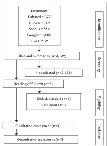
ESULTS Study selectionA low diagram demonstrating the selection process is outlined in Figure 1. In all, 2,129 titles and summaries were screened and ive original articles were selected for full-text analysis. One study was excluded, which is explained in Figure 1. Four original articles were included from quality assessment.10-13 We also analyzed 79 titles from the
refer-ence lists of the four studies above, but we did not ind any additional article.
CASTRO AA ETAL.
280 REV ASSOC MED BRAS 2017; 63(3):278-283
that ultrasonography is a safe method to rule out the diagnosis of DVT.10,11,13 One study evaluated the agreement
between ultrasonography and magnetic resonance imag-ing.12 This study was not included in quantitative
analy-sis (Figure 1).12 In all, 486 participants were enrolled in
these four studies. Characteristics of the studies included can be seen in details in Table 1.
Quality assessment
The methodological assessment chart shows the percent-age of low, high and unclear results for quality domain (Figure 2). The graph shows some potential areas of con-cern. Considering risk of bias, three out of four studies were classiied as high risk of bias for the low and timing domains due to participants being excluded from analy-sis.10,11,13 Other domains were classiied as unclear or low.
One study did not report if the sample was consecutive or random;10 three studies did not report if the reference
standard results were interpreted without knowledge of the results of the index test.10,11,13 Considering
applicabil-ity, one study reported the agreement between ultrasound and magnetic resonance imaging.12
Outcomes
Three of the studies could be used in the analysis.10,11,13
They did not report true-positive cases and false-positive FIGURE 1 Flow diagram summarizing the selection process.
Excluded article (n=1)
Case report (n=1)
Quantitative assessment (n=3) Reading of full text (n=5)
Eligibility
Scr
eening
Titles and summaries (n=2,129)
Identiication
Not selected (n=2,124)
Qualitative assessment (n=4) Inclusion Databases
Pubmed = 377 LILACS = 159 Scopus = 554 Google = 1,000
SIGLE = 39
FIGURE 2 Graphical representation of quality assessment.
Patient selection
Patient selection Index tests
Index tests Reference standard
Reference standard Flow and timing
QU
AD
AS – 2 Risk of bias
QU
AD
AS – 2 Applicability
High Unclear Low
25 25
50 50
75 75
(%) (%)
O F U LT R AS O U N D TO D ET EC T TH R O M B O SIS IN PR EG N AN CY : A SY ST EM AT IC R EV IE W M ED B R AS 2 0 1 7 ; 6 3 (3 ):2 7 8 -2 8 3 2 8 1
Author Year Type of study
Participant Diagnostic criteria Responsible for diagnostic
Follow-up Main result Remark
Le Gal et al.10
2006 Retrospective Pregnant and postpartum women; mean age of all women was 29 ± 5 years; mean age of pregnant women was 29 ± 5 years and mean age of postpartum women 30 ± 5 years
Number of participants: 8 in 1st
trimester, 29 in 2nd trimester, 43 in
3rd trimester, and 82 in postpartum
The US compression test was considered negative when the veins were fully compressible, with no thrombus visualized, and when the Doppler signal at the common femoral vein was phasic during spontaneous respiration
Several specialists including senior and junior examiners
12 weeks 44 out of the 162 had positive test results; 118 had negative test
results and 107 out of them were found during the follow-up time remaining negative
118 participants completed the follow-up
Only women with negative results were included
Three participants who received heparin were excluded from analysis
Ratiu et al.11
2010 Prospective 87 consecutive pregnant women with clinical suspicion of DVT; mean age was 24.68 ± 3.99 years; gestational age was 35.32 ± 2.81 weeks
Positive results were based on lack of complete luminal obliteration following compression, low void on color Doppler scans and lack of low detection on spectral analysis
Not reported 6 weeks 30 (34.48%) had positive test results and two (2.29%) had a high clinical suspicion of DVT; 55 (63.21%) had negative test results and repeated the US exam after 7 days remaining negative
Participants who were positive on US or negative on US but had high clinical probability of DVT underwent venography and were excluded
12 participants who were lost to follow-up were excluded from analysis
Torkzad et al.12
2010 Prospective 27 pregnant women with diagnosed DVT from 23rd gestational week
until 37th gestational week; mean
age of the patients was 30.2 years (range 21-46), the average gestational age at DVT was 29.5 (range 23-39) weeks
Direct examination of the thrombus based on gray-scale ultrasound, compression technique and color low Doppler, and indirect imaging based on low measurements with spectral and color-Doppler
Specialists in clinical physiology with 15-17 years of experience in vascular ultrasound
Not reported Three out of the 27 cases (11.5%) of DVT in the pelvic veins were missed on ultrasound but detected by magnetic resonance
Participants received heparin Magnetic resonance was used in this study to see the agreement between diagnostic tests
Le Gal et al.13
2012 Prospective Pregnant and postpartum women; mean age of all women was 33 years (interquartile range 28-37 years)
Number of participants: 20 in 1st
trimester, 51 in 2nd trimester, 96 in
3rd trimester, and 43 in postpartum
Deep-vein thrombosis was diagnosed with lack of compressibility of a deep vein and, for the iliac vein, in the absence of Doppler low or direct visualization of a thrombus
Vascular medicine specialists with at least 10 years of experience in vascular ultrasound imaging
12 weeks 22 out of the 210 participants (10.5%) had positive test results; two out of the 177 who had negative test results were positive during follow-up
Participants who were positive on US were excluded
10 participants who received heparin were excluded from analysis One participant who was lost to follow-up was excluded from analysis
CASTRO AA ETAL.
282 REV ASSOC MED BRAS 2017; 63(3):278-283
cases.10,11,13 Meta-analysis was not possible. We could not
determine accuracy, sensibility, speciicity and positive predictive value. Data from the three studies were used to calculate negative predictive value.10,11,13 The negative
predictive value was 99.39%.
D
ISCUSSIONOur hypothesis was not conirmed in this systematic review. Although four accuracy studies were identiied, in three of them only patients with negative results were analyzed,10,11,13
and in one of them there was an agreement between ultra-sonography and magnetic resonance imaging.12 We
deter-mined predictive negative value that was 99.39%.
Our main problem was analyzing the negative cases without positive cases. Positive cases received anticoagu-lant therapy and were excluded from the analysis.10,11,13
One systematic review published in 2006 that analyzed DVT and pulmonary embolism concluded that only two studies gave support to treat pregnant women but only if there was a high clinical suspicion and normal results from serial plethysmography.14 The absence of data on
true-positive and false-positive cases resulted in 100% speciicity, but this value is not true. Accuracy, sensitivity and positive predict value could not be estimated in this systematic review.
The studies included analyzed pregnant and postpar-tum women with high suspicion of DVT. None of the studies included reported the prevalence of DVT. Blind-ing of outcome assessment was not reported and the authors reported more than one specialist analyzing the participants. We do not know if these specialists analyzed different participants alone or together. The authors did not report agreement between specialists. One author classiied his study as prospective, but the follow-up time was not reported.12 In this study, more positive cases could
be diagnosed if the time of the follow-up was described and adequate.
The QUADAS-2 is a tool for systematic review and helps to evaluate the quality of accuracy of studies.9 Two
domains were problematic considering the risk of bias: reference standard and low and timing. It was unclear if the results of the reference standard were interpreted without knowledge of the index test results. Blinding of this process is necessary to determine the best accuracy of the ultrasonography. Some participants could not be analyzed because they were lost during the follow-up. These participants could be positive cases and the out-comes of this systematic review could be misleading. We considered low concerns about applicability.
Negative predictive value totaled 100% in two of the studies10,11 and 98.87% in another one.13 Our result was
99.39%. Systematic reviews and meta-analyses aim to summarize the accuracy of diagnostic tests.15 We cannot
determine other parameters and the accuracy of ultraso-nography for DVT in pregnant women is unknown.
In current clinical practice, pregnant women who have a positive diagnosis of DVT based on ultrasonog-raphy are treated with heparin, which is considered more conservative than other treatments. The studies includ-ed in our review analyzinclud-ed lower-limbs of pregnant and postpartum women, yielding negative results based on ultrasonography alone, disregarding other tests such as D-dimer. No deinite conclusion can be drawn from negative ultrasonography results. It is more conservative follow the women during pregnancy when a negative result and a high clinical suspicion are present.
Further studies of low risk of bias are needed to de-termine the diagnostic accuracy of ultrasound to detect deep-vein thrombosis in pregnant patients. In particular, authors have to analyze patients with positive results from ultrasonography with other diagnostic tests such as D-dimer, plethysmography and clinical presentation.
C
ONCLUSIONThe accuracy of the ultrasonography to diagnose deep--vein thrombosis in pregnant women was not established due to lack of data from positive results. Negative predic-tive value was 99.39%. Further studies of low risk of bias are needed to determine the diagnostic accuracy of ultra-sonography in this scenario.
R
ESUMOAcurácia da ultrassonograia para detectar trombose na gravidez: uma revisão sistemática
Objetivo: Determinar a acurácia diagnóstica da ultras-sonograia para detectar trombose venosa profunda (TVP) em pacientes grávidas.
Resultados: Títulos e resumos de 2.129 artigos foram identiicados. Quatro estudos que avaliaram trombose venosa profunda em grávidas foram incluídos. No total, 486 participantes foram selecionadas. Alto risco de viés
foi visto em três dos quatro estudos incluídos conside-rando o domínio luxo e tempo do QUADAS-2. O valor preditivo negativo foi 99,39%.
Conclusão: A acurácia da ultrassonograia para diagnos-ticar trombose venosa profunda em mulheres grávidas não pôde ser determinada em razão da ausência de dados de resultados positivos. Estudos com baixo risco de viés são necessários para determinar a acurácia diagnóstica da ultrassonograia nesse cenário clínico.
Palavras-chave: gravidez, ultrassonografia, trombo-se venosa.
R
EFERENCES1. James AH. Pregnancy and thrombotic risk. Crit Care Med. 2010; 38(2 Suppl):S57-63.
2. James AH. Thrombosis in pregnancy and maternal outcomes. Birth Defects Res C Embryo Today. 2015; 105(3):159-66.
3. Simcox LE, Ormesher L, Tower C, Greer IA. Pulmonary thrombo-embolism in pregnancy: diagnosis and management. Breathe (Sheff). 2015; 11(4):282-9. 4. Chunilal SD, Bates SM. Venous thromboembolism in pregnancy: diagnosis,
management and prevention. Thromb Haemost. 2009; 101(3)428-38.
5. Khalafallah AA, Morse M, Al-Barzan AM, Adams M, Dennis A, Bates G, et al. D-dimer levels at different stages of pregnancy in Australian women: a single centre study using two different immunoturbidimetric assays. Thromb Res. 2012; 130(3):e171-7.
6. To MS, Hunt BJ, Nelson-Piercy C. A negative D-dimer does not exclude venous thromboembolism (VTE) in pregnancy. J Obstet Gynaecol. 2008; 28(2):222-3.
7. Mathis G. [Ultrasound in thromboembolism]. Praxis (Bern 1994). 2015; 104(19):1013-8.
8. Moher D, Liberati A, Tetzlaff J, Altman DG; PRISMA Group. Preferred reporting items for systematic reviews and meta-analyses: the PRISMA statement. PLoS Med. 2009; 6(7):e1000097.
9. Whiting PF, Rutjes AW, Westwood ME, Mallett S, Deeks JJ, Reitsma JB, et al. QUADAS-2: a revised tool for the quality assessment of diagnostic accuracy studies. Ann Intern Med. 2011; 155(8):529-36.
10. Le Gal G, Prins AM, Righini M, Bohec C, Lacut K, Germain P, et al. Diagnostic value of a negative single complete compression ultrasound of the lower limbs to exclude the diagnosis of deep venous thrombosis in pregnant or postpartum women: a retrospective hospital-based study. Thromb Res. 2006; 118(6):691-7. 11. Ratiu A, Navolan D, Spătariu I, Biriş M, Miculiţă M, Motoc A. Diagnostic value of a negative single color duplex ultrasound in deep vein thrombosis suspicion during pregnancy. Rev Med Chir Soc Med Nat Iasi. 2010; 114(2):454-6.
12. Torkzad MR, Bremme K, Hellgren M, Eriksson MJ, Hagman A, Jörgensen T, et al. Magnetic resonance imaging and ultrasonography in diagnosis of pelvic vein thrombosis during pregnancy. Thromb Res. 2010; 126(2):107-12. 13. Le Gal G, Kercret G, Ben Yahmed K, Bressollette L, Robert-Ebadi H, Riberdy
L, et al.; EDVIGE Study Group. Diagnostic value of single complete compression ultrasonography in pregnant and postpartum women with suspected deep vein thrombosis: prospective study. BMJ. 2012; 344:e2635. 14. Nijkeuter M, Ginsberg JS, Huisman MV. Diagnosis of deep vein thrombosis and pulmonary embolism in pregnancy: a systematic review. J Thromb Haemost. 2006; 4(3):496-500.