Syzygium Cumini (L.) Seeds Extract
Ameliorates Cisplatin Induced
Hepatotoxicity in Male Wistar Rats
R.Maheswari1* and S.Manohari1 1*
Head of the Department of Biochemistry, K.M.G College of Arts and Science Gudiyattam – 635 803, TN, India
*E-Mail : rmaheswari@gmail.com
Abstract
The discovery of cisplatin, cis-[Pt(II)(NH(3))(2)Cl(2)] ([PtCl2(NH3)2] or CDDP), was a corner stone which triggered the interest in platinum(II)-and other metal-containing compounds as potential anticancer drugs. Cisplatin, is one of the most potent chemotherapy drugs widely used for cancer treatment. In our present study, an attempt has been made to study the effect of Cisplatin on biochemical and histopathological parameters and ameliorating effects of the Syzygium cumini (L.) aqueousseeds extract or Eugena Jambolana in male wistar rats. Adult male wistar rats were divided into four different groups. Group I Served as vehicle treated normal saline (Control), Group II Rats received single intra-peritoneal (Ip) injection of cisplatin (7mg/kg bw), Group III received Syzygium cumini (L.) aqueousseeds extract400mg/kg/bw orally for 7 days beginning one day prior to cisplatin (CP) injection. Group IV Rats received alone Syzygium cumini (L.) aqueousseeds extract (400mg/kg bw) treated. Cisplatin exposure leads to adverse effects on hematological, hepatotoxic parameters including Erythrocytes (RBCs). Cisplatin induction leads to reduction in the levels of Enzymic and Non-Enzymic antioxidants levels. However, on treatment with Syzygium cumini (L.) aqueous seeds extractnormalized the levels of all the biochemical and hematological parameters. These findings highlight the efficacy of Syzygium cumini (L.) aqueousseeds extract as protective effects Cisplatin induced hepatotoxicity.
Key Words: Cisplatin, Syzygium cumini (L.) seeds, Hepatotoxicity, Hepatoprotective, Free radicals, Antioxidants.
Introduction
Liver
The largest organ in human body is the Liver and the chief site for intense metabolism and excretion. Weighing in at around 3 pounds, the liver is the body’s second largest organ. It has a surprising role in the maintenance, performance and regulating homeostasis of the body. It is involved with almost all the biochemical pathways to growth, fight against disease, nutrient supply, energy provision and reproduction. These platinum complexes react in vivo, binding to and causing cross linking of DNA, which ultimately triggers apoptosis (programmed cell death).
and to cure ulcers. The fruits are acrid and sweet, cooling, dry and astringent to bowels. They increase “Vata” and remove bad smell from the mouth. As per Unani system of medicine they acts as liver tonic, enriches blood, strengthens teeth and gums and forms good lotion for removing ringworm infection of the head.
Powdered seeds are used as a remedy in diabetes [2,3,4,5] and in metrorrhagia. Seed powdered in combination with mango kernels were administered with curd to overcome the problem of diarrohea and dysentery, enlargement of spleen and as diuretic in scanty or suppressed urine and oil of leaves is useful in skin diseases. Seeds of Eugenia jambolana contain glycosides, a trace of pale yellow essential oil, fat, resin, albumin, chlorophyll, an alkaloid- jambosine, gallic acid, ellagic acid, corilagin and related tannin, 3,6- hexa hydroxyl diphenoylglucose and its isomer 4,6- hexa hydroxyl diphenoylglucose, 1- galloyl glucose, 3- galloylglucose, quercetin and elements such as zinc, chromium, vanadium, potassium and sodium. Unsaponifiable matter of seed fat contains β-sitoterol. Dry seeds of Eugenia jambolana have been reported with 11.67% alcohol soluble extractive, 3.397% inorganic, 40% of water-soluble gummy fiber and 15% of water insoluble neutral detergent fibers.
Free Radicals
The term ‘free radical’ is used in a broad sense and also includes related reactive species such as ‘excited states’ that lead to free radical generation or those species that result from free radical reactions. In general, free radicals are very short lived, with half lives in milli-, micro-, or nanoseconds. Free radicals have been implicated in the etiology of several human diseases as well as ageing [6,7].
ROS and RNS are both produced in a well regulated manner to help maintain homeostasis at the cellular level in the normal healthy tissues and play an important role as signaling molecules. Most cells can produce superoxide (O2°-), hydrogen peroxide (H2O2) and nitric oxide (NO) on demand.
Antioxidants
Antioxidants are substances that neutralize free radicals or their actions [8]. Nature has endowed each cell with adequate protective mechanisms against any harmful effects of free radical, superoxide dismutase, (SOD), glutathione peroxidase glutathione reductase, thioredoxin, thiols and disulfide bonding are buffering systems in every cell. α-Tocopherol (Vitamin E) is an essential nutrient which functions as a chain breaking antioxidant which prevents the propagation of free radical reactions in all cell membranes in the human body. Antioxidants, capable of neutralizing free radicals or their actions, act at different stages. They act at the levels of prevention, interception and repair. Preventive antioxidants attempt to stop the formation of ROS. These include superoxide dismutase (SOD) that catalyses the dismutation of superoxide to H2O2 and catalase that breaks it down to water [8,9]. Interception of free radicals is mainly by radical scavenging, while at the secondary level scavenging of peroxyl radicals are effected. The effectors include various antioxidants like Vitamin C, Vitamin E glutathione, other thiol compounds, carotenoids flavonoids, etc., at the repair and reconstitution level, mainly repair enzymes are involved [8,9].
Materials and Methods Chemicals
All the fine chemicals were purchased from Sigma Chemical Co., USA. Cisplatin (CP) was procured from Dabur Pharma Ltd., New Delhi, India. All other chemicals used were of good quality and analytical grade. Eugena Jambolana Seeds Extract Preparation
Fresh fruits of Syzygium cumini (L.) were collected from the local market. All Syzygium cumini (L.) fruits were washed with double distilled water (1/10 w/v). The ripe fruits of S.cumini are de-pulped and the obtained seeds are sun dried. These seeds are pulverized and kept in air tight glass container. 20g of seed powder is soaked in 1000ml of deionized water overnight and centrifuged at 4000g for 20min at 4°C the supernatant was collected. Aqueous extract was used because most of the antioxidant components are extracted in water. During the experiment Syzygium cumini (L.) aqueousseeds extract was daily prepared and administrated to rats.
Animal Model
Male albino rats of Wistar strain (200±10g) procured from Tamil Nadu University for Veterinary and Animal Sciences, (TANUVAS) Chennai, India were used for the study. Animals were fed with commercially available standard rat pelleted feed (M/s Pranav Agro Industries Ltd., India) under the trade name Amrut rat/mice feed and water was provided ad libitum. The rats were housed under conditions of controlled temperature (25±2°C) and acclimatized to 12-h light, 12-h dark cycle. Animal experiments were conducted according to the guidelines of institutional animal ethical committee.
Experimental Design Seggregation of Groups
Group II : Rats received single intra-peritoneal (Ip) injection of cisplatin (7 mg/kg bw).
Group III : Rats received Cisplatin (CP) (7 mg/kg bw) as in group II
and Syzygium cumini (L.) aqueous seeds extract(400 mg/kg bw) orally for 7 days beginning one day prior to cisplatin (CP) injection.
Group IV : Rats received alone Syzygium cumini (L.) aqueous seeds extract
(400 mg/kg bw).
Collection of Samples for Biochemical Analysis
After the experimental period, the animals were anaesthetized by intra-peritoneal injection of phenobarbital sodium (30mg/kg body weight) and were sacrificed. Blood was collected in sterile tubes. Liver tissues were immediately excised and immersed in ice-cold physiological saline. A section of the liver ventricle was set aside for the microscopic studies.
Serum Separation
The blood samples collected in plain centrifuge tubes were kept in inclined position to allow complete clotting of blood and then centrifuged at 2500 rpm for 10 min. The resultant clear supernatant was pipetted out and preserved in small vials in the freezer for the purpose of biochemical investigations.
Preparation of Tissue Homogenate
The liver tissue was excised, rinsed in ice-cold physiological saline and homogenized in 0.1 M Tris-HCl buffer (pH 7.4) using a tissue homogenizer with a teflon pestle at 4°C. The resultant supernatant was kept under refrigeration until further biochemical analysis. All the assay procedures were carried out with in 48 hr of the sample collection.
Histopathological Study
Haematoxylin and Eosin Staining
A portion of hepatic (liver) tissue was fixed in 10 % formalin. The washed tissue was dehydrated in descending grades of isopropanol and cleared in xylene. The tissue was then embedded in molten paraffin wax. Sections were cut at 5-μm thickness and stained with haematoxylin and eosin (H&E).
Statistical Analysis
The values are expressed as mean ± SD for six rats in each group. All of the grouped data were analyzed with SPSS/13.0 student software. Hypothesis testing method included one way analysis of variance (ANOVA) followed by post hoc testing performed with least significant difference (LSD) test. The ‘p’ value of less than 0.05, 0.01, were considered to indicate statistical significance.
Results and Discussions
In our present study, an attempt has been made to study the effect of Cisplatin on biochemical and histopathological parameters and ameliorating effects of the Syzygium cumini (L.) aqueous seeds extract in male wistar rats. In view of our findings, it is possible to conclude that Syzygium cumini (L.) aqueous seeds extract administration results in pronounced oxidative stress and hepatic damage. Syzygium cumini (L.) aqueous seeds extract treatment would protect hepato toxicity against cisplatin induced toxicity through decreasing ROS, hydroxyl radicals, lipid peroxidation, preventing protein degradation, DNA fragmentation because Syzygium cumini (L.) aqueous seeds extract are instrumental for the bioactive compounds, antioxidant properties, above results may be important mechanisms underlying the protective effects of Syzygium cumini (L.) aqueous seeds extract observed in cisplatin hepato toxicity. Thereby, we provide the mechanistic basis of Syzygium cumini (L.)
aqueous seeds extract clinical application to protect patients from cisplatin-induced hepato toxicity. Syzygium cumini (L.) aqueous seeds extract increased the hepato and serum levels of oxidative stress markers such as lipid peroxidation etc., However, Syzygium cumini (L.) aqueous seeds extract treatment significantly inhibited the formation of lipid peroxides in cisplatin exposed rats. This could be due to potential antioxidant effect of
shown the protective efficacy of Syzygium cumini (L.) aqueous seeds extract on cisplatin induced hepato toxicity. Along with biochemical and molecular alterations we have also observed histological changes in hepatic tissues. Histopathological studies revealed alterations of the hepatic tissues caused by cisplatin exposure were prevented by Syzygium cumini (L.) aqueous seeds extract administration. Epidemiological evidence links high antioxidant status with low risk of degenerative disease including tumor promotion and cancer in humans. The increased consumption of fresh vegetables and fruits is usually associated with the decreased use of fish, meats and fats. Furthermore, supplementation of bioavailable and safe antioxidants are essential because we do not get enough antioxidant vitamins and minerals from foods and beverages we consume daily. These research studies demonstrate Syzygium cumini (L.) aqueous seeds extract as a safe, novel, highly potent and bioavailable free radical scavenger and antioxidant possessing a broad spectrum of health benefits. Syzygium cumini (L.)
aqueous seeds extract functions at the genetic level and promotes therapeutic efficacy. Further mechanistic and clinical studies are in progress to unveil the mechanism of this novel natural antioxidant.
Graph 1. Effect of cisplatin and Syzygium cumini (L.) seedson the activities of lipid peroxidation in the serum of control and experimental group of rats.
Results are expressed as mean ± SD for 6 different sets of experiments. Values are considered significantly different at P < 0.05 with post-hoc LSD test.
Graph 2. Effect of cisplatin and Syzygium cumini (L.) seedson the level of GSH, Vitamin C and E in plasma of control and experimental group of rats.
*
*
NS
0 1 2 3 4 5 6 7 8 9 10
Lipid peroxides
nmoles of MDA
liberated/
mg
pr
otei
n
Control
Cisplatin Induced
Cisplatin Induced + Syzygium.C Treated Syzygium Cumini Alone
a
a
a b
b
b NS
NS
NS
0 10 20 30 40 50 60 70 80
GSH Vit - C Vit - E
mg/D
L
Results are expressed as mean ± SD for 6 different sets of experiments. Values are considered significantly different at P < 0.05 with post-hoc LSD test. Statistically significant variations are compared as follows a
Cisplatin-induced vs control. bCisplatin- induced + Syzygium Cumini treated vs Cisplatin-induced. : a,bindicates P < 0.05 and NS indicates non-significant.
Table 1. Effect of cisplatin and Syzygium cumini (L.) aqueous seeds extracton the activities of SOD, CAT, GPx, GST and GR in the serum of control and experimental rats.
Particulars Control Cisplatin induced
Cispatin induced + Syzygium cumini (L.) aqueous seeds extract
treated
Syzygium cumini (L.) aqueous seeds
extract alone
SOD 3.47±0.07 1.65±0.04 2.96±0.05 3.58±0.03
CAT 155.63±6.77 96.65±3.54 124.63±4.534 159.35±5.85
GPx 4.67±0.24 2.74±0.36 3.64±0.35 4.26±0.63
GST 0.98±0.012 0.39±0.014 0.74±0.02 0.97±0.019
GR 1.75±0.06 1.15±0.03 1.54±0.04 1.78±0.06
Results are expressed as mean ± SD for 6 different sets of experiments. Values are considered significantly different at P < 0.05 with post-hoc LSD test.
Table 2. Effect of cisplatin and Syzygium cumini (L.) aqueousseeds extracton the hepatic marker enzymes in control and experimental group of rats.
Particulars Control Cisplatin induced
Cisplatin induced + Syzygium cumini (L.) aqueous seeds extract
treated
Syzygium cumini (L.) aqueous seeds extract alone
ALP 4.63±0.12 9.36±0.08 3.43±0.09 4.35±0.42
SGOT 167.83±6.86 278.34±8.65 196.65±3.86 173.63±6.54
SGPT 88.75±4.63 156.65±6.55 108.45±3.64 90.45±4.34
Results are expressed as mean ± SD for 6 different sets of experiments. Values are considered significantly different at P < 0.05 with post-hoc LSD test.
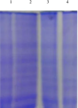
Fig 1. Effect of cisplatin and Syzygium cumini (L.) aqueous seeds extracton the SDS-PAGE pattern of the hepatic tissue of control and experimental groups.
1 2 3 4
Protein fragmentation analysis by SDS-PAGE electrophoresis in rat liver tissue homogenate. Lane 1: Control
Lane 2: Cisplatin– Induced
Lane 3: Cisplatin + Syzygium cumini (L.) aqueous seeds extract Lane 4: Syzygium cumini (L.) aqueous seeds extract.
Summary
The protective effect of Syzygium cumini (L.) aqueous seeds extracton Cisplatin induced hepatotoxicity poisoning in male wistar albino rats model was studied. Oxidative stress was proposed to be an important/vital reason for the hepatotoxic activity. Hence this work was designed to biochemically evaluate the protective effect of Syzygium cumini (L.) aqueous seeds extract against Cisplatin induced hepatotoxicity by assessing the Biochemical parameters, Antioxidant Enzymes status, Non-Enzymatic antioxidant Enzymes, Lipid peroxidation [10], proteins etc.,
Exposure to cisplatin significantly decreased the serum and hepatic antioxidant enzymes like superoxide dismutase, catalase, glutathione peroxidase [11], glutathione-S-transferases [12] & glutathione reductase (Table 1) and non enzymic antioxidants like reduced glutathione, vitamin C & vitamin E (Graph 2) and the activities of
Syzygium cumini (L.) aqueous seeds extract treatment improved the antioxidant status by increasing the activities/levels of these enzymic and non-enzymic antioxidants.
Exposure to cisplatin showed significant increase in the activities of hepatic marker enzymes such as SGOT, SGPT, alkaline phosphatase (Table 2) in serum and a subsequent decrease in these enzyme activities in liver after supplementation of Syzygium cumini (L.) aqueous seeds extract.
The electrophoretic pattern of proteins by SDS-PAGE (Fig 1) also showed the protective role of Syzygium cumini (L.) aqueous seeds extract on cisplatin induced protein fragmentation. Along with biochemical and molecular alterations we have also observed histological changes in hepatic tissue. Histopathological studies revealed alterations of the hepatic tissue caused by cisplatin exposure were prevented by Syzygium cumini (L.)
aqueous seeds extract administration.
Conclusion
The liver in normal physiological conditions is resistance to oxidative damage because of their efficient protective mechanisms. However, under oxidative stress, the liver (hepatocytes) and their mechanisms are very sensitive to oxidative damage due to their content of enzymes which are continuously exposed to high concentration of oxygen. The work was designed to evaluate the protective effect of Syzygium cumini (L.) aqueous seeds extract against cisplatin induced oxidative damage/injury in hepatocytes.
In our present study, Cisplatin induced shows the decrease in the levels of antioxidants and Non-Enzymic antioxidant Enzymes levels, increase in the levels of lipid peroxidations (Graph 1). Cisplatin exposure alters the biochemical parameters viz., liver antioxidant and Non-Enzymatic antioxidants. However, Syzygium cumini (L.)
aqueous seeds extractnormalized the levels of antioxidant and non enzymatic antioxidant Enzymes.
Our findings highlight the efficacy of Syzygium cumini (L.) aqueous seeds extract as protective effects against cisplatin induced oxidative damage to the hepatic cells which induced toxicity. Thus it is concluded that Syzygium cumini (L.) aqueous seeds extract provide a protective effect in the cisplatin induced hepatotoxicity.
Cisplatin exposure leads to adverse effects on hematological, hepatotoxic parameters including Erythrocytes (RBCs). Cisplatin induction leads to reduction in the levels of Enzymic and Non-Enzymic antioxidants levels. However, on treatment with Syzygium cumini (L.) aqueous seeds extract normalized the levels of all the biochemical and hematological parameters. These findings highlight the efficacy of Syzygium cumini (L.)
aqueous seeds extractas protective effects Cisplatin induced hepatotoxicity and further molecular studies should be done in near future and some studies are under progress.
Acknowldegement
References
[1] Wang D, Lippard SJ. 2005. Cellular processing of platinum anticancer drugs. Nat Rev Drug Discov; 4: 307–320.
[2] Murray-Lyon, I.M. Eddleston, A.L. Williams, R. Brown, M. Hogbin, B.M. Bennett, A. Edwards, J.C. Taylor, K.W., Treatment of multiple-hormone-producing malignant islet-cell tumour with streptozotocin, Lancet, 2 (7574): 895–8 (1968)
[3] Rerup, C.C., Drugs producing diabetes through damage of the insulin secreting cells, Pharmacol. Rev., 22: 485–518 (1970)
[4] Schnedl, W.J. Ferber, S. Johnson, J.H. Newgard, C.B., STZ transport and cytotoxicity. Specific enhancement in GLUT2-expressing cells, Diabetes, 43 (11): 1326–33 (1994)
[5] Vavra, J.J. Deboer, C. Dietz, A. Hanka, L.J. Sokolski, W.T., Streptozotocin, a new antibacterial antibiotic, Antibiot. Annu., 7: 230–5 (1959)
[6] Harman D. Ageing: a theory based on free radical and radiation chemistry.1957. J Gerontol, 11: 298-300. [7] Halliwell B, Gutteridge JMC, (eds) 1997, Free radicals in Biology and Medicine, Oxford University Press, Oxford. [8] Sies H. (ed) 1996, Antioxidants in Disease, Mechanisms and Therapy, Academic Press, New York.
[9] Cadenas E and Packer L, (eds) Hand Book of Antioxidants. Plenum Publishers, New York, 1996.
[10] Ohkawa H, Ohishi N, Yagi K. Assay for lipid peroxidation in animal tissues by thiobarbituric acid reaction. Anal Biochem, 1979;195:351–8.
[11] Rotruck JT, Pope AL, Ganther HE, Swanson AB, Hafeman DG, Hoekstra WG.Selenium: biochemical role as a component of glutathione peroxidase. Science, 1973;79:588–90.
[12] Habig WH, Pabst MJ, Jakoby WB. Glutathione-S-transferases: the first enzymatic step in mercapturic acid formation. J Biol Chem 1974;249:7130–9.
[13] King J. The dehydrogenases or oxidoreductases-lactacte dehydrogenase. In:Van D, editor. Practical clinical enzymology. London: Nostrand Company Limited; 1965. p. 83–93.
[14] King J. The hydrolases-acid and alkaline phosphatases. In: Van D, editor. Practical Clinical Enzymology. London: Nostrand Co; 1965. p. 191–208.