* Study carried out in the Pulmonology Sector of the Department of Clinical Medicine at the Universidade Estadual de Campinas – UNICAMP, State University at Campinas – School of Medical Sciences, Campinas, Brazil.
1. Resident in Pulmonology in the Department of Clinical Medicine at the Faculdade de Ciências Médicas – Universidade Estadual de Campinas – FCM/UNICAMP, State University at Campinas School of Medical Sciences – Campinas, Brazil.
2. Associate Professor of Pulmonology in the Department of Clinical Medicine at the Faculdade de Ciências Médicas – Universidade Estadual de Campinas – FCM/ UNICAMP, State University at Campinas School of Medical Sciences – Campinas, Brazil.
3. PhD Assistant Professor of Pulmonology in the Department of Clinical Medicine at the Faculdade de Ciências Médicas – Universidade Estadual de Campinas – FCM/UNICAMP, State University at Campinas School of Medical Sciences – Campinas, Brazil.
4. PhD Professor at the Campinas Pontifícia Universidade Católica – PUC, Pontifical Catholic University – School of Medicine, Campinas, Brazil. Correspondence to: Tiago de Araujo Guerra Grangeia. Rua Benedito Florêncio, 94, Taquaral, CEP 13090-190, Campinas, SP, Brasil.
Tel 55 19 3788-7948. E-mail: [email protected]
Submitted: 31 July 2006. Accepted, after review: 20 December 2006.
Acute respiratory failure as a manifestation of eosinophilia-myalgia
syndrome associated with L-tryptophan intake*
Tiago de Araujo Guerra Grangeia1, Marcelo Schweller1, Ilma Aparecida Paschoal2,
Lair Zambon3, Mônica Corso Pereira4
Abstract
Eosinophilia-myalgia syndrome was described in 1989 in patients who presented progressive and incapacitating myalgia and eosinophilia in blood, fluids and secretions. Most patients report previous L-tryptophan intake. Respiratory manifestations are found in up to 80% of the cases, occasionally as the only manifestation. Treatment includes drug discontinuation and administration of corticosteroids. Here, we describe the case of a 61-year-old female admitted with acute respiratory failure after using L-tryptophan, hydroxytryptophan and other drugs. The patient presented eosinophilia, together with elevated eosinophil counts in the bronchoalveolar lavage and pleural effusion. After discontinuation of the drugs previously used, corticosteroids were administered, resulting in clinical and radiological improvement within just a few days.
Keywords: Respiratory insufficiency; Tryptophan; Eosinophilia-myalgia syndrome.
Introduction
Eosinophilia-myalgia syndrome (EMS) was described in 1989, appearing in an epidemic manner when several patients presented incapacitating myalgia and peripheral eosinophilia after L-tryptophan intake. This led to the removal of L-tryptophan from the market.(1) Since then,
more than 5000 cases have occurred, the majority in female white patients with a mean age of 49 years.(1-4)
L-tryptophan is an amino acid found in lean meats, fish, milk, cheese, nuts, and legumes. It is a natural precursor of serotonin and has been used to raise the organic levels of serotonin, to improve cognitive activities, and to treat depression, as well as to regulate sleep, sexual appetite, sensitivity to pain, and body temperature.(5)
L-tryptophan intake has been implicated as causal agent of EMS.(1,6,7) Some patients might present respiratory
complaints and radiological alterations, and occasionally, they present without the involvement of other organs.
Here, we report a case of acute respiratory failure, presumably related to L-tryptophan intake.
Case report
bronchograms), peripheral consolidations, and areas of ground-glass attenuation in the right lung;
• Chest ultrasound: left pleural effusion, without septations, which extended from the base to the upper third of the left hemithorax; • Thoracocentesis: bright yellow fluid, with
protein: 3.7 g/dL (pleural protein/serum = 0.6), LDH = 242 U/L (LDH pleural/serum = 0.72), glucose = 104 mg/dL, 1960 leukocytes/mm3
(47.5% neutrophils, 25.5% lymphocytes, 24.5% eosinophils, and 1% basophils) and 3000 erythocytes/mm3.
• Testing and culture for fungi and bacteria: negative;
• Bronchoscopy: bronchoalveolar lavage (BAL) with 53,000 cells/mL, with negative results of testing and culture for fungi and mycobac-teria, and positive culture for Staphylococcus aureus;
• Cytology (BAL): purulent active chronic inflam-matory exudate with frequent eosinophils, siderophores, and multinucleated giant cells of foreign-body type; and
• Transbronchial biopsy: focus of incipient non-necrotic granulomatous reaction, in the bronchovascular region, with negative cultures.
The patient was initially treated for severe community-acquired pneumonia, with antibiotic Upon physical examination, she presented
regular overall health status and fever, without alterations in cardiac auscultation. She presented dyspnea and tachypnea (respiratory rate between 28 and 40 breaths/min). Upon inspection and palpation of the chest, reduced and symmetric expansibility was observed. Pulmonary auscultation revealed bilateral coarse crackles and rhonchi, which were predominant in the left hemithorax.
Complementary tests showed the following: • Arterial blood gas analysis (room air):
pH = 7.55; arterial oxygen tension = 53 mmHg; arterial carbon dioxide tension = 23 mmHg; peripheral oxygen saturation (SpO2) = 91%; • Blood workup: 17,180 leukocytes/mm3, 9% of
which were eosinophils (1540 cells/mm3);
• Erythrocyte sedimentation rate: 120 mm in the first hour;
• Electrolytes, urea, creatinine, bilirubin, hepatic enzymes, lactate dehydrogenase (LDH): normal;
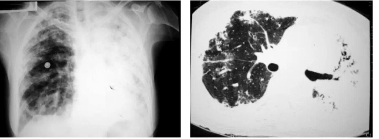
• Blood cultures, urine culture, serologic testing for HIV, parasitological tests: negative; • Initial chest X-ray (Figure 1): extensive areas
of consolidation throughout the left lung and with predominantly peripheral distribution in the right lung;
• Initial computed tomography scan of the chest (Figure 2): diffuse areas of consolidation in the left lung (with volumetric loss and air
Figure 1 - Initial chest X-ray. Extensive areas of
consolidation, involving the entire left lung and presenting predominantly peripheral distribution in the right lung.
together with elevated numbers of eosinophils in the blood, in the BAL, and in the pleural effusion. In the differential diagnoses of pulmonary eosinophilia, the following were considered not very probable: chronic eosinophilic pneumonia (based on the acute clinical presentation); Churg-Strauss syndrome (since there were no signs of peripheral neuropathy and no history of asthma); simple pulmonary eosi-nophilia (due to the negative parasitological test results and the severe clinical presentation); allergic bronchopulmonary aspergillosis (given the lack of a history of asthma, bronchiectasis, or cystic fibrosis); and hypereosinophilic syndrome (based on the absence of extrapulmonary involvement).
There was a clear temporal relationship between the onset of the clinical symptoms and the use of several substances, making the hypothesis of drug-induced pulmonary eosinophilia more likely. Among the substances used, L-tryptophan and hydrox-ytryptophan might be associated with pulmonary infiltrate and eosinophilia.(1,8) Fluoxetine might be
associated with subacute interstitial pneumonia, although there have been no reports of pulmonary eosinophilia.(9)
By the time EMS was described, the diagnostic criteria included incapacitating myalgia, periph-eral eosinophilia > 1000 cells/mm3, and absence
of infection or neoplasia that would explain the clinical symptoms.(2,8,10-12) Although L-tryptophan
intake was not a diagnostic criterion, 86 to 97% of the patients reported it, with doses ranging from 500 to 12,000 mg/day, and a four-week to six-year interval between the initial intake and the onset of the clinical symptoms.(2-4) The association described
suggests a causal relationship between the drug intake and EMS, although such a relationship could not be confirmed.(6,7,13) Other authors believe that
the accumulation of L-tryptophan metabolites is responsible for such clinical manifestations.(1)
The numerous cases of EMS described have widened the range of clinical signs and symptoms, allowing us to state that the disease is characterized by an acute phase, with an approximate duration of four months, in which there might be incapacitating myalgia, fever, weight loss, weakness, dyspnea, cough, chest pain, peripheral edema, arthralgia, exanthema, paresthesia, hyperesthesia, cutaneous thickening, pruritus, dyspepsia, and palpitations, as well as other symptoms.(2,8,11,14) In the chronic
therapy (ceftriaxone and levofloxacin) and oxygen therapy. After one week, she still presented fever, hypoxemia, and dyspnea upon minimal exer-tion, without radiological improvement. There was an increase in eosinophilia in the blood (2670 cells/mm3, 16%), which, in conjunction with
the pleural eosinophilia and eosinophilia in the BAL, motivated the treatment with 125 mg of methyl-prednisolone every 6 hours.
The evolution after the introduction of corti-costeroid treatment was excellent: there was resolution of the fever and the eosinophilia within three days; after one week, there was improvement of the hypoxemia (SpO2 = 94% on room air) and of the radiological profile, with only a cystic image remaining in the left lung which was no longer detected in subsequent X-rays, being therefore interpreted as pneumatocele (Figure 3).
After 15 days, the patient presented no complaints and maintained SpO2 = 96% on room air. She was discharged with a prescription for oral prednisone (60 mg), gradually reduced until its discontinuation after four months. At the time of this writing, the patient presented fatigue of the proximal muscles of the lower limbs, with no respiratory complaints.
Discussion
The patient presented acute respiratory failure accompanied by bilateral pulmonary consolidations,
Figure 3 - Chest X-ray after one week treatment, no
longer showing the areas of consolidation or the linear, heterogeneous opacities in the right lung base. Although the cystic image (pneumatocele) is still visible in the left hemithorax, it was not observed on subsequent chest
treatment imperative, and the rapid response to the treatment made the biopsy unnecessary.
The treatment for EMS consists of drug discon-tinuation and administration of corticosteroids: 5 to 60 mg of oral prednisone a day for four weeks, with subsequent gradual reduction of the dose. Approximately 80% of patients present symptom improvement, the majority within the first three days of treatment, as was observed in this patient. The most refractory symptoms are fatigue and neurological (cognitive) symptoms. The estimated mortality is 2 to 6%, and the majority of deaths are due to ascending polyneuropathy with respiratory failure.(1)
Based on the clinical profile of acute respiratory failure accompanied by peripheral and pulmonary eosinophilia, in a context in which other eosinophilic diseases would be less probable, it is possible to suspect a causal relationship between the acute disease presented and L-tryptophan intake.
L-tryptophan is a substance widely used in Brazil. It is used indiscriminately, and without medical prescription control, since it is part of several ‘formulas’ used in so-called ‘alternative’ treatments, as nutritional supplements, or in ‘ orthomolecular medicine’. Therefore, it is important that we be attentive to the possibility of the appearance of this broad spectrum of clinical manifestations, which, at an extreme level of severity, might evolve to acute respiratory failure and death.
References
1. Ahmed MM, Mubashir E, Sairam S, Lisse JR. Eosinophilia-Myalgia Syndrome. [monograph on the Internet]. e-medicine; WebMD; c1996-2006 [cited 2006 May 5]. Available from: http://www.emedicine.com/med/topic693. htm#section~workup
2. Herrick MK, Chang Y, Horoupian DS, Lombard CM, Adornato BT. L-tryptophan and the eosinophilia-myalgia syndrome: pathologic findings in eight patients. Hum Pathol. 1991;22(1):12-21.
3. Tazelaar HD, Myers JL, Drage CW, King TE Jr, Aguayo S, Colby TV. Pulmonary disease associated with L-tryptophan-induced eosinophilic myalgia syndrome. Clinical and pathologic features. Chest. 1990;97(5):1032-6.
4. Campagna AC, Blanc PD, Criswell LA, Clarke D, Sack KE, Gold WM, Golden JA. Pulmonary manifestations of the eosinophilia-myalgia syndrome associated with tryptophan ingestion. Chest. 1992;101(5):1274-81.
5. Ballone GJ. Serotonina. [monograph on the Internet] Psiqweb; c2005 [cited 2006 Oct 4]. Available from: http:// virtualpsy.locaweb.com.br/index.php?sec=61&art=295 6. Shapiro S. L-tryptophan and eosinophilia-myalgia syndrome.
Lancet. 1994;344(8925):817-9. phase, fatigue and neurological symptoms (deficits
of attention and memory) predominate.(12)
Respiratory symptoms are common in EMS and might be present in up to 80% of the patients,(14) even
as the only manifestation. In a study of 1531 cases,(15)
cough or dyspnea were present in 611 (59%). In another study,(16) involving 118 patients, respiratory
symptoms were found in 64%, radiological altera-tions in 16%, and pleural effusions in 15%. The pulmonary involvement confers a worse prognosis, and some patients might evolve to acute respiratory failure.(3,4,17)
Complementary tests, in general, show leukocytosis and eosinophilia (values above 3000 cells/mm3 are more suggestive).(11) The chest
X-ray might be normal or might present linear or reticulonodular interstitial opacities, diffuse pulmo-nary consolidations, and pleural effusion.(3,4,10) The
pleural effusions might appear in up to one-third of the cases, and generally, they are eosinophilic exudates.(4,10,16)
Of a total of 12 cases reported, 5 were submitted to a tomography scan of the chest.(4,17,18) Consolidations,
in general focal and of basal predominance, were detected in four patients; pleural effusion was detected in one patient; and peribronchial nodules in isolation were described in one patient. No exten-sive consolidations were reported. The small number of patients does not allow us to establish a pattern, although consolidations were found in 80% of the patients.
The spirometry might be normal or might show obstructive or restrictive ventilatory disorder.(3,10,14)
The syndrome is defined on the basis of clinical and radiological criteria, and the biopsy findings consistent with the diagnosis are not obligatory, especially since there is no typical pattern. Various are the histopathologic alterations described: intersti-tial pneumonia with predominance of lymphocytes; transmural vasculitis and perivascular inflammatory infiltrate(14); granulomatous vasculitis(2,3); fibrous
pleuritis; and follicular bronchiolitis.(3) In the case
reported, the transbronchial biopsy fragment showed a focus of incipient non-necrotic granulomatous reaction in the bronchovascular region, findings consistent with granulomatous vasculitis similar to the case described in a study.(3) Open lung biopsy,
13. Margolin L. Non-L-tryptophan related eosinophilia-myalgia syndrome with hypoproteinemia and hypoalbuminemia. J Rheumatol. 2003;30(3):628-9.
14. Silver RM. Pathophysiology of the eosinophilia-myalgia syndrome. J Rheumatol Suppl. 1996;46:26-36.
15. Swygert LA, Maes EF, Sewell LE, Miller L, Falk H, Kilbourne EM. Eosinophilia-myalgia syndrome. Results of national surveillance. JAMA. 1990;264(13):1698-703.
16. Centers for Disease Control (CDC). Clinical spectrum of eosinophilia-myalgia syndrome--California. MMWR Morb Mortal Wkly Rep. 1990;39(6):89-91.
17. Banner AS, Borochovitz D. Acute respiratory failure caused by pulmonary vasculitis after L-tryptophan ingestion. Am Rev Respir Dis. 1991;143(3):661-4.
18. Strumpf IJ, Drucker RD, Anders KH, Cohen S, Fajolu O. Acute eosinophilic pulmonary disease associated with the ingestion of L-tryptophan-containing products. Chest. 1991;99(1):8-13.
7. Shapiro S. Epidemiologic studies of the association of L-tryptophan with the eosinophilia-myalgia syndrome: a critique. J Rheumatol Suppl. 1996;46:44-58; discussion 58-9.
8. Clauw DJ, Pincus T. The eosinophilia-myalgia syndrome: what we know, what we think we know, and what we need to know. J Rheumatol Suppl. 1996;46:2-6.
9. de Kerviler E, Trédaniel J, Revlon G, Groussard O, Zalcman G, Ortoli JM, et al. Fluoxetin-induced pulmonary granulomatosis. Eur Respir J. 1996;9(3):615-7.
10. Williamson MR, Edison M, Rosenberg RD, Williamson SL. Eosinophilia-myalgia syndrome: findings on chest radiographs in 18 patients. Radiology. 1991;180(3):849-851.
11. Hertzman PA. Criteria for the definition of the eosinophilia-myalgia syndrome. J Rheumatol Suppl. 1996;46:7-12. 12. Pincus T. Eosinophilia-myalgia syndrome: patient status