Monti’s Procedure as an Alternative Technique in Complex
Urethral Distraction Defect
Jalil Hosseini, Ali Kaviani, Mohammad M. Mazloomfard, Ali R. Golshan
Reconstructive Urology, Shohada Tajrish Hospital, Shaheed Beheshti Medical Sciences University,
Tehran, Iran
ABSTRACT
Purpose: Pelvic fracture urethral distraction defect is usually managed by the end to end anastomotic urethroplasty. Surgi-cal repair of those patients with post-traumatic complex posterior urethral defects, who have undergone failed previous surgical treatments, remains one of the most challenging problems in urology. Appendix urinary diversion could be used in such cases. However, the appendix tissue is not always usable. We report our experience on management of patients with long urethral defect with history of one or more failed urethroplasties by Monti channel urinary diversion.
Materials and Methods: From 2001 to 2007, we evaluated data from 8 male patients aged 28 to 76 years (mean age 42.5) in whom the Monti technique was performed. All cases had history of posterior urethral defect with one or more failed procedures for urethral reconstruction including urethroplasty. A 2 to 2.5 cm segment of ileum, which had a suitable blood supply, was cut. After the re-anastomosis of the ileum, we closed the opened ileum transversely surrounding a 14-16 Fr urethral catheter using running Vicryl sutures. The newly built tube was used as an appendix during diversion.
Results: All patients performed catheterization through the conduit without dificulty and stomal stenosis. Mild stomal incontinence occurred in one patient in the supine position who became continent after adjustment of the catheterization intervals. There was no dehiscence, necrosis or perforation of the tube.
Conclusion: Based on our data, Monti’s procedure seems to be a valuable technique in patients with very long complicated urethral defect who cannot be managed with routine urethroplastic techniques.
Key words: urethra; urethral stricture; urinary diversion Int Braz J Urol. 2010; 36: 317-26
INTRODUCTION
Strictures and defects of the posterior urethra
in men is one of the most signiicant clinical compli -cations concerning urologists (1). Posterior urethral injuries in pelvic fracture were estimated at 5 to 10 percent in previous studies (2). Anastomosis is usu-ally performed for defects of the posterior urethra. However, in some cases the urethral defect is so long that it cannot be negotiated with vigorous releasing of urethra from surrounding tissue, inferior pubectomy
doi: 10.1590/S1677-55382010000300008
and even re-routing maneuvers (1,3). Based on the location and length of the stricture, various techniques have been used in such cases including onlay repairs, stricture excision with augmented anastomosis, a
tubularized lap of sigmoid colon, and free or vascu
-larized skin lap, etc. However, many complications
more severe urethral injuries (8); although, the ap-pendix is not always usable (9). The apap-pendix may
be absent or insuficient in length or quality. It may
have a precarious blood supply, a short mesentery
or histopathologic changes, such as chronic inlam
-mation or ibrous lumen obstruction (9). Regarding
these situations, the technique which was originally proposed by Monti et al. is a good alternative method when the appendix is unavailable, atretic or used con-currently with another procedure (10). We reviewed our results regarding this surgical technique in eligible patients.
MATERIALS AND METHODS
From 2001 to 2007, we evaluated data from 8 male patients aged 28 to 76 years (mean age 42.5) on whom we performed the Monti technique at
Tajr-ish Hospital, Tehran, Iran. All patients had a previous
history of urethral distraction defect and a history of at least one failed urethroplasty and a defect longer than 10 centimeters in distal prostatic, membranous, bulbar and some part of penile urethra. Due to a very long urethral defect that could not be repaired by ure-throplasty, a Monti urinary diversion was performed
in the patients. Informed consents were signed by
all enrolled patients. The study was approved by the Ethics Committee of our hospital.
Surgical Technique
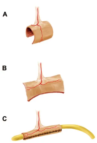
After isolating a 2 to 2.5 cm segment of ileum, with a suitable blood supply, we opened the ileal segment along its anti-mesenteric border by Metzenbaum scissors, and then closed the opened ileum transversely surrounding a 14-16 Fr urethral catheter using running Vicryl sutures (Figure-1). The length of small intestine which was resected did not determine the length of the newly built tube, but rather its diameter. Therefore, using 1 or 2 cm segment of the small intestine, leads to a narrow and wide tube, respectively. The 15 cm of terminal ileum was not routinely used for this type of procedure.
The double tube technique was used in obese
patients. In this procedure, a 5 cm segment of the il
-eum was isolated, cut into two halves and tabularized, each one exactly as described previously. The two segments were anastomosed to each other using an interrupted 3-0 Vicryl sutures to build a single tube.
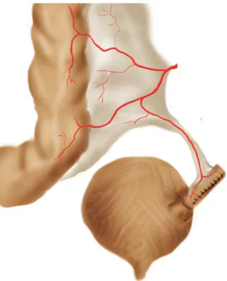
After the reconstruction of a new appendix, anastomosis was performed on the superior part of the postero-lateral junction of the bladder. The Mi-trofanoff principle was not used; the bladder wall was opened and anastomosed to the new appendix using 3-0 Vicryl sutures (Figure-2). The stoma was made at level which was located proximally relative to the bladder in order that gravity can help the patient’s continence. A cystostomy tube was performed for all the patients to increase the safety measures.
Figure 1 – Isolating a 2 to 2.5 cm segment of ileum (A) and
All patients were discharged 5-6 days postoperatively as soon as they could tolerate solid food. The diversion catheter was removed 3 weeks post-operatively. All patients were put on a clean
intermittent catheterization (CIC) regimen using a
14 or 16 Fr nelaton catheter every 3 hours. Presence of urinary leakage during the interval was considered as the patient being incontinent. The cystostomy tube
was removed 7 days later, if there was no dificulty
in catheterization.
Demographic characteristics, distraction defect length, previous surgical procedures, time of operation and hospitalization, estimated blood loss, and complications such as peri-operative bleeding (need for blood transfusion), adjacent organ damage, hematoma and wound infection were recorded.
The patients were regularly followed-up at 3,6,18 and 24 months postoperatively, with special attention to any problems with catheterization and incontinence. Follow-up plan consisted of physi-cal examination including stoma evaluation; upper
urinary tract sonography and determining of post catheterization urine residue; and serum creatinine level and catheter size assessment.
RESULTS
Eight patients were included in this study. Causes of urethral injury and pelvic fracture consisted of 4 motor vehicle accidents, 2 falls and one shot gun injury. The time interval between injury and Monti procedure ranged from 23 to 48 months (mean 31.4). Patients’ general data, previous operative procedures and outcome are listed in Table-1. Sonographic as-sessment of upper urinary tract did not reveal any
pathologic indings, and mean serum creatinine level
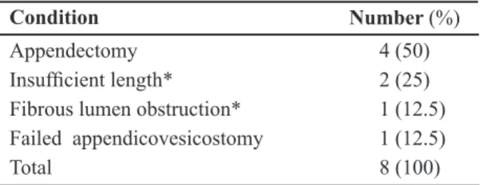
was 1.3 mg/dL (0.6 to 1.7) pre-operatively. The pa-tients did not have an available or suitable appendix (Table-2).
Seven patients underwent single tube tech-nique and in the obese patient, double tube procedure was performed. Mean surgical time was 4.5 hours (range 3 to 8) with defect lengths of 11.75 cm (10 to 14). Average estimated blood loss was around 350 cc (ranged 200 to 800). There was no need for blood transfusion or adjacent organ damage. All patients were discharged 5-6 days post operatively.
Follow-up ranged from 24 to 30 months
(mean 25.75). Immediate post-operative complica -tions such as hematoma and wound infection were not detected. All patients performed
catheteriza-tion through the conduit without dificulty every 3
hours. Catheter size ranged from 14 to 16 Fr. None of the 8 patients had stomal stenosis during the fol-low-up period. Mild stomal incontinence occurred in one patient in the supine position which became continent after some adjustments of the catheteriza-tion intervals. This patient had previous history of urethroplasty and failed appendicovesicostomy at another surgical center. There was no dehiscence, necrosis, or perforation of the tube during the fol-low-up period.
Also, there was no signiicant difference be -tween pre-operative and post-operative serum creati-nine levels and upper tract sonographic data, which were evaluated at the time of scheduled surgery as well as 3,6,18 and 24 months post-operatively. Figure 2 – Anastomosis of newly build tube on the superior part
COMMENTS
In 1989 Turner-Warwick explained some fea -tures of complex urethral distraction defect including
long urethral gap between tow ends (11). In severe
urethral injuries with long strictures or urethral
de-fects especially in patients who have undergone failed previous surgical treatments, various methods have been used to obtain urethral continuity (4). Surgical options are offered based on the location and length of
the stricture. One-stage vascularized scrotal skin lap
urethroplasty and a two-stage Johanson’s procedure
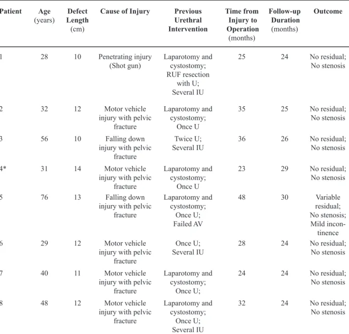
Patient Age
(years)
Defect Length
(cm)
Cause of Injury Previous
Urethral Intervention
Time from Injury to Operation
(months)
Follow-up Duration
(months)
Outcome
1 28 10 Penetrating injury (Shot gun)
Laparotomy and cystostomy;
RUF resection with U; Several IU
25 24 No residual; No stenosis
2 32 12 Motor vehicle injury with pelvic
fracture
Laparotomy and cystostomy;
Once U
35 25 No residual; No stenosis
3 56 10 Falling down injury with pelvic
fracture
Twice U;
Several IU 36 26 No residual;No stenosis
4* 31 14 Motor vehicle injury with pelvic
fracture
Laparotomy and cystostomy;
Once U
23 29 No residual; No stenosis
5 76 13 Falling down injury with pelvic
fracture
Laparotomy and cystostomy;
Once U;
Failed AV
48 30 Variable residual; No stenosis; Mild
incon-tinence 6 29 12 Motor vehicle
injury with pelvic fracture
Once U;
Several IU 28 24 No residual;No stenosis
7 40 11 Motor vehicle injury with pelvic
fracture
Laparotomy and cystostomy;
Once U;
24 24 No residual; No stenosis
8 48 12 Motor vehicle injury with pelvic
fracture
Laparotomy and cystostomy;
Once U; Several IU
32 24 No residual; No stenosis
* The only double Monti channel; RUF = recto-urethral istula; U = urethroplasty; IU = internal urethrotomy; AV = appendicovesi -costomy.
were two surgical examples for treatment of complex
lengthy urethral strictures (12). Skin lap urethroplasty
can lead to some complications such as recurrent stricture, troublesome post void dribbling, and
di-verticulum formation (4). In the last decade, buccal
mucosa urethroplasty has increased in popularity be-cause of its feasibility, good functional outcome, and low morbidity at the reconstructed urethra. However, treatment of long, complicated urethral strictures by buccal mucosal graft may not be useful, because of limited material (4,5).
Recently some investigators have described
novel surgical techniques for male long segment
urethral defect. In 2006, Yue-Min Xu et al. reported
a new technique for treatment of men with long ure-thral defect after pelvic trauma using the intact and pedicled pendulous urethra to replace the bulbar and membranous urethra, followed by reconstruction of the anterior urethra (12). Buyukunal et al. developed a new treatment modality in a rabbit model, using appendix interposition for substitution of severe pos-terior urethral injuries (13). This technique was also used by Aggarwal et al. in recurrent urethral strictures (14).
Other options such as perineostomy or supra-pubic tube could also be used as a salvage procedure in such situations. Suprapubic tube is a safe and simple treatment of acute or chronic urinary retention but has some complications especially in long-term such as
infection, dificulty in changing of catheter and risk of
malignancy (6). Barbagli et al. evaluated the clinical outcome of patients with complex urethral pathology who were treated with perineal urethrostomy. These authors showed that success rate of urethroplasty after perineal urethrostomy is lower in younger patients with traumatic urethral stricture (7).
In 1980, Mitrofanoff irst described the use of
the appendix as a continent urinary stoma (15). The major indications for constructing a urinary diver-sion are patients with a low leak-point pressure and neurogenic bladder, an unreconstructable bladder (e.g. exstrophy), an unreconstructable urethral disease or the inability to catheterize the urethra in a neurogenic bladder (8).
With this concern, we use a urinary diver-sion in patients with unreconstructable long urethral defect, in order to empty their bladder. As Monti et al. described in 1997 (10), a continent catheterizable conduit using short segments of the small intestine was used for this aim. The use of this technique allows
us to obtain some beneits. Only 2 to 2.5 cm segment
of the ileum is required. The caliber of such a tube allows catheterization with a 16F to 18F catheter, and the mucosal folds of the ileum are aligned with its longitudinal axis. These tubes have an abundant supply of blood and are able to be used anywhere inside the abdomen (9,10).
It is important to note that the length of the
segment can be adjusted by using a double tube or using a section of the large bowel, allowing applica-tion of this technique in adults or obese patients (9). A 2.0-2.5 cm segment of bowel will usually result in a tube of 6-7 cm in length, when re-tubularized
trans-versely. If a longer channel is needed, two consecu -tive segments can be cut, and anastomosed together to form a tube twice as long but with mesentery only
in the central portion of the tube. In our study, one
patient was candidate for the double tube technique. No stenosis or incontinence occurred during his fol-low-up.
One of the best characteristics provided by
Monti’s procedure is urinary continence. In the series
with longer follow-up periods, continence main-tenance is always greater than 90% and shows no considerable changes with time (16,17). Narayanas-wamy et al. reported their results with 94 Mitrofanoff procedures, of which 25 were Monti channels. Overall 23 of 25 patients were successfully catheterized at the time of the report and only 3 of 25 had stomal leakage
(18). In another large series Castellan et al. reported
a comparison among different types of channels for urinary and fecal incontinence, including 45 Monti urinary channels, with a mean follow-up of 38 months.
Condition Number (%)
Appendectomy 4 (50)
Insuficient length* 2 (25)
Fibrous lumen obstruction* 1 (12.5) Failed appendicovesicostomy 1 (12.5)
Total 8 (100)
* Finding on operation room.
Four of these channels were double Monti channels, while the others were single Monti channels. Channel replacement was performed in three patients (7%)
due to complete ibrosis, and 3 cases (7%) had stomal
incontinence (16).
We did not use the Mitrofanoff principle to create an anti-incontinent submucosal tunnel. Only anastomosis was performed on the superior part of
the postero-lateral junction of the bladder. Yang et al. (19) evaluated the pressure proile of the channel tube,
and detected two high-pressure zones: one in the sub mucosal tunnel and the other at the point at which the muscle layer of the abdominal wall is crossed. These data suggest that the muscle layer of the abdominal wall is a major factor in preserving of continence (9).
Our study shows that Monti’s procedure, even without the use of the Mitrofanoff principle, is a reliable technique with low incontinence and stricture rate. Obviously, we are not proposing that
the Monti’s procedure be the deinitive treatment for
complicated posterior urethral injuries. Moreover, it can be performed in patients with very long urethral stricture that cannot be corrected with the urethroplas-tic techniques, and who also do not have a suitable ap-pendix for apap-pendix diversion techniques. However, evaluation of patient’s satisfaction and the choice of eligible cases need more investigations with larger number of patients.
CONCLUSION
Based on our data, Monti’s procedure is a valuable technique in patients with very long com-plicated urethral defect who lack a suitable appendix for appendicovesicostomy technique.
CONFLICT OF INTEREST
None declared.
REFERENCES
1. Hosseini J, Tavakkoli Tabassi K: Surgical repair of posterior urethral defects: review of literature and
pre-sentation of experiences. Urol J. 2008; 5: 215-22. 2. Cass AS, Godec CJ: Urethral injury due to external
trauma. Urology. 1978; 11: 607-11.
3. Andrich DE, Mundy AR: What is the best technique for urethroplasty? Eur Urol. 2008; 54: 1031-41. 4. Xu YM, Qiao Y, Sa YL, Wu DL, Zhang XR, Zhang J,
et al.: Substitution urethroplasty of complex and long-segment urethral strictures: a rationale for procedure
selection. Eur Urol. 2007; 51: 1093-8; discussion
1098-9.
5. Barbagli G, Lazzeri M: Surgical treatment of anterior
urethral stricture diseases: brief overview. Int Braz J Urol. 2007; 33: 461-9.
6. Scorer CG: The suprapubic catheter; a method of treat-ing urinary retention. Lancet. 1953; 265: 1222-5.
7. Barbagli G, De Angelis M, Romano G, Lazzeri M:
Clinical outcome and quality of life assessment in patients treated with perineal urethrostomy for anterior
urethral stricture disease. J Urol. 2009; 182: 548-57. 8. Freitas Filho LG, Carnevale J, Melo Filho AR, Vicente
NC, Heinisch AC, Martins JL: Posterior urethral
in-juries and the Mitrofanoff principle in children. BJU Int. 2003; 91: 402-5.
9. Monti PR, de Carvalho JR, Arap S: The Monti proce
-dure: applications and complications. Urology. 2000;
55: 616-21.
10. Monti PR, Lara RC, Dutra MA, de Carvalho JR: New
techniques for construction of efferent conduits based
on the Mitrofanoff principle. Urology. 1997; 49:
112-5.
11. Turner-Warwick R: Prevention of complications
resulting from pelvic fracture urethral injuries--and
from their surgical management. Urol Clin North Am.
1989; 16: 335-58.
12. Wu DL, Jin SB, Zhang J, Chen Y, Jin CR, Xu YM:
Staged pendulous-prostatic anastomotic urethroplasty followed by reconstruction of the anterior urethra: an effective treatment for long-segment bulbar and
membranous urethral stricture. Eur Urol. 2007; 51:
504-10; discussion 510-11.
13. Büyükünal SN, Cerrah A, Dervisoglu S: Appendix interposition in the treatment of severe posterior
ure-thral injuries. J Urol. 1995; 154: 840-3.
14. Aggarwal SK, Goel D, Gupta CR, Ghosh S, Ojha H:
The use of pedicled appendix graft for substitution of urethra in recurrent urethral stricture. J Pediatr Surg. 2002; 37: 246-50.
15. Mitrofanoff P: Cystostomie continente trans-appen-diculiaire dans le traitement des vessies neurologiques. Chir Pediatr. 1980; 21: 297-305.
Clinical applications of the Monti procedure as a
conti-nent catheterizable stoma. Urology. 1999; 54: 152-6.
17. Leslie JA, Dussinger AM, Meldrum KK: Creation of continence mechanisms (Mitrofanoff) without
ap-pendix: the Monti and spiral Monti procedures. Urol
Oncol. 2007; 25: 148-53.
18. Narayanaswamy B, Wilcox DT, Cuckow PM, Duffy
PG, Ransley PG: The Yang-Monti ileovesicostomy: a problematic channel? BJU Int. 2001; 87: 861-5. 19. Yang WH: Yang needle tunneling technique in creating
antirelux and continent mechanisms. J Urol. 1993;
150: 830-4.
Accepted after revision: November 3, 2009
Correspondence Address:
Dr. Mohammad Mohsen Mazloomfard Shohada Tajrish Hospital
Shaheed Beheshti Medical Sciences University Tehran, Iran
Fax: + 98 21 8852-6901
E-mail: [email protected]
EDITORIAL COMMENT
The authors report their experience on the management of eight patients with long urethral de-fects already submitted to at least one unsuccessful urethroplasty. All of them received continent cutane-ous urinary diversion using as efferent catheterizable conduit transversely tubularized ileal segments with direct implantation into the bladder wall without
antirelux technique. After two years of minimum
follow up all subjects were continent with easy cath-eterization. The ileal tube was created to replace the appendix when unavailable to construct a urinary
diversion based on the Mitrofanoff principle. Until
that the proposed technical alternatives (around 20) showed clearly inferior results compared to the ap-pendix technique and were based on the use of ure-teral segments, longitudinally tapered ileal segments,
gastric tubes, tubularized cecum laps, fallopian tube, skin tubes (preputial penile or clitoral skin laps, labia minora laps), vas deferens, tubularized bladder lap,
Meckel’s diverticulum, hipogastric artery segment,
human umbilical vein, rectus abdominis muscle,
aponeurosis lap. The long term follow up of ileal
tube technique application provided equivalent results to those of the appendix related to function, durabil-ity and low complications index (1,2). For the tube construction, some technical points matter. The tube made from 2.5 cm isolated segment allows 14F to 16F catheters inside and the measurement should be performed with the bowel at rest, without stretching it. The tubularization is done with running suture of Vicryl 3-0 in adults and 4-0 in children and preceded by resection of lateral mucosal excess of the open
intestinal plate. In the case of double tube, the suture
between the plates should be done with simple inter-rupted stitch, which makes the tubularization easier.
You can also use the double spiral tube, as proposed by
Casale (3). The passage of the tube to the skin should be straight and as short as possible. Very long tubes
vicryl 3-0 interrupted stitch to stabilize the structure. The stoma can be done in a simple way or with skin
laps interposition. It is noteworthy the author’s op -tion for direct implanta-tion of the tube into the blad-der wall trusting just in the resistance offered by the abdominal muscle layer when the tube pass through it. Since the Mitrofanoff’s pioneer publication in 1980 (reference 15) there were rare descriptions of direct implantation of the conduit into the reservoir without
antirelux technique and with short periods of conti
-nence. Yang himself quoted by the authors (reference 19 in the article) utilized the antirelux technique in
his unique case with ileal tube and interprets literally
the pressure proile study of the tube: “The results show that although there are 2 high pressure proile
zones for the continent ileal tube, the skeletal muscle pressure zone has a lesser role in the continence mechanism than the submucosal portion of the ileal tube”. Stress tests show an equal increased pressure
inside the reservoir and in the antirelux tunnel but
not in the skeletal muscle zone. This conclusion is the current stand-point and it seems risky to dismiss
the use of an antirelux technique mainly in cases in
which the tube implantation was done into the bladder wall, a structure that offers the best results among the available options. Long term studies show that the continent cutaneous urinary diversion made by the
Mitrofanoff technique with appendix or reconigured
ileal tube offers consistent and lasting results besides the use of technical principles of easier execution
already widely known and used in Urology.
REFERENCES
1. Lemelle JL, Simo AK, Schmitt M: Comparative study
of the Yang-Monti channel and appendix for continent
diversion in the Mitrofanoff and Malone principles. J
Urol. 2004; 172: 1907-10.
2. Cain MP, Dussinger AM, Gitlin J, Casale AJ, Kaefer M,
Meldrum K, et al.: Updated experience with the Monti catheterizable channel. Urology. 2008; 72: 782-5.
3. Casale AJ: A long continent ileovesicostomy using a
single piece of bowel. J Urol. 1999; 162: 1743-5.
Dr. Paulo R. Monti
Section of Urology Federal University of Minas Triangle Uberaba, Minas Gerais, Brazil E-mail: [email protected]
EDITORIAL COMMENT
Traumatic posterior urethral strictures (better
deined as “pelvic fracture related urethral injuries”)
as well as non-traumatic posterior strictures are rare conditions (1,2). As mentioned by the authors, most of these strictures can be managed by anastomotic
repair. However, reports on “what to do” after failed
urethroplasty are very scarce. The Monti-procedure
was irst described in 1997 (3) in an animal (dog)
model and quickly found clinical applications as a continent catheterizable stoma in adult and paediatric patients (4), in case the appendix could not been used.
This paper is the irst to describe this technique for
posterior urethral strictures after failed urethral
recon-struction. The major importance of this paper is that it shows the feasibility of the procedure in these situ-ations. Although it is explained in the text, the title is somewhat misleading. Monti’s procedure must not be regarded as an alternative to other procedures (such as anastomotic repair, substitution urethroplasty, perin-eostomy) in complex urethral distraction defects. One or even more attempts to restore urethral continuity must always be performed for these often young
patients. If these attempts failed however, a strategy
that abandons the urethral outlet can be proposed.
rather than the term “an alternative technique” for the
Monti’s procedure in these patients. The authors did not apply the Mitrofanoff principle for implantation at the bladder. One patient out of 8 suffered from stomal incontinence. The authors state that this technique has thus a low continence rate. However, this conclusion
is drawn on a small number of patients. Unless larger
series can prove the opposite, there is at the present no reason to abandon the Mitrofanoff principle for prevention of stomal incontinence. Patients must also be informed about the long-term complications
related to the Monti’s procedure dificult catheterisa -tion, stomal stenosis and incontinence and it has been reported that 23-27.5% will need revision surgery at the Monti’s tube (5,6). There is no reason to assume that these complication and revision rate will be dif-ferent in patients with traumatic urethral distraction defects.
REFERENCES
1. Lumen N, Hoebeke P, Troyer BD, Ysebaert B, Oost -erlinck W: Perineal anastomotic urethroplasty for
posttraumatic urethral stricture with or without previ-ous urethral manipulations: a review of 61 cases with
long-term followup. J Urol. 2009; 181: 1196-200.
2. Lumen N, Oosterlinck W: Challenging non-traumatic posterior urethral strictures treated with urethroplasty:
a preliminary report. Int Braz J Urol. 2009; 35:
442-9.
3. Monti PR, Lara RC, Dutra MA, de Carvalho JR: New
techniques for construction of efferent conduits based
on the Mitrofanoff principle. Urology. 1997; 49:
112-5.
4. Castellan MA, Gosalbez R Jr, Labbie A, Monti PR:
Clinical applications of the Monti procedure as a
conti-nent catheterizable stoma. Urology. 1999; 54: 152-6.
5. Leslie JA, Dussinger AM, Meldrum KK: Creation of continence mechanisms (Mitrofanoff) without
ap-pendix: the Monti and spiral Monti procedures. Urol
Oncol. 2007; 25: 148-53.
6. Leslie JA, Cain MP, Kaefer M, Meldrum KK,
Duss-inger AM, Rink RC, et al.: A comparison of the Monti and Casale (spiral Monti) procedures. J Urol. 2007;
178: 1623-7; discussion 1627.
Dr. Nicolaas Lumen
Department of Urology Ghent University Hospital Ghent, Belgium E-mail: [email protected]
EDITORIAL COMMENT
In his commentary, recently, Barbagli un -derlined that the management of posterior urethral strictures, in patients after pelvic fracture urethral
distraction defects (PFUDD), has evolved over time
(1). Forty, thirty years ago, in the ‘70s and the ‘80s,
the transpubic urethroplasty was considered the gold standard in the majority of adults and children
suffer-ing from PFUDD. Since ‘90s, thank to Webster and Ramon’s work, an elaborated perineal approach to the posterior urethra was suggested (2). It used ancillary
maneuvers, such as separation of the corporeal body, inferior pubectomy and retrocrural urethral rerouting,
in order to reduce the gap between the bulbar urethra and the prostatic apex, to remove scar tissue and to perform a tension-free anastomosis.
The management of failed posterior
ure-throplasty after PFUDD remains challenging and its surgery demanding. In this issue of International Brazil Journal of Urology, Hosseini et al. reported
procedure is generally used in children. The reader should be aware that failed posterior urethroplasty, in adults, may require urinary diversion just like in primary reconstructive surgery for children. Adults
and children are two different populations. In children, PFUDD may evolve into complex urethral strictures
because it involves a not-yet-developed proximal urethra (prostatic tract and bladder neck) as well as rudimentary gland and pubo-prostatic ligaments (3,4).
Furthermore, prepubescent boys may have insuficient
vascular connections in the glans, which is smaller than in adults, resulting in inadequate retrograde blood
low to the distally-based bulbar urethral lap (as a
result of bulbar urethral transection and full
mobiliza-tion). This compromises retrograde blood low to the
anastomotic site may explain the lower success rate of anastomotic urethroplasty in prepubescent boys compared to the adult population (5).
Recently, we compared the spectrum of posterior urethral strictures following PFUDD in de -veloping countries and in Western countries, in order to evaluate if the differences in etiopathogenesis and
early treatment of PUFDD might inluence the out -come (6). We found remarkable differences in
patho-genesis and early treatment of patients with PFUDD. In developing countries, the majority of patients with PFUDD developed an obliterative complex posterior
stricture as a consequence of a more serious trauma and delayed primary treatment, which was done by the general surgeon. Hosseini et al.’s paper could
conirm this suggestion and it pushes us to relect upon
the following matter. Due to increasing migration rates, the urologists, working in Western countries, will most likely once again encounter the forgotten
complicated posterior urethral strictures after PFUDD,
in the migrants who have been previously managed in their original country that may require complex
perineal/transpubic access or urinary diversion. The implications are evident. Surgical training for urethral reconstruction surgery should be done within inter-national approved surgical training programs which deal with complex, challenging and forgotten situa-tions such those Hosseini and colleagues described and treated in their work.
REFERENCES
1. Barbagli G: History and evolution of transpubic ure-throplasty: a lesson for young urologists in training.
Eur Urol. 2007; 52: 1290-2.
2. Webster GD, Ramon J: Repair of pelvic fracture pos -terior urethral defects using an elaborated perineal
approach: experience with 74 cases. J Urol. 1991;
145: 744-8.
3. Wu DL, Jin SB, Zhang J, Chen Y, Jin CR, Xu YM:
Staged pendulous-prostatic anastomotic urethroplasty followed by reconstruction of the anterior urethra: an effective treatment for long-segment bulbar and
membranous urethral stricture. Eur Urol. 2007; 51:
504-10; discussion 510-11.
4. Chapple C, Barbagli G, Jordan G, Mundy AR, Ro -drigues-Netto N, Pansadoro V, McAninch JW:
Con-sensus statement on urethral trauma. BJU Int. 2004;
93: 1195-202.
5. Flynn BJ, Delvecchio FC, Webster GD: Perineal repair of pelvic fracture urethral distraction defects:
experi-ence in 120 patients during the last 10 years. J Urol.
2003; 170: 1877-80.
6. Kulkarni SB, Barbagli G, Kulkarni JS, Romano G, Lazzeri M: The Spectrum of Posterior Urethral Stric
-tures Following Pelvic Fracture Urethral Distraction Defects (PFUDD) in Developing and Developed Countries, and Implications in the Choice of Surgical Technique. J. Urol. 2010; in press.