INSTITUTO SUPERIOR DE CIÊNCIAS DA SAÚDE
EGAS MONIZ
MESTRADO INTEGRADO EM CIÊNCIAS FARMACÊUTICAS
THE ROLE OF HUMAN PARAOXONASE 1 (PON-1) IN VIRAL
INFECTIONS
Trabalho submetido por
Nuno de Sousa Paulino Neves
para a obtenção do grau de Mestre em Ciências Farmacêuticas
INSTITUTO SUPERIOR DE CIÊNCIAS DA SAÚDE
EGAS MONIZ
MESTRADO INTEGRADO EM CIÊNCIAS FARMACÊUTICAS
THE ROLE OF HUMAN PARAOXONASE 1 (PON-1) IN VIRAL
INFECTIONS
Trabalho submetido por
Nuno de Sousa Paulino Neves
para a obtenção do grau de Mestre em Ciências Farmacêuticas
Trabalho orientado por
Doutora Maria Gabriela Almeida
e coorientado por
Doutora Sofia Azeredo Pereira
ACKNOWLEDGEMENTS
A todos os que contribuíram para a realização deste trabalho e cujo apoio e dedicação foi indispensável à sua concretização, nomeadamente a toda a equipa do GB2
do CIIEM-Egas Moniz, especialmente à Doutora Célia Silveira pela preciosa ajuda. Um agradecimento à equipa de Translational Pharmacology do CEDOC da Universidade Nova de Lisboa, por toda a informação prestada, especialmente à Doutora Sofia Azeredo Pereira e à Doutora Aline Teixeira Marinho pelas revisões e opiniões.
À Doutora Maria Gabriela Almeida, por me ter aceite como orientando e por me ter apoiado e incentivado ao longo destes últimos anos.
RESUMO
De acordo com a Organização Mundial da Saúde, o Vírus da Imunodeficiência
Humana, o vírus da Hepatite B e o vírus da Hepatite C são uma das causas mais comuns
de doença crônica com origem viral em todo o mundo.
A enzima Paraoxonase-1 é uma enzima dependente do cálcio associada às
lipoproteínas de alta densidade, com várias atividades, incluindo paraxaonase, arilesterase
e lactonase. A capacidade de hidrolisar vários substratos parece dar à enzima propriedades
antioxidantes e sensibilidade ao estresse oxidativo.
Este trabalho faz uma revisão dos estudos mais recentes e relevantes na área e
tenta demonstrar as interligações e importância da enzima Paraoxonase-1, que pode
fornecer informações clínicas significativas.
Vários estudos apontam para alterações significativas que ocorrem na atividade
enzimática da Paraxonase-1 em pacientes infetados com vírus da Imunodeficiência
Humana, vírus da Hepatite B e vírus da Hepatite C. Estas modificações parecem ocorrer
devido as alterações das atividades da enzima causados pelas infeções no organismo,
conduzindo a várias perturbações nas vias anti-oxidantes e anti-inflamatórias, que
parecem envolver a Paraoxonase-1.
Sendo as alterações na atividade da Paraoxonase-1 aparentemente motivadas
por causas malignas, podem ser retiradas informações clínicas úteis através da avaliação
de tais alterações. A enzima Paraoxonase-1 possivelmente poderá ser usada, com mais
pesquisas no futuro, como biomarcador para as infeções virais.
ABSTRACT
According to the World Health Organization, Human Immunodeficiency Virus,
Hepatitis B virus and Hepatitis C virus are one of the most common causes of chronic
viral related disease.
The Paraoxonase-1enzyme is a calcium-dependent enzyme associated to high
density lipoprotein, with various activities, including paraxaonase, arylesterase and
lactonase. This ability to hydrolase various subtracts seems to give the enzyme
anti-oxidant properties and sensibility to oxidative stress.
This review cross references the information of most of the recent and available
studies done in the area and tries to demonstrate the connections and the importance of the Paraoxonase-1 enzyme that could provide invaluable clinical information.
Several studies conclude that significant alterations occur on the enzymatic
activity of Paraoxonase-1 in Human Immunodeficiency Virus, Hepatitis B virus and
Hepatitis C virus infected patients. This modifications seem to occur due to the increased
levels caused by the infections on the organism, leading to multiple disruption on
antioxidant and anti-inflammatory pathways that were demonstrated to involve
Paraoxonase-1.
Although Paraoxonase-1 activity alterations seem to be caused by malignant
causes, positive and useful clinical information might be extracted by measuring such
alterations. Paraoxonase-1 possibly will be used with more research in future as a
biomarker for some viral infections.
INDEX
Page(s) ACKNOWLEDGEMENTS
RESUMO 2
ABSTRACT 3
INDEX 4
PICTURE INDEX 5
TABLE INDEX 6
ABREVIATIONS LIST 7
1. INTRODUCTION 10
1.1 – HUMAN IMMUNODEFIECIENCY VIRUS (HIV)
INFECTIONS 11
1.2 – HEPATITIS B INFECTIONS 15
1.3 – HEPATITIS C INFECTIONS 18
1.4 – BIOMARKERS FOR VIRAL DISEASE 20
1.5 – PARAOXONASE-1 21
2. DEVELOPMENT 26
2.1– PON-1 AND HIV INFECTIONS 26
2.1.1 – PON-1 ENZYME AND HDL 26
2.1.2 – PON-1 IN HIV INFECTED PATIENTS 27
2.1.3 – PON-1 ACTIVITY, CD4+ AND CD8+ T-CELLS
IN HIV INFECTED PATIENTS 32
2.1.4 THE ANTIOXIDANT AND
ANTI-INFLAMATORY ROLE OF PON-1 IN HIV INFECTED PATIENTS
33 2.1.5 PON-1 HAS A BIOMARKER FOR HIV
INFECTION 34
2.2– PON-1 AND VIRAL HEPATITIS (HBV AND HCV) 35 2.2.1 – PON-1 ACTIVITY AS A BIOMARKER FOR
LIVER DISEASE 35
2.2.2 – PON-1 ACTIVITY AND OXIDATIVE STRESS
IN THE LIVER 39
2.2.3 – PON-1 ACTIVITY AND HBV INFECTIONS 42
3CONCLUSIONS 44
PICTURE INDEX
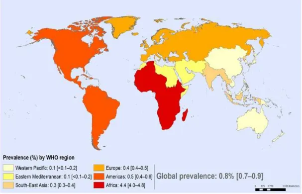
Page(s) Fig. 1 - Adult HIV prevalence (15-49 years) in 2015 by WHO region. 11
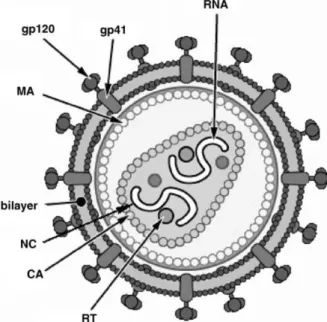
Fig. 2 - Diagram of HIV-1 mature virion. 12
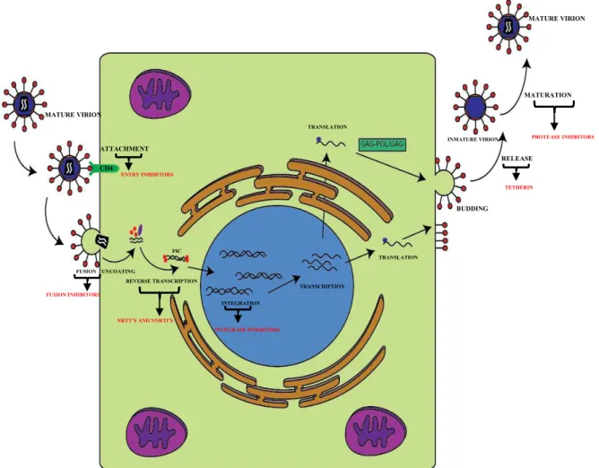
Fig. 3 - HIV-1 replicative cycle and main antiretroviral therapeutical targets. 13
Fig. 4 - Diagram of HBV virion. 15
Fig. 5 - HBV replicative cycle and targets for antiviral therapy. 16
Fig. 6 - Diagram of HCV virion. 18
Fig. 7 - Schematic representation of the HCV replicative cycle and targets
for antiviral therapy. 19
Fig. 8 - Schematic representation of overall structure of PON-1. 21 Fig. 9 - Schematic representation of PON-1 anchored to HDL. 22 Fig. 10 - Hydrolysis reaction of paraoxon by PON-1. 22 Fig. 11 - Lactonase activity of PON-1 in the homocysteine metabolism. 23 Fig. 12 - PON-1 activity HIV in infected individuals. 30 Fig. 13 - Diagnostic accuracy of serum biomarkers in the diagnosis of chronic
TABLE INDEX
Page(s)
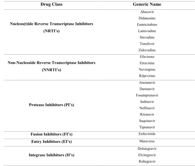
Table 1 - Antiretroviral drugs used against HIV. 14
Table 2 - Clinical study comparing HDL levels in human serum between healthy subjects, infected HIV patients with or without ART
and between the most used ART’s.
28
Table 3 - Comparison between biochemical variables between Caucasian HIV-infected patients without treatment and HIV-infected patients treated with efavirenz.
28
Table 4 - Comparison between biochemical variables in healthy subjects
and treated and non-treated HIV+ patients. 29 Table 5 - Variations of PON-1 activity and concentration, HDL
cholesterol ApoA-1 and oxidized LDL with gender, presence of HCV co-infection, lipodystrophy, CD4+ cell count and viral load.
29
Table 6 - Comparison between PON-1 activity and HIV markers of
infection. 32
Table 7 - Comparison of demographic variables, standard liver function tests and basal PON-1 activity in chronic hepatitis cases and healthy controls.
ABREVIATIONS LIST
AE Serum Arylesterase
AIDS Acquired Immune Deficiency Syndrome
ALP Alkaline Phosphate
ALT Alanine Aminotransferase
Apo A-1 Apolipoprotein A-1
APPs Acute Phase Proteins
ART Antiretroviral Therapy
ARV Antiretroviral
AST Aspartate Aminotransferase
AUC Area Under the Curve
BHMT Betaine-Hcy Methyltransferase
cART combined Antiretroviral Therapy
CBS Cystathionineβ- synthase
cccDNA Covalently Closed Circular Deoxyribonucleic acid
CRP C reactive protein
CSE Cystathio- nine -lyase
Cys Cysteine
Cyst Cystathionine
DEP Dietil-fosfate
EI’s Entry Inhibitors
EMA European Medicines Agency
FDA Food and Drug Administration
FI’s Fusion Inhibitors
HBV Hepatitis B Virus
HCTL Homocysteine-thiolactone
HCV Hepatitis C Virus
Hcy Homocysteine
HDL High Density Lipoprotein
HIV Human Immunodeficiency Virus
HIV-2 Human Immunodeficiency Virus type 2
HT Hcy-thiolactone
HTase Hcy-thiolactonase
IFN-α Interferon-alfa
II’s Integrase Inhibitors
LDL Low Density Lipoproteins
LPS Lipopolysaccharide
MA Matrix Protein
MAT Methionine Adenosyltransferase
Met Methionine
MetRS Methionyl-tRNA Synthase
MDA Malonaldehyde
MPO Myeloperoxidase
MS Methionine Synthase
MT Methyltransferase
NC Nucleocapsid Protein
NNRTI’s Non-Nucleoside Reverse Transcriptase Inhibitors
NO Nitric Oxide
NPV Negative Predictive Value
NUC’s Nucleotide/nucleoside analogues
NRTI’s Nucleoside Reverse Transcriptase Inhibitors
PAF-AH Platelet Activating Factor Acetyl-hydrolase
pegIFN-α Polyethyleno Glycol interferon-alfa
PI’s Protease Inhibitors
PNP p-nitrofenol
PON´s Paraoxonases
PON-1 Paraoxonase 1
PON-2 Paraoxonase 2
PON-3 Paraoxonase 3
PPV Positive Predictive Value
RNA Ribonucleic Acid
ROS Reactive Oxygen Species
RT Reverse Transcriptase
RT-PCR Reverse Transcriptase Polymerase Chain Reaction Technique
SAA Serum Amyloid A
SAHH S-Adenosyl-Hcy hydrolase
S-Adenosyl-Hcy S-adenosyl-homocysteine
S-Adenosyl-Met S-adenosyl-methionine
TNF Tumor Necrosis Factor
VL Viral Load
VLDL Very Low Density Lipoproteins
1. INTRODUCTION
Several clinical studies and authors have stated that some viral infections, namely the Human Immunodeficiency Virus (HIV), Hepatitis B (HBV) and Hepatitis C (HCV) viruses, are somehow related with the paraxonase-1 (PON-1) enzyme that could provide invaluable clinical information.
1.1 – HUMAN IMMUNODEFIECIENCY VIRUS (HIV) INFECTIONS
According to World Health Organization (WHO), over 35 million people have globally died from HIV-related causes to date. Almost 1.1million of those deaths were in 2015 alone (WHO, 2016). According to the report of the Joint United Nations Program on HIV and AIDS (UNAIDS) of 2016, there are still 36,7 million people infected around the globe (figure 1) with only 17 million of then been able to access combined antiretroviral therapy (cART) (UNAIDS, 2016). It is considered a major pandemic with no cure to date and the only way to keep the disease controlled is throughout the chronic use of cART which is expensive and not absent of adverse reactions. (Carr & Cooper, 2000; Cost Considerations and Antiretroviral Therapy | Adult and Adolescent ARV Guidelines | AIDSinfo, 2016).
The known causing agents of Acquired Immune Deficiency Syndrome (AIDS) are HIV-1 (figure 2) and HIV-2. Though they share some similarities (in key proteins like Gag or Pol), they differ in almost 50% of their genome. The differences in the viral genotype translate in several symptomatic disparities such as the latency period, which is increased in the HIV-2 strain, or the increased infection rate observed in HIV-1 strains, which remains to date the major culprit of HIV infections all over the world. (Simon, Ho
Fig. 1 - Adult HIV prevalence (15-49 years) in 2015 by WHO region. Adapted from Global Health
In Portugal, HIV infections are caused not only by the 1 variant but by the HIV-2 genotype due to the geopolitical and historical ties to Guinea-Bissau and Cape Verde, were the majority of HIV-2 infections have been recorded (Fernández‐Hidalgo, Almirante & Pahissa, 2009).
The worldwide spread of HIV indicates that the virus has one of the most effective infection mechanisms, taking advantage of cellular pathways while neutralizing and hiding from the immune system. The primary target are CD4+ T lymphocyte cells, which
are crucible for induction of specific humoral and cell-mediated immune response. In the long term, an untreated host will develop opportunist diseases that otherwise would remain at bay with an uncompromised immune response and eventually lead to death (Sierra, Kupfer & Kaiser, 2005). With access to ART, it is possible to extend the life expectancy but with severe complications such as dyslipidemias, atherosclerosis, neurodegenerative diseases, to name a few (Depairon et al., 2001; Marsillach, Parra, Coll, Joven, & Camps, 2008; Pereira et al., 2009).
The HIV replicative cycle (figure 3) is very complex and its duration and outcome varies according to the target cell and cell activation (Coffin, Hughes & Varmus, 1997).
Fig. 2 – Diagram of HIV-1 mature virion. MA (matrix protein); CA (capsid protein); NC
(nucleocapsid protein); RT (reverse transcriptase); RNA (ribonucleic acid); gp120 and gp41 (glycoprotein receptors). Adapted from Sierra et al. (2005).
Over the years several strategies were established to decrease the viral load hoping to increase the quality of life of infected patients and even develop a possible cure. Several antiretroviral drugs have been created that exploited numerous key stages in the replicative cycle of the virus, such as fusion inhibitors, entry inhibitors, reverse transcriptase inhibitors (nucleotide based and non-nucleotide based), protease inhibitors and integrase inhibitors (table 1) (FDA, 2016).
Fig. 3 – HIV-1 replicative cycle and main antiretroviral therapeutical targets. Once a mature virion
attaches itself to the CD4 membrane receptor of the lymphocyte T cell, it begins a fusion process in with it releases all the viral machinery into the cell, including the viral RNA and reverse transcriptase. Then it starts to reverse transcript the RNA into the pre-integration complex, which is incorporated in the cell’s DNA via integrase. Once integrated in the nuclear DNA, it begins the transcription and translation process, taking advantage of the cells own machinery. Once all the viral component are fully formed, they start to assembly and release themselves from the cell creating a new immature virion. For a time this virion undergoes a maturation process by the viral protease to produce all the mature viral particles. Adapted from Sierra et al. (2005).
MATURE VIRION
FUSION UNCOATING
REVERSE TRANSCRIPTION INTEGRATION TRANSCRIPTION TRANSLATION TRANSLATION BUDDING RELEASE INMATURE VIRION MATURATION MATURE VIRION ATTACHMENT CD4
NRTT’S AND NNRTI’S
Drug Class Generic Name
Nucleos(t)ide Reverse Transcriptase Inhibitors
(NRTI’s) Abacavir Didanosine Emtricitabine Lamivudine Stavudine Tenofovir Zidovudine
Non-Nucleoside Reverse Transcriptase Inhibitors
(NNRTI’s)
Efavirenz
Etravirine
Nevirapine
Rilpivirine
Protease Inhibitors (PI’s)
Atazanavir Darunavir Fosamprenavir Indinavir Nelfinavir Ritonavir Saquinavir Tipranavir
Fusion Inhibitors (FI’s) Enfuvirtide
Entry Inhibitors (EI’s) Maraviroc
Integrase Inhibitors (II’s)
Dolutegravir
Elvitegravir
Raltegravir
Adapted from FDA (2016).
HIV infections are prone to cause lipid and acute phase protein (APP) alterations (Treitinger et al., 2001), especially during the initial stages of infection. Some lipid modifications might be related to the host’s reaction to infection intermediated by cytokines, seen as an increase in serum Tumor Necrosis Factor (TNF), interleukins and decrease in High Density Lipoprotein (HDL) cholesterol (Feingold & Grunfeld, 1992), which can be linked to viral replication and consequent cholesterol membrane metabolism (Ono & Freed, 2001; Rose et al., 2008). The acute phase response has been shown to be correlated to the increase in plasma triglyceride concentrations and the increased turn-over and loss of protein mass (Treitinger et al., 2001).
1.2- HEPATITIS B INFECTIONS
Hepatitis B is also a disease caused by a viral infection. The causing agent is the Hepatitis B Virus (HBV) which is a small, enveloped DNA virus that belongs to the
Hepadnaviridae family. It targets liver cells and can cause acute or chronic liver disease.
According to the WHO, in 2016, more than 240 million people were chronically infected with HBV, with 686,000 people dying every year from complications related to the disease (WHO, 2016a). This infection can be easily prevented by vaccination, however high costs and lack of awareness of HBV infection risks hamper the successful outcome of immunization. Also, the high prevalence in some regions (e.g. Asia) is due to vertical transmission of HBV virus, from chronically infected mothers (Grimm, Thimme & Blum 2011).
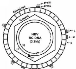
HBV is a small, enveloped DNA virus (figure 4). There are currently eight known different genotypes (A-H), being the A and D the most common in Europe. The virus itself does not seem to be cytopathic. The cytotoxicity in HBV infection comes from immune mediated responses (Grimm et al., 2011).
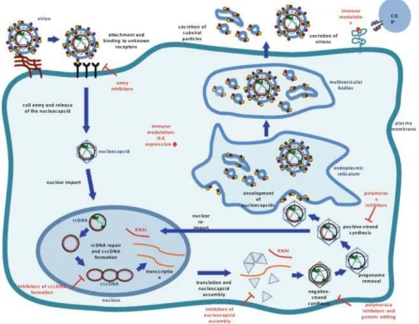
Currently, there are seven drugs approved by both the Food and Drug Administration (FDA) and European Medicines Agency (EMA) for hepatitis B infections (figure 5).
Fig. 4 – Diagram of HBV virion. Envelope proteins, S, M, L, with their S, preS2 and preS1 domains.
The conventional interferon-alfa (IFN-α), the PEGylated interferon-alfa (pegIFN-α) and the nucleotide/nucleoside analogues (NUC’s), such as adefovir, entecavir, telbivudine and tenofovir and lamivudine, also both used in HIV infections (EASL, 2012; Stein & Loomba, 2009).
The aims of cART are preventing disease progression to cirrhosis, end-stage liver disease or death and to improve the quality of life of chronically HBV-infected patients (Wang, 2012).
Fig. 5 – HBV replicative cycle and targets for antiviral therapy. HBV uses reverse
More than 95% of patients with acute HBV infection recover spontaneously and without the necessity for ART (Tassopoulos et al., 1987). However, patients with fulminant or severe hepatitis must be evaluated for liver transplantation (Castaldo & Chari, 2006).
1.3– HEPATITIS C INFECTION
Hepatitis C is also a disease caused by a RNA virus (HCV) (from the Flaviridae
family), with more than 86% chance of developing chronic infection. By the WHO assessments, it is estimated to affect at least 150 million people chronically worldwide, a significant proportion prone to develop complications like cirrhosis or liver cancer (WHO, 2016b). Although drugs can cure approximately 90% of people, the access to diagnosis and treatment is highly ineffective. This is due mainly to the high cost of treatment (WHO, 2016b).
Hepatitis C can be found worldwide but the most affected regions are Africa and Central Asia (WHO, 2016b).
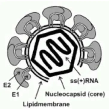
Fig. 6 – Diagram of HCV virion. HCV adopts an icosahedral scaffold; glycoproteins E1 and E2 are
The diagnosis of hepatitis C is divided in two steps, the first constitutes the screening for anti-HCV antibodies with a serological test. The second part is a confirmation test which screens for RNA. This is because some people are able to fight the initial infection successfully and no longer have the virus but still test positive for the anti-bodies of HCV (WHO, 2016b).
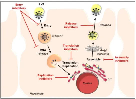
Nowadays, the available ART options are very effective (figure 7). However, as above mentioned, they can be extremely expensive. To date, there are no vaccines available has for preventing HCV infection (“Hepatitis B and C - Hepatitis B and C Treatments,” β016.;
“WHO | Hepatitis C,” β016).
HBC and HCV are both prone to cause chronic liver disease and increase the occurrence of reactive oxygen species and hepatic lipid peroxidation. This causes an accumulated oxidative stress that leads to liver cell apoptosis (Zeisel, Crouchet, Baumert & Schuster, 2015).
Fig. 7 – Schematic representation of the HCV replicative cycle and targets for antiviral
therapy. HCV interacts with the basolateral membrane of hepatocytes, resulting in viral entry into the host cell. The virus is internalized via endocytosis and translation of the HCV RNA occurs in the cytoplasm following viral fusion and uncoating. Viral replication takes place within the cytoplasm in perinuclear endoplasmic reticulum (ER)-derived membranes called the “membranous
web”. Progeny virions are assembled on cytosolic lipid droplets and subsequently transported along
1.4– BIOMARKERS FOR VIRAL DISEASE
A biomarker is a quantifiable biological or biochemical variable which
indicates, when measured objectively, the functioning of biological processes, pathogenic
or a pharmacological response to a given therapy (Strimbu & Tavel, 2010). It is normally
found in blood, urine, tissue biopsy, saliva or other biological samples. Continued research into a number of urinary, serologic and genetic biomarkers, will guide clinicians with diagnosis, prognosis, and treatment. (Vasan, 2006; Downes & Shah, 2012). Ideally, the means of analysis of a clinical biomarker of interest must meet certain requirements
such as high reproducibility, precision and specificity (Laborde et al., 2012).
In viral infections such as HIV, HBV or HCV infections, early diagnosis is
critical to improve not only the therapeutic outcomes, but also the survival rate and the
prognosis. PON-1 enzyme appears to be sensitive in early stages of some viral infections
and to the course of their progression due to a close association of the enzyme with
oxidative stress (Rose et al., 2006; Gangadharan, Antrobus, Dwek & Zitzmann, 2007;
Marsillach, Parra, Coll, Joven & Camps, 2008; Ali, Shehata, Ali-Labib & Zahra, 2009;
Downes & Shah, 2012). If a relation can be establish, a new biomarker to better evaluate
1.5– PARAOXONASE-1
Paraoxonases (PON´s) are a family of enzymes, called PON-1, PON-2 and PON-3. Their genes are located in humans at the chromosome 7q21-22 (Kulka, 2016; Primo-Parmo, Sorenson, Teiber & La Du, 1996).
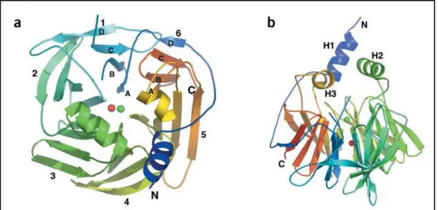
PON-1 was first described by Abraham Mazur, in 1946, who reported the existence of an enzyme capable of hydrolyzing organophosphates in animal tissues (Mazur, 1946). This eventually led to the discovery of PON-1 in the early 1950’s (Aldridge, 1953). Described as a calcium dependent serum HDL-associated serum aryldialkylphosphatase (Aldridge, 1953), the structure of PON-1 (figure 8) consists of a six-bladed -propeller,
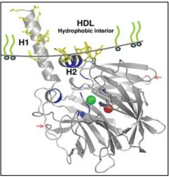
each blade consisting of four -sheets. In the active center of the PON-1 enzyme there is a tunnel containing two calcium atoms that is crucial for structural stability and catalytic activity. The enzyme also has an anchoring portion for the High Density Lipoprotein (HDL) particle that is located at the top of the propeller which has three α-helices (figure 9) (Harel et al., 2004).
Fig. 8 – Schematic representation of overall structure of PON-1. a) View of the six-bladed β
-propeller from above. The top of the -propeller is, by convention, the face carrying the loops connecting the outer β-strand of each blade (strand D) with the inner strand (A) of the next blade. Shown are the N and C termini, and the calcium atoms in the central tunnel of the propeller (Ca1, green; Ca2, red); b) A
The enzyme was named after studying its first known subtract, paraoxon, an active metabolite of the organophosphate parathion (Lessire et al., 1996; van Himbergen, van Tits, Roest & Stalenhoef, 2006). The ability of PON1 to hydrolyze paraoxon has been largely employed over the years for quantifying the activity of different enzyme variants and their tissue concentrations (figure 10) (Shih etal., 1998; Camps, Marsillach & Joven, 2009). Over the years it became clear that the activity of PON-1 was highly variable according to the genetic background of the individual, making possible to associate certain alleles with the activity of the enzyme. It was also showed that the presence of low-activity alleles varied considerably between each ethical group, with elevated frequency being perceived in Caucasian populations (Eckerson, Wyte & La Du, 1983).
Fig. 9 – Schematic representation of PON-1 anchored to HDL. Adapted from Harel et al. (2004).
Fig. 10 –Hydrolysis reaction of paraoxon by PON-1. DEP – dietil-fosfate; PNP –p-nitrofenol.
PON-1 has demonstrated a strong lactonase activity for homocysteine-thiolactone and other xenobiotic lactones (Jakubowski, 2000; Ng et al., 2005). Being evolved in the homocysteine metabolism via homocysteine-thiolactone (figure 11), the impact of the study of PON-1 increases, specially due to its possible role in the prevention of cardiovascular diseases (mainly atherosclerosis) (Shih et al., 1998) and involvement in
Alzheimer’s disease (Jakubowski, 1997; Cervellati et al., 2015).
Fig. 11 – Lactonase activity of PON-1 in the homocysteine methabolism. Homocysteine (Hcy) is
maintained at very low levels in serum due to the methionine cycle, which leads to a rapid methyl transfer reaction using the following enzyme substrates: methionine (Met), S-adenosyl-methionine (S-Adenosyl-Met), and S-adenosyl-homocysteine (S-Adenosyl-Hcy). The methionine cycle is controlled by methionine synthase (MS), betaine-Hcy methyltransferase (BHMT), methionine adenosyltransferase (MAT), methyltransferase (MT), and S-Adenosyl-Hcy hydrolase (SAHH). Met and Hcy are also metabolized by methionyl-tRNA synthase (MetRS) to become Met- tRNA and Hcy-thiolactone (HT). Under normal conditions, Hcy-thiolactone is rapidly converted to Hcy by Hcy-thiolactonase (HTase), in this case PON-1. HT reacts spontaneously with protein ε-lysine residues, leading to formation of N-Hcy-protein. Alternatively, Hcy can enter the transsulfuration pathway. It is then converted to cystathionine (Cyst) by cystathionine β - synthase (CBS), which is further converted to cysteine (Cys) by cystathio- nine -lyase (CSE). The transsulfuration pathway ends with sulfate. Adapted from Usuki (2012)
According to Aviram et al., (1998), PON-1 might have two different active sites that give it asterolytic and hydroxyperoxide activities - the first one metabolizes substrates such as esterified lipids, while the lather is involved in the reduction of hydroperoxides. It has been suggested that both catalytic activities may act in sequence, as seen with p -nithophenol 13-hydroperoxylinoleate when used as PON-1 subtract. The reduction mechanism paralleled hydrolysis, indicating an esterolytic activity requirement before the esterified fatty acid hydroperoxide is reduced (Karabina, Lehner, Frank, Parthasarathy & Santanam, 2005).
Human PON-1 is thought to be mainly synthetized and secreted by the liver. In fact, Northern-blot analysis of hepatic tissue samples presented only positive results for PON-1 mRNA (Hassett et al., PON-199PON-1). However, recent evidences suggested that other tissues such as lungs, brain, pancreas, small-intestine and kidney have the same capacity; the presence of PON-1 mRNA in such tissues was detected, using reverse transcriptase polymerase chain reaction technique (RT-PCR) (Eckerson et al., 1983). Nevertheless, the liver is, the most likely source of PON-1 due to the high PON-1 gene expression; besides, it is here that HDL is synthesized and secreted into the circulation.
Human HDLs particles form a heterogeneous group of lipoproteins, classified according to their increasing size and lipid and protein composition: HDL3c, HDL3b,
HDL3a, HDL2a, HDL2b (Rosenson et al., 2011). In bovine serum, 85% of the total PON-1
activity has been associated with HDL fractions. Less than 5% of the total PON-1 activity was identified in very low density lipoproteins (VLDL) and low density lipoprotein (LDL) (Miyamoto, Takahashi, Oohashi, Sato, & Oikawa, 2005).
There is no consensus about the major role of PON-1 in the human organism. The first hypothesis was proposed by Mackness, Arrol & Durrington (1991). This research work highlighted the safeguards against copper-induced LDL oxidation provided by HDL and PON-1. It was observed that PON-1 prevented lipoperoxide generation during the process of LDL oxidation, which indicated that this enzyme might be involved in a protective function of HDL. Of notice is that a PON-1 free sulfhydryl group in cysteine 284 was shown to be essential for the enzyme’s activity against lipid peroxides but not for its activity against paraoxon or other xenobiotics. This suggests that the mechanisms of hydrolysis are somewhat different (Aviram et al., 1998).
simultaneously depleted (Lenten et al., 1995). It appears that under inflammatory conditions, a displacement of the Apo A-1 by the positive acute phase proteins (APPs) serum amyloid A (SAA) in HDL might occur, thus leading to a decrease in PON-1 activity. However, in mice with acute infection a decrease in PON-1 and platelet activating factor acetyl-hydrolase (PAF-AH) activities was concomitant with unchanged Apo A-1 levels (Lenten et al., 2001). Acute phase response induced by endotoxin, lipopolysaccharide (LPS), caused a decrease in PON-1 serum activity (over 60%) within 48 hours in Syrian Hamsters and initiated a rapid decrease in PON-1 mRNA concentrations, persisting for at least 48 hours, in hamster hepatic cell cultures. Moreover, it was observed that mediators such as TNF and IL-1 diminished PON-1 activity in a similar manner (Feingold, Memon, Moser & Grunfeld, 1998).
2. DEVELOPMENT
2.1– PON-1 AND HIV INFECTIONS
2.1.1 – PON-1 ENZYME AND HDL
As mentioned before PON-1 is an esterase/lactonase enzyme, present in HDL, with a broad spectrum of substrates. Yet, its physiological function is not completely understood. However, a considerable amount of evidence suggests that, among other roles, PON-1 may hydrolyze oxidized lipid peroxides from LDL and HDL and, thus, possess antioxidant and anti-inflammatory properties (Parra et al., 2007).
Several studies have shown significant alterations on the enzymatic activity of PON-1 in HIV infected patients. Severe dyslipidemias and low levels of HDL (Riddler et al., 2007; Stein et al., 2008) cause long-term infected HIV patients to develop increased risk of cardiovascular diseases (Lang et al., 2010).
Low levels of HDL cholesterol is widely prevalent in Western countries, and is an independently predictive risk of cardiovascular disease, even in individuals with low concentrations of LDL. However, low concentrations of HDL are frequently concomitant with increased concentrations of small, cholesterol-depleted LDL particles and increased concentrations of cholesterol enriched triglyceride remnants. Thus, the cardiovascular risk associated with low HDL values is difficult to distinct from that of other associated lipoprotein abnormalities (Assmann, Schulte, Von Eckardstein & Huang, 1996; Otvos, Jeyarajah & Cromwell, 2002).
2.1.2 – PON-1 IN HIV INFECTED PATIENTS
Viral replication and some clinical manifestations of HIV infection involve an imbalance in the reduction–oxidation status and free radical production (Schwarz, 1996). It is likely that the HIV-induced oxidized environment could result in an increased binding of free radicals to PON-1, resulting in a less active enzyme in circulation; in agreement to the reports of oxidized lipids reacting with a free (–SH) group at PON’s cysteine-284 site leading to inactivation of the enzyme. Another reason for this decrease could be the lower concentrations of HDL cholesterol and Apo A-1 (Aviram et al., 1999). HIV infected patients frequently develop long-term pro-atherogenic metabolic modifications. These problems may be explained by the infection itself or by secondary effects of the ART, mainly protease inhibitors (Périard et al., 1999). These findings gained additional clinical relevance since the introduction of effective therapeutic measures, which have gradually changed HIV infections from a life threatening to a chronic disease. Furthermore, the combination of classical cardiovascular risk factors with some genetic polymorphisms appears to predispose these individuals to a higher risk of premature arteriosclerosis (Alonso-Villaverde et al., 2005; Coll et al., 2005; Coll et al., 2006). There are changes in lipoprotein metabolism in the course of HIV infection, including increased lipid peroxidation, hypocholesterolemia, hypertriglyceridemia and low HDL concentration (Rose et al., 2006). Also, patients with higher HDL concentrations appear to have a better disease course than patients with lower HDL concentrations (Alonso-Villaverde et al., 2003).
When PON-1´s activity is compared between HIV infected patients and healthy subjects, it is possible to conclude that the activity is decreased in HIV infected patients (44.3 nmol.min-1.mL-1 ± 38.6 vs 71.8 nmol.min-1.mL-1 ± 37.4 ; p=0,001) (Daminelli et
Controls HIV+ N ART HIV+ PI VL+ HIV+ PI VL- HIV+ NNRTI
HDL (mg/dl) 56 45.1 54.4 56.1 54.1
ART – antiretroviral therapy; VL ± - viral load above or below detection limit (20 copies/ml); adapted from Siegel et al. (2015).
Parameters Untreated Treated (Efavirenz)
HDL (mg/dl) 48 46
PON-1 paraxaonase activity (U/L) 58,8 77,35
PON-1 activity /HDL ratio 1,3 1,88
Adapted from Pereira et al. (2009).
The evidence provided by this study is reinforced in a work presented by Marinho et al. (2016) in which is demonstrated once again that PON-1 activity increases when using cART containing a different NNRTI, Nevirapine (Marinho et al., 2016).
In a study published by Daminelli et al. (2008) (table 4), the HIV+ group of patients showed that the LDL cholesterol and triglyceride levels in plasma are decreased than those of the control group, while HDL levels were identical. HIV+ patients frequently show reduced LDL, but triglycerides are generally described as increased (Treitinger et al., 2001), in contrast with the results of this study. However, increased triglyceride concentration leaded to a HDL cholesterol decrease, and the fact that triglycerides were not increased in the HIV+ group ultimately facilitated the analysis of HDL. According to Daminelli et al. (2008), the three subgroups of HIV-infected patients did not differ among them in respect to LDL. Regarding HDL, however, its levels were higher in the subgroup treated with NRTI plus NNRTI combined therapy, when compared with the subgroup treated with NRTI plus PI therapy, with non-treated HIV+ patients or with the values of the control group. The diminution of PON-1 activity in HIV-infected patients confirms the findings of Parra et al. (2007), in both treated and non-treated patients.
Table 2 -Clinical study comparing HDL levels in human serum between healthy subjects, infected
HIV patients with or without ART and between the most used ART’s.
Table 3 -Comparison between biochemical variables between Caucasian HIV-infected patients
Adapted from Daminelli et al. (2008)
In fact, Parra et al. (2007) found a reduction in PON-1 activity with an increase in its concentration in HIV infected patients, which could be explained by the addition of a higher quantity of free radicals to PON-1, by lower serum concentrations of HDL and Apo A-1 and by changes in the liver that would influence the serum concentration of PON-1 (table 5).
Reprinted from Parra et al. (2007)
The results from this study indicated that the co-infection with HCV was associated with a significantly lower PON-1 activity and decreased amount of oxidized LDL. There were no significant differences regarding PON-1, HDL or Apo A-1 concentrations. Patients with an active viral replication had higher PON-1 concentrations and lower HDL cholesterol levels. It was not found any significant difference in PON-1
Parameters Controls Untreated NRTI +
NNRTI NRTI + PI
HDL (mg/dl) 41 39 53 38
PON-1 (paraoxonase) (nmol.min-1.ml-1) 72 46 55 34
LDL (mg/dl) 127 100 106 97
Triglycerides (mg/dl) 116 98 103 98
Table 4 - Comparison between biochemical variables in healthy subjects and treated and
non-treated HIV+ patients.
lymphocyte count, did not present any significant differences in PON-1 status, HDL-cholesterol, Apo A-1 or oxidized LDL levels. The presence of lipodystrophy was associated with lower HDL-cholesterol concentrations and higher oxidized LDL levels. CD4+ T and CD8+ T lymphocyte cell counts showed weak negative correlations with PON-1 concentration (Parra et al., 2007).
In regards to ethnicity, specifically between Caucasian and Black patients, Pereira et al. (2009) demonstrated that it is also important to take into account the genetic variability specifically those of individuals of different ethnicities, when evaluating PON-1 activity. Other studies do not take the genetic variability into account (Alonso-Villaverde et al., 2003; Maselli et al., 2014; Parra et al., 2007).
Only a few studies have been published to date regarding the association between HIV infection and PON levels, and these have produced diverse results (Parra et al., 2007; Daminelli et al., 2008; Pereira et al., 2009). Daminelli et al. (2008) verified a significant decreased activity of PON-1 in HIV+ patients compared to healthy controls; ART
Fig. 12 - PON-1 activity HIV in infected individuals. PON-1 activity was lower in untreated [58.80
U.L-1 (51.36, 66.23); n = 18] than in treated [77.35 U.L-1 (65.66, 89.04); n = 18]; Caucasian patients
and both were lower than Black patients despite treat- ment (*P < 0.05, **P < 0.01 and ***P < 0.001, one-way ANOVA plus Bonfer-roni’s multiple comparison test); No treatment ; treated with Efavirenz (?) . Adapted from Pereira et al. (2009).
2
treatment did not reverse this deficiency. Parra et al. (2007) also found decreased PON-1 activity but also identified an increased PON-1 concentration in HIV+ patients compared
to heathy controls.
2.1.3 – PON-1 ACTIVITY, CD4+ AND CD8+ T-CELLS IN HIV INFECTED PATIENTS
The Maselli et al. (2014) work showed lower serum PON-1 activity in HIV+ patients
with no ART (table 6). PON-1 activity showed a significant positive correlation with the number of CD4+ T-cells in HIV+ patients with no ART, suggesting that the immune status
may directly affect the activity of PON-1. Other studies have shown that enzyme levels are reduced in HIV+ patients with no ART who have a reduced number of CD4+ T-cells (Le Moing et al., 2007). Thus, it might be proposed the existence of a relationship between the number of CD4+ T-cells and PON-1 activity, which would be directly related to a good outcome in a HIV infection. It remains uncertain whether the replication of the HIV virus itself would be related to PON-1. According to Maselli et al. (2014) the study established the factor that likely determines the reduction of PON-1 activity is the suppression of the immune system by HIV (Maselli et al. 2014).
Adapted from Maselli et al. (2014)
According to Pereira et al., (2009) there is also a possible relation between PON-1 activity and CD4 cell count, corroborated by other studies (Parra et al., 2007; Parra et al., 2010). It should be taken in consideration that in early studies there was no differentiation between HIV patients with and without co-infection by Hepatitis C virus, that can largely affect the outcomes on PON-1 activity, since this enzyme is mainly synthetized in the liver and a marker of hepatic function (Ferré et al., 2006).
Parameters Controls Untreated Efavirenz/Nevirapine Lopinavir/Ritonavir
CD4+ T-cells (cells/mm3) ND 404 483 486
CD8+ T-cells (cells/mm3) ND 955 859 952
CD4:CD8 ratio ND 0.4 0.5 0.5
HIV-1 RNA (copies/ml) - 89.266 14.121 16.301
PON-1 paraoxonase activity
(U/L) 117 81 109 106
2.1.4 THE ANTIOXIDANT AND ANTI-INFLAMATORY ROLE OF PON-1 IN HIV INFECTED PATIENTS
2.1.5 PON-1 HAS A BIOMARKER FOR HIV INFECTION
According to Parra et al. (2007), serum PON-1 concentrations showed a strong correlation with -2-microglobulin concentration [B=75.19 (50.32–100.06); p<0.001].
Given that -2-microglobulin is a known effective marker of infection by HIV (Savès et al., 2001), this association might be worth exploring and could lead into new ways to better monitor the infection at multiple levels.
Maselli et al. (2014) demonstrated that the enzymatic activity of PON-1 is correlated with CD4+ T-cell count in patients not receiving ART. The demonstration that PON-1 activity was reduced in untreated patients, but not in individuals receiving ART, suggested that the activity of PON-1 is associated with the immune status of HIV-1 infected individuals, making the enzymatic activity a good candidate for an alternative biomarker (Maselli et al., 2014).
2.2– PON-1 AND VIRAL HEPATITIS (HBV AND HCV)
2.2.1 – PON-1 ACTIVITY AS A BIOMARKER FOR LIVER DISEASE
Chronic hepatitis is included in a group of liver disorders where the hepatic inflammation and necrosis lasts for more than six months. The severity of the disease might vary from minor forms, in which the progression is very slow and might go undetected for an extended period of time, to more serious forms which are linked with liver parenchymal scarring and structural disorders that progress to cirrhosis (Fauci, Kasper, Longo, Hauser & Jameson, 2015).
In chronic liver diseases of slow progression the traditional liver function tests often remain unaltered, until more severe forms of hepatic disorder becomes apparent.
In order to diagnose such slow progressive liver diseases before reaching advanced states such as hepatocellular necrosis and fibrosis, aside the outdated biochemical tests, alternative parameters become necessary to evaluate liver damage (Fauci, Kasper, Longo, Hauser & Jameson, 2015).
Currently histopathological analysis of liver biopsy samples are considered the best method to accurately diagnose liver disease. However, liver biopsy is an invasive technique with inevitable complications (Sanai & Keeffe, 2010). Once more, the need for a trustworthy and accurate diagnostic tool that does not require invasive and painful procedures emerges. In this regard, it would be convenient to find a hepatic biomarker, preferably one that can be easily and quickly assessed (Gangadharan, Antrobus, Dwek & Zitzmann, 2007; Marsillach et al., 2008).
The pathogenesis of numerous liver diseases are often associated with oxidative stress (Tanikawa & Torimura, 2006). The oxidative stress is strongly linked to the progression of alcoholic liver disease, non-alcoholic steatohepatitis and hepatitis B and C virus infection, which can lead to the development of acute hepatitis, chronic hepatitis, hepatocellular carcinomas and liver cirrhosis (Gil, Pla, Gonzalvo, Hernández & Villanueva, 1993).
serum PON-1 activity in patients with chronic hepatitis (Ferré et al., 2002; Ferré et al., 2005; Kilic, Aydin, Kilic, Erman, Aydin & Celik, 2005).
Recently, Pyati (2015) showed that serum basal PON-1 activity is considerably reduced (p < 0.001) in patients with chronic hepatitis when compared to healthy controls. These findings are in agreement with the results from the studies reported by Başkol,
Başkol, Deniz, Ozbakir, & Yücesoy, (2005); N. Ferré et al., (2002), (2005); Kilic, Aydin, Kilic, Erman, Aydin, Suleyman, et al., (2005) and Ali, Shehata, Ali-Labib, & Zahra, (2009).
According to Pyati (2015) (table 6), the activity of PON-1 is positively correlated with total serum protein levels and albumin levels, and are negatively correlated with serum bilirubin, alanine aminotransferase (ALT), and alkaline phosphate (ALP), with a great statistical significance (p< 0.001). No such correlation has been demonstrated between standard liver function tests and basal PON-1 activity in healthy controls. These results are consistent with previous works (Ferré et al., 2002; Ferré et al., 2005; Kilic, Aydin, Kilic, Erman, Aydin, Suleyman, et al., 2005; Başkol et al., β005; Ali, Shehata, Ali-Labib, & Zahra, 2009).
Adapted from Pyati (2015)
According to Pyati (2015), basal PON-1 activity seems to be a better marker for diagnosis of chronic hepatitis than total protein, albumin and ALP with a sensitivity of 68% and specificity of 100%. It also demonstrated a positive predictive value of 100%
Parameters Controls Chronic Hepatitis p-value
Age 45.6 ± 11.8 41.8 ± 9.8 0.080
Total Bilirubin (%) 0.74 ± 0.14 4.05 ± 1.74 < 0.001
Total Protein (%) 6.83 ± 0.72 6.2 ± 0.34 < 0.001
Albumin (g/dl) 3.72 ± 0.55 3.54 ± 0.34 0.35
ALT (U/L) 21.18 ± 6.87 135.92 ± 41.86 < 0.001
ALP (U/L) 88.78 ± 35.48 164.98 ± 47.42 < 0.001
Pon-1 activity (nmol/ml/min) 176.68 ± 25.88 101.88 ± 23.08 < 0.001
and negative predictive value of 75%. However, although PON-1 activity seems to be a more reliable predictor of liver disease compared to total protein, albumin and ALP levels, when bilirubin and ALT are taken into account they still show better results in terms of predictability of disease than basal PON-1 activity. The analysis of thereceiver operating characteristic (ROC) curve (figure 13) demonstrated that, among the study parameters, ALT demonstrated highest diagnostic accuracy (AUC=0.999) followed by total bilirubin (AUC=0.977), PON-1 (AUC=0.990), ALP (AUC=0.904), total protein (AUC=0.790) and albumin (AUC=0.595) (Pyati, 2015). These findings were similar to those observed in the study conducted by (Ferré et al. 2002).
The measurement of PON-1 activity may represent a significant improvement in liver disease detection. The analysis of the data provided by the ROC curves (Ferré et al., 2002; Pyati, 2015) demonstrated that serum PON-1 activity is, in fact, worth exploring. The ROC analysis also supports that the measurement of baseline serum PON-1 activity is an excellent candidate to become a biomarker for liver disease. Although bilirubin and ALT seem to be more accurate biomarkers than PON-1, they are only indicatives of some types of liver injuries (Pyati, 2015).
a)
Fig. 13 –Diagnostic accuracy of serum biomarkers in the diagnosis of chronic hepatitis. a) bilirubin
(BLUE), ALT (GREEN), ALP (YELLOW); b) total protein (BLUE), Albumin (GREEN) and PON-1
(YELLOW) ; reference line (PURPLE). Adapted from Pyati, (2015)
Specificity Specificity
ROC CURVES
Sensitivity Sensitivity
2.2.2 – PON-1 ACTIVITY AND OXIDATIVE STRESS IN THE LIVER
Hepatocytes are constantly exposed to reactive oxygen species (ROS), but protected from oxidative injury by a range of antioxidant pathways. The defenses against free radical-mediated injury include enzymatic deactivation and direct reaction with free radicals (Osman, Gabr, Lotfy, & Gabr 2007).
Although free radicals are normally produced by many reactions essential for cell metabolism and energy production, they are implicated also in the pathogenesis of several different human diseases, including viral infections such as HBV or HBC (Schwarz, 1996).
In patients with viral or alcoholic liver disease, the consequent alternation of cellular redox state is potentiated by a decrease in the antioxidant enzymes and an increase of free radical-mediated damage and apoptosis of liver cells (Loguercio & Federico, 2003).
Several studies suggested that free radical generation and oxidative stress play an important role in the mechanisms developed by HCV to survive and progress in the infected host (Cardin et al., 2001; Thorén, Romero, Lindh, Dahlgren & Hellstrand, 2004; Waris, Turkson, Hassanein & Siddiqui, 2005).
Lipid peroxidation is caused by free radicals leading to oxidative destruction of polyunsaturated fatty acids constitutive of cellular membranes. The destruction of polyunsaturated fatty acids leads to the production of toxic and reactive aldehyde metabolites such as malonaldehyde (MDA) (Levent et al., 2006). MDA content usually reflects the level of lipid peroxidation and indirectly reflects the extent of hepatocellular injury in vivo (Osman et al., 2007).
In the study by Ali et al. (2009), it was established that serum MDA levels were higher in patients with HCV than the healthy ones. This result considers the consequence of lipid peroxidation in the pathophysiology of patients with HCV. Serum MDA concentration in patients with chronic active hepatitis C as well as transaminase and bilirubin levels were found to be increased in many other studies (Mansurova, Mutikhova, & Olimova, 2005; Levent et al., 2006; Osman et al., 2007). MDA was showed to be elevated in the liver and the blood of patients with HCV as reported by De Maria et al. (1996). Moreover, Boya et al. (1999) reported that, the peripheral blood mononuclear cells separated from chronic hepatitis C patient had increased MDA concentrations.
Some recent studies are in agreement with the present investigation such as Gangadharan, Antrobus, Dwek, & Zitzmann, (2007) who found a decreasing in serum AE and PON-1 activities in chronic and cirrhotic HCV patients compared with healthy controls. The study proposed that serum AE and PON-1 carried in circulation bounded to HDL particles protect LDL from peroxidation, supported by Mackness, Durrington, & Mackness, (2004). Thus, there are two possible explanations for decreased PON-1 and AE activities in chronic hepatitis. Firstly, the decrease in PON-1 and AE enzymatic activities or gene expression could be the consequence of the hepatic dysfunction. The second is that serum PON and AE activities could be decreased as a consequence of an altered synthesis and/or secretion of HDL (Ali et al., 2009).
PON-1 and AE act as antioxidants that protect LDL from oxidative modifications and can reduce oxidized lipid in oxidized lipoproteins (Mackness et al., 2004; Kilic, Aydin, Kilic, Erman, Aydin, & Celik, 2005). Other important observation in the study by Ali et al., (2009) was that the activities of both enzymes have significantly decreased in cirrhotic more than in chronic HCV patients, indicating that the level of decrease in AE and PON-1 activity in the serum of patients with chronic liver diseases is probably a consequence of liver dysfunction. That could emphasize the prognostic value of AE and PON-1 in HCV infected patients and give clinicians a signal for deterioration of the liver function with the progression of the disease. The results also demonstrated that serum AE was negatively correlated with serum ALT and aspartate aminotransferase (AST) activities; whereas serum PON-1 was negatively correlated with serum ALT, AST and ALP activities and serum total bilirubin and direct bilirubin levels in HCV patients group. Kilic, Aydin, Kilic, Erman, Aydin, & Celik, (2005) are in agreement with Ali et al., (2009) that serum AE and PON-1 activities were also negative correlated with serum ALT and AST activities and albumin and bilirubin concentrations as well as between serum PON-1 and ALP activity.
PON-1 has an active role in the regulation of oxidative stress, fibrosis and hepatic cell apoptosis in chronic liver diseases (Ferré et al., 2006). Marsillach et al. (2007) reported that, PON-1 activity measurement would be very useful in discriminating between alcoholic patients and healthy subjects.
When studying the ROC curves of both AE and PON-1, it was found that the areas under the curves were 0.968 and 0.966 respectively, and best cutoff values of serum AE and PON-1 were 764 and 482 nmol/min/ml respectively. Applying these cutoff values, the maximum sensitivity, specificity, positive predictive and negative predictive values of (AE and PON-1) were (86% and 79.5%), (90% and 100%), (95% and 100%) and (76% and 70%) respectively (Ali et al., 2009).
The study by Marsillach et al., (2007) reported that, PON-1 activity measurement would be very useful in discriminating between alcoholic patients and healthy subjects; on studying the ROC curve of PON-1 activity, the area under the curve of PON-1 activity
was ≥0.90, the increased sensitivity up to 8γ.5–96.6% and specificity up to 95.8–99.6%. AE was the hepatitis C biomarker marker which showed the highest sensitivity, when measured as a single marker without any combination with others, while MDA or PON-1 was the marker which gave the highest specificity, when each of them was measured alone without combination. But in combination, the best combined ones for sensitivity, specificity, positive predictive value (PPV) and negative predictive value (NPV) were MDA plus albumin, PON-1 plus albumin and PON-1 plus AST (Marsillach et al., 2007).
The over release of the oxidative free radicals [MDA, nitric oxide (NO) and myeloperoxidase (MPO)] and the decrement of the antioxidants (PON-1 and AE) might play a role in the pathogenesis of viral hepatitis. Moreover, these markers are beneficial in testing liver dysfunction and detecting high-risk patients of cirrhotic from chronic HCV liver disease patients (Ali et al., 2009; Pyati, 2015).
2.2.3 – PON-1 ACTIVITY AND HBV INFECTIONS
Karsen et al. (2012) proposed that, serum PON-1 concentration was considerably lower in patients with chronic hepatitis B than in the control group. The results presented that patients with chronic hepatitis B are exposed to oxidative stress and showed decreased PON-1 activity. The study proposes that predisposal factors may also, in part, play a role in the pathogenesis of atherosclerosis in patients with chronic hepatitis B.
There is a well-established relation between the enzyme PON-1 in cardiovascular diseases and atherosclerosis (Michael I Mackness et al., 2004; Yilmaz, 2012; Kim, Marsillach, Furlong, & Jarvik, 2013; Kulka, 2016). (Draganov & La Du, 2004). HDL PON-1 activity is shown to be associated with modulation of endothelial functions and regulation of coronary vasomotor tone. (Malin et al., 2001; Pasqualini et al., 2005) Increased incidence of subclinical atherosclerosis in carotid arteries has been reported in patients with chronic hepatitis B (Alkhouri et al., 2010). On the other hand, other studies have been unable to demonstrate any difference between chronic hepatitis B and controls and indicated that HBs Ag seropositivity was not related with increased mortality risks of atherosclerosis related cardiovascular diseases (Wang, Chen, Lee, Yang & Hsiao, 2010). The reduction of PON-1 activity under oxidative stress is an independent risk factor for coronary arterial disease and mostly attributed to changes in the redox status of the free sulfhydryl groups of proteins since sulfhydryl prevent the inhibition of PON-1 activity (Mackness et al., 2001). Free sulfhydryl groups of proteins constitute the main antioxidant component of serum and have been shown to be associated with the coronary heart disease (Rozenberg & Aviram, 2006).
Free radicals are produced in metabolic and physiological processes, and harmful oxidative reactions may occur in organisms. Organisms are protected against oxidative stress via enzymatic and non-enzymatic antioxidative mechanisms. Under normal conditions, a balance exists between free radical formation and their removal by antioxidant enzymes and other molecules (Mackness et al., 2001).
groups in plasma, and total antioxidant capacity levels were significantly lower in chronic hepatitis B subjects than observed in the control group, while LOOH, total oxidant status levels and oxidative stress index were significantly higher. These results represent the severe oxidative stress in patients with chronic hepatitis B infection. On one hand, chronic hepatitis B is the most common cause of hepatocellular carcinoma but on the other hand it could contribute to the development of atherosclerosis (Karsen et al., 2012).
CONCLUSIONS
Several studies conclude that significant alterations occur on the enzymatic activity of PON-1 in HIV, HBV and HCB infected patients. This modifications seem to occur due to the increased levels caused by the infections on the organism, leading to multiple disruption on antioxidant and anti-inflammatory pathways that seem were demonstrated to involve PON-1.
Although PON-1 activity alterations seem to be caused by malignant causes, positive and useful clinical information might be extracted by measuring such alterations. PON-1 possibly will be used with more research in future as a biomarker.
In HIV infections, there are well established markers for evaluating the infection status, however the insight provided by the additional monitoring of PON-1 activity could, in fact, be extremely useful. This additional assessment might lead to the prevention of the most common complications due to the chronic use of ART, such as cardiovascular disease and dyslipidemias. This prevention can be achieved by obtaining a more suitable diagnostic tool, for the disease progression and all its complications and by doing so, applying preventive measures that could avoid such complications (Coll et al., 2006; Parra et al., 2007; Pereira et al., 2009; Dias et al., 2014).
In regards to clarifying whether PON-1 could be used as a biomarker for HBV or HCV infections, several works including those of Ali et al., (2009), Pyati, (2015) Marsillach et al., (2007) demonstrated that the precision of PON-1 as a marker for disease is similar to those of ALT or bilirubin, already well accepted and used parameters of analysis (Pyati, 2015).
The enzyme PON-1, in addition to its possible biomarker applications in both HIV, HBV and HCV infections, revealed an extremely important role in the oxidative stress caused by those infections. As an enzyme with anti-oxidant proprieties and closely involved in regulating LDL peroxidation, in both cases the activity of the enzyme was closely associated with the progression of the disease which can definitely provide useful analytic tools for the diagnose and evaluation of those patients (Ferré et al., 2006; Mackness, Arrol, Abbott, & Durrington, 1993; Valyi-Nagy & Dermody, 2005).
3. REFERENCES
ALDRIDGE, W. N. (1953). Serum esterases. II. An enzyme hydrolysing diethyl p-nitrophenyl phosphate (E600) and its identity with the A-esterase of mammalian sera. The Biochemical Journal, 53(1), 117–24. Retrieved from http://www.ncbi.nlm.nih.gov/pubmed/13032042
Ali, E. M. M., Shehata, H. H., Ali-Labib, R., Esmail Zahra, L. M. & Zahra, L. M. E. (2009). Oxidant and antioxidant of arylesterase and paraoxonase as biomarkers in patients with hepatitis C virus. Clinical Biochemistry, 42(13–14), 1394–1400. http://doi.org/10.1016/j.clinbiochem.2009.06.007
Alkhouri, N., Tamimi, T. A.-R., Yerian, L., Lopez, R., Zein, N. N. & Feldstein, A. E. (2010). The inflamed liver and atherosclerosis: a link between histologic severity of nonalcoholic fatty liver disease and increased cardiovascular risk., 55(9). http://doi.org/10.1007/s10620-009-1075-y
Alonso-Villaverde, C., Coll, B., Gómez, F., Parra, S., Camps, J., Joven, J. & Masana, L. (2005). The efavirenz-induced increase in HDL-cholesterol is influenced by the multidrug resistance gene 1 C3435T polymorphism. AIDS (London, England),
19(3), 341–2. Retrieved from http://www.ncbi.nlm.nih.gov/pubmed/15718846 Alonso-Villaverde, C., Segues, T., Coll-Crespo, B., Perez-Bernalte, R., Rabassa, A.,
Gomila, M., … Masana, L. (2003). High-density lipoprotein concentrations relate to the clinical course of HIV viral load in patients undergoing antiretroviral therapy.
AIDS (London, England), 17(8), 1173–1178.
http://doi.org/10.1097/01.aids.0000060360.78202.05
Assmann, G., Schulte, H., Von Eckardstein, A., & Huang, Y. (1996). High-density lipoprotein cholesterol as a predictor of coronary heart disease risk. The PROCAM experience and pathophysiological implications for reverse cholesterol transport.
Aviram, M., Billecke, S., Sorenson, R., Bisgaier, C., Newton, R., Rosenblat, M., … La
Du, B. (1998). Paraoxonase active site required for protection against LDL oxidation involves its free sulfhydryl group and is different from that required for its arylesterase/paraoxonase activities: selective action of human paraoxonase allozymes Q and R. Arteriosclerosis, Thrombosis, and Vascular Biology, 18(10), 1617–1624. http://doi.org/10.1161/01.ATV.18.10.1617
Aviram, M., Rosenblat, M., Billecke, S., Erogul, J., Sorenson, R., Bisgaier, C. L., … La
Du, B. (1999). Human serum paraoxonase (PON 1) is inactivated by oxidized low density lipoprotein and preserved by antioxidants. Free Radical Biology &
Medicine, 26(7–8), 892–904. Retrieved from
http://www.ncbi.nlm.nih.gov/pubmed/10232833
Aviram, M., Rosenblat, M., Bisgaier, C. L., Newton, R. S., Primo-Parmo, S. L. & La Du, B. N. (1998). Paraoxonase inhibits high-density lipoprotein oxidation and preserves its functions: A possible peroxidative role for paraoxonase. Journal of Clinical
Investigation, 101(8), 1581–1590. http://doi.org/10.1172/JCI1649
B, J. J. C. P. R. S., & Aldridge, B. Y. W. N. (1953). Serum Esterases 1., (1949), 0–1.
Başkol, M., Başkol, G., Deniz, K., Ozbakir, O., & Yücesoy, M. (β005). A new marker for lipid peroxidation: serum paraoxonase activity in non-alcoholic steatohepatitis.
The Turkish Journal of Gastroenterology : The Official Journal of Turkish Society
of Gastroenterology, 16(3), 119–23. Retrieved from
http://www.ncbi.nlm.nih.gov/pubmed/16245219
Boya, P., de la Peña, A., Beloqui, O., Larrea, E., Conchillo, M., Castelruiz, Y., … Prieto, J. (1999). Antioxidant status and glutathione metabolism in peripheral blood mononuclear cells from patients with chronic hepatitis C. Journal of Hepatology,
31(5), 808–14. Retrieved from http://www.ncbi.nlm.nih.gov/pubmed/10580577 Çakatay, U., Kayali, R. & Uzun, H. (2008). Relation of plasma protein oxidation
parameters and paraoxonase activity in the ageing population. Clinical and
Experimental Medicine, 8(1), 51–57. http://doi.org/10.1007/s10238-008-0156-0
Camps, J., Marsillach, J., & Joven, J. (2009). The paraoxonases: role in human diseases and methodological difficulties in measurement. Critical Reviews in Clinical
Laboratory Sciences, 46(2), 83–106. http://doi.org/Pii 909183322\rDoi
10.1080/10408360802610878
Cardin, R., Saccoccio, G., Masutti, F., Bellentani, S., Farinati, F., & Tiribelli, C. (2001). DNA oxidative damage in leukocytes correlates with the severity of HCV-related liver disease: validation in an open population study. Journal of Hepatology, 34(4), 587–92. Retrieved from http://www.ncbi.nlm.nih.gov/pubmed/11394660
Carr, A., & Cooper, D. A. (2000). Adverse effects of antiretroviral therapy. Lancet
(London, England), 356(9239), 1423–30.
http://doi.org/10.1016/S0140-6736(00)02854-3
Castaldo, E. T., & Chari, R. S. (2006). Liver transplantation for acute hepatic failure.
HPB : The Official Journal of the International Hepato Pancreato Biliary
Association, 8(1), 29–34. http://doi.org/10.1080/13651820500465741
Cervellati, C., Romani, A., Bergamini, C. M., Bosi, C., Sanz, J. M., Passaro, A., & Zuliani, G. (2015). PON-1 and ferroxidase activities in older patients with mild
cognitive impairment, late onset Alzheimer’s disease or vascular dementia. Clinical
Chemistry and Laboratory Medicine, 53(7), 1049–56.
http://doi.org/10.1515/cclm-2014-0803
Chambers, J. E. (2008). PON1 multitasks to protect health. Proceedings of the National
Chevaliez, S., & Pawlotsky, J.-M. (2006). HCV Genome and Life Cycle. Hepatitis C
Viruses: Genomes and Molecular Biology. Horizon Bioscience. Retrieved from
http://www.ncbi.nlm.nih.gov/pubmed/21250393
Coffin, J. M., Hughes, S. H., & Varmus, H. E. (1997). Retroviruses. Retroviruses. Cold
Spring Harbor Laboratory Press. Retrieved from
http://www.ncbi.nlm.nih.gov/pubmed/21433340
Coll, B., Alonso-Villaverde, C., Parra, S., Montero, M., Tous, M., Joven, J., & Masana, L. (2005). The stromal derived factor-1 mutated allele (SDF1-γ’A) is associated with a lower incidence of atherosclerosis in HIV-infected patients. AIDS (London,
England), 19(16), 1877–83. Retrieved from
http://www.ncbi.nlm.nih.gov/pubmed/16227796
Coll, B., Parra, S., Alonso-Villaverde, C., de Groot, E., Aragonés, G., Montero, M., … Masana, L. (2006). HIV-infected patients with lipodystrophy have higher rates of carotid atherosclerosis: the role of monocyte chemoattractant protein-1. Cytokine,
34(1–2), 51–5. http://doi.org/10.1016/j.cyto.2006.03.013
Cost Considerations and Antiretroviral Therapy | Adult and Adolescent ARV Guidelines | AIDSinfo. (n.d.). Retrieved from https://aidsinfo.nih.gov/guidelines/html/1/adult-and-adolescent-arv-guidelines/459/cost-considerations-and-antiretroviral-therapy Daminelli, E. N., Spada, C., Treitinger, A., Oliveira, T. V., Latrilha, M. D. C., &
Maranhão, R. C. (2008). Alterations in lipid transfer to high-density lipoprotein (HDL) and activity of paraoxonase-1 in HIV+ patients. Revista Do Instituto de
Medicina Tropical de Sao Paulo, 50(4), 223–227.
http://doi.org/10.1590/S0036-46652008000400007
De Maria, N., Colantoni, A., Fagiuoli, S., Liu, G. J., Rogers, B. K., Farinati, F., … Floyd, R. A. (1996). Association between reactive oxygen species and disease activity in chronic hepatitis C. Free Radical Biology & Medicine, 21(3), 291–5. Retrieved from http://www.ncbi.nlm.nih.gov/pubmed/8855439
Depairon, M., Chessex, S., Sudre, P., Rodondi, N., Doser, N., Chave, J. P., … Mooser,