Expression, mutation and copy number analysis of platelet-derived
growth factor receptor A (PDGFRA) and its ligand PDGFA in
gliomas
O Martinho1, A Longatto-Filho1,2, MBK Lambros3, A Martins1, C Pinheiro1, A Silva4, F Pardal4, J Amorim5, A Mackay3, F Milanezi1,6, N Tamber3, K Fenwick3, A Ashworth3, JS Reis-Filho3, JM Lopes6,7and RM Reis*,1 1
Life and Health Sciences Research Institute (ICVS), School of Health Sciences, University of Minho, 4710 Braga, Portugal;2Instituto Adolfo Lutz, 355-01246-902 Sa˜o Paulo, Brazil;3The Breakthrough Breast Cancer Research Centre, Institute of Cancer Research, London SW3 6JB, UK;4Department of Pathology, S. Marcos Hospital, 4710 Braga, Portugal;5Department of Oncology, S. Marcos Hospital, 4710 Braga, Portugal;6IPATIMUP, 4200 Porto, Portugal;7Medical Faculties of Porto University, 4200 Porto, Portugal
BACKGROUND:Malignant gliomas are the most prevalent type of primary brain tumours but the therapeutic armamentarium for these tumours is limited. Platelet-derived growth factor (PDGF) signalling has been shown to be a key regulator of glioma development. Clinical trials evaluating the efficacy of anti-PDGFRA therapies on gliomas are ongoing. In this study, we intended to analyse the expression of PDGFA and its receptor PDGFRA, as well as the underlying genetic (mutations and amplification) mechanisms driving their expression in a large series of human gliomas.
METHODS:PDGFA and PDGFRA expression was evaluated by immunohistochemistry in a series of 160 gliomas of distinct World Health Organization (WHO) malignancy grade.PDGFRA-activating gene mutations (exons 12, 18 and 23) were assessed in a subset of 86 cases by PCR—single-strand conformational polymorphism (PCR-SSCP), followed by direct sequencing. PDGFRA gene amplification analysis was performed in 57 cases by quantitative real-time PCR (QPCR) and further validated in a subset of cases by chromogenicin situhybridisation (CISH) and microarray-based comparative genomic hybridisation (aCGH).
RESULTS:PDGFA and PDGFRA expression was found in 81.2% (130 out of 160) and 29.6% (48 out of 160) of gliomas, respectively. Its expression was significantly correlated with histological type of the tumours; however, no significant association between the expression of the ligand and its receptor was observed. The absence of PDGFA expression was significantly associated with the age of patients and with poor prognosis. AlthoughPDGFRAgene-activating mutations were not found,PDGFRAgene amplification was observed in 21.1% (12 out of 57) of gliomas. No association was found between the presence ofPDGFRAgene amplification and expression, excepting for grade II diffuse astrocytomas.
CONCLUSION: The concurrent expression of PDGFA and PDGFRA in different subtypes of gliomas, reinforce the recognised significance of this signalling pathway in gliomas.PDGFRAgene amplification rather than gene mutation may be the underlying genetic mechanism driving PDGFRA overexpression in a portion of gliomas. Taken together, our results could provide in the future a molecular basis for PDGFRA-targeted therapies in gliomas.
British Journal of Cancer(2009)101,973–982. doi:10.1038/sj.bjc.6605225 www.bjcancer.com Published online 25 August 2009
&2009 Cancer Research UK
Keywords: PDGFA; PDGFRA; expression; mutations; amplification; gliomas
Malignant gliomas are highly heterogeneous and invasive tumours and account for approximately 70% of all primary brain tumours (Louiset al, 2007). Histologically, gliomas are classified into several entities, with astrocytic tumours being the most prevalent type, followed by oligodendroglial and mixed oligoastrocytic tumours, and less frequently ependymomas (Louiset al, 2007). According to the World Health Organisation (WHO), tumours are classified into four grades of malignancy: grade I generally behave in a benign
fashion, whereas grade II – IV are biologically malignant, diffusely infiltrating the adjacent brain tissues and ultimately progressing to glioblastoma (WHO grade IV) (Louis et al, 2007). Although relatively uncommon, malignant gliomas are associated with disproportionately high morbidity and mortality, with a median survival time of 12 to 15 months for glioblastomas and 24 to 60 months for patients with anaplastic gliomas (Lacroixet al, 2001). Despite advances in understanding glioma molecular pathogenesis and treatment improvements, little is known about the cause of this disease and strategies which may result in effective treatment (Louis, 2006; Wen and Kesari, 2008). Therefore, further investiga-tion of the molecular basis of gliomagenesis is essential for the identification of new therapeutic targets for these tumours.
The platelet-derived growth factor receptor A (PDGFRA) is a transmembrane protein with five immunoglobulin-like repeats in Revised 21 May 2009; accepted 8 July 2009; published online 25 August
2009
*Correspondence: Dr RM Reis, Life and Health Sciences Research Institute (ICVS), School of Health Sciences, University of Minho, Campus de Gualtar, 4710-057 Braga, Portugal. E-mail: [email protected]
www.bjcancer.com
Molecular
the extracellular domain and with a split intracellular tyrosine kinase domain. PDGFRA belongs to class III family of receptor tyrosine kinases (RTKs) that also includes PDGFRB, KIT, the macrophage colony-stimulating-factor receptor and Fl cytokine receptor (Blume-Jensen and Hunter, 2001). Ligand-activated receptors trigger downstream signal transduction pathways, including MAP kinase, PI3-kinase/AKT and JAK/STAT and have pivotal roles in proliferation, differentiation, invasion and survival (Blume-Jensen and Hunter, 2001). PDGFRA and its main ligand PDGFA are key regulators of glial cells proliferation, mainly oligodendrocytes, and have an important role in normal develop-ment of the central nervous system (Richardsonet al, 1988).
Platelet-derived growth factor (PDGF) has also been implicated in cancer, including central nervous system tumours (Shih and Holland, 2006). PDGF and PDGF receptors are commonly coexpressed in gliomas, suggesting that autocrine PDGF receptor stimulation may contribute to their growth (Hermanson et al, 1992; Westermark et al, 1995). Glioma-like tumours can be induced in mice after overproduction of PDGF in mouse brain (Uhrbom et al, 1998). Taken together, these findings provide strong circumstantial evidence to suggest that PDGFR signalling may be a driver of gliomagenesis. Given that PDGFRA is a transmembrane tyrosine kinase receptor and that these receptors have been shown to be amenable to exploitation as therapeutic targets, it seems reasonable to hypothesise that PDGFRA may constitute a potential target for anticancer therapy in gliomas.
The interest in PDGFR as a cancer drug target has increased with the availability of clinically useful small-molecule inhibitors, such as imatinib mesylate (Glivec) and sunitinib (Sutent) (Pietraset al, 2003). Imatinib is an orally available RTK inhibitor, which, in addition to PDGFRs, also inhibits KIT, c-Abl, Bcr – Abl and Arg
(Capdeville et al, 2002). The clinical efficacy of imatinib is well demonstrated in chronic myeloid leukaemia and in gastrointest-inal stromal tumours (GISTs), which are driven by activated forms of BCR – ABL and mutated KIT or PDGFRA genes, respectively (Drukeret al, 2001; Demetriet al, 2002). In addition, clinical trials are ongoing using imatinib for the treatment of recurrent glioblastoma patients (Reardon et al, 2005; Raymond et al, 2008). However, the molecular alterations underlying PDGF overexpression and response to PDGFR antagonists in gliomas remain poorly understood. Thus, the aim of this study was to define the frequency of PDGFRA and PDGFA expression in a large series of gliomas and to determine whether expression of PDGFRA is driven byPDGFRAgene mutations and/ or amplification.
MATERIALS AND METHODS
Tissue samples
Representative formalin-fixed paraffin-embedded blocks from one hundred and sixty consecutive craniotomies for gliomas were retrieved from pathology archives of the Department of Pathology of Hospital S Joa˜o, Porto and of Hospital S Marcos, Braga, Portugal. Cases were classified according to the WHO criteria (Louiset al, 2007). This series (Table 1) includes 83 astrocytic, 68 oligodendroglial and 9 oligoastrocytic tumours. The mean age of patients at diagnosis was 45.9±17.6 (range, 2 – 79 years), with a female/male ratio of 0.93. Follow-up data were available in 108 patients (range: 0 – 210 months, mean: 38.4±42.1 months). The procedures followed in the present study were in accordance with the institutional ethical committees. All the samples enrolled in
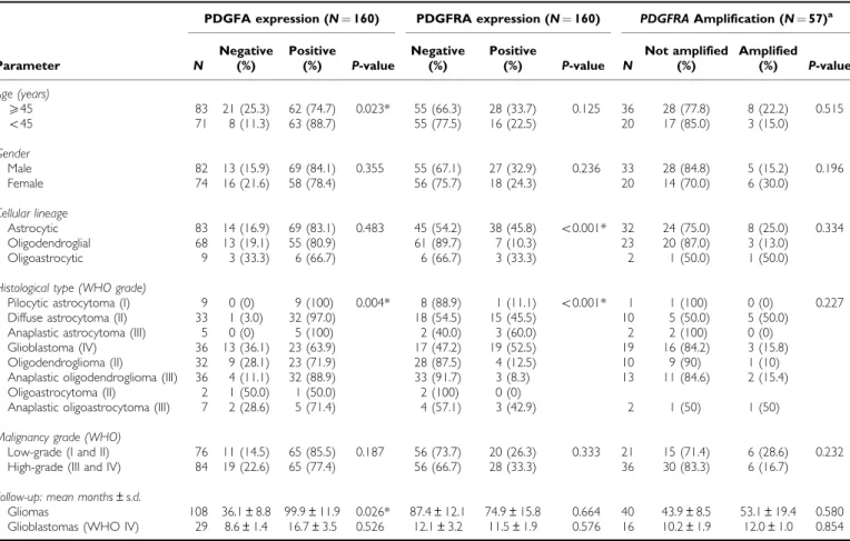
Table 1 PDGFA /PDGFRA expression andPDGFRAamplification in glioma patients and correlation with clinical – pathological data
PDGFA expression (N¼160) PDGFRA expression (N¼160) PDGFRAAmplification (N¼57)a
Parameter N
Negative (%)
Positive
(%) P-value
Negative (%)
Positive
(%) P-value N
Not amplified (%)
Amplified (%) P-value Age (years)
X45 83 21 (25.3) 62 (74.7) 0.023* 55 (66.3) 28 (33.7) 0.125 36 28 (77.8) 8 (22.2) 0.515
o45 71 8 (11.3) 63 (88.7) 55 (77.5) 16 (22.5) 20 17 (85.0) 3 (15.0)
Gender
Male 82 13 (15.9) 69 (84.1) 0.355 55 (67.1) 27 (32.9) 0.236 33 28 (84.8) 5 (15.2) 0.196 Female 74 16 (21.6) 58 (78.4) 56 (75.7) 18 (24.3) 20 14 (70.0) 6 (30.0)
Cellular lineage
Astrocytic 83 14 (16.9) 69 (83.1) 0.483 45 (54.2) 38 (45.8) o0.001* 32 24 (75.0) 8 (25.0) 0.334 Oligodendroglial 68 13 (19.1) 55 (80.9) 61 (89.7) 7 (10.3) 23 20 (87.0) 3 (13.0) Oligoastrocytic 9 3 (33.3) 6 (66.7) 6 (66.7) 3 (33.3) 2 1 (50.0) 1 (50.0)
Histological type (WHO grade)
Pilocytic astrocytoma (I) 9 0 (0) 9 (100) 0.004* 8 (88.9) 1 (11.1) o0.001* 1 1 (100) 0 (0) 0.227 Diffuse astrocytoma (II) 33 1 (3.0) 32 (97.0) 18 (54.5) 15 (45.5) 10 5 (50.0) 5 (50.0) Anaplastic astrocytoma (III) 5 0 (0) 5 (100) 2 (40.0) 3 (60.0) 2 2 (100) 0 (0) Glioblastoma (IV) 36 13 (36.1) 23 (63.9) 17 (47.2) 19 (52.5) 19 16 (84.2) 3 (15.8) Oligodendroglioma (II) 32 9 (28.1) 23 (71.9) 28 (87.5) 4 (12.5) 10 9 (90) 1 (10) Anaplastic oligodendroglioma (III) 36 4 (11.1) 32 (88.9) 33 (91.7) 3 (8.3) 13 11 (84.6) 2 (15.4) Oligoastrocytoma (II) 2 1 (50.0) 1 (50.0) 2 (100) 0 (0)
Anaplastic oligoastrocytoma (III) 7 2 (28.6) 5 (71.4) 4 (57.1) 3 (42.9) 2 1 (50) 1 (50)
Malignancy grade (WHO)
Low-grade (I and II) 76 11 (14.5) 65 (85.5) 0.187 56 (73.7) 20 (26.3) 0.333 21 15 (71.4) 6 (28.6) 0.232 High-grade (III and IV) 84 19 (22.6) 65 (77.4) 56 (66.7) 28 (33.3) 36 30 (83.3) 6 (16.7)
Follow-up: mean months±s.d.
Gliomas 108 36.1±8.8 99.9±11.9 0.026* 87.4±12.1 74.9±15.8 0.664 40 43.9±8.5 53.1±19.4 0.580 Glioblastomas (WHO IV) 29 8.6±1.4 16.7±3.5 0.526 12.1±3.2 11.5±1.9 0.576 16 10.2±1.9 12.0±1.0 0.854
aAssessed by QPCR; N¼number of cases; (*) Statistically significant values (P o0.05). 974
Molecular
Diagnostic
this study were completely anonymised after retrieval of follow up information.
PDGFA and PDGFRA immunohistochemistry
Representative 3-mm thick sections were cut from formalin-fixed
and paraffin-embedded samples and subjected to immunohisto-chemical analysis. Immunohistochemistry was carried out using a LabVision Autostainer (LabVision Corporation, Fremont, CA, USA) and the streptavidin—biotin – peroxidase complex techni-que, with rabbit polyclonal antibodies raised against human PDGFA (clone N-30, dilution 1:80; Santa Cruz Biotechnology, Santa Cruz, USA), and PDGFRA (dilution 1:175; LabVision Corporation) as previously described (Carvalhoet al, 2005; Reis et al, 2005). In brief, deparaffinised and rehydrated sections used to study PDGFA expression were pre-treated by microwaving in 10 mMcitrate buffer (pH 6.0) three times for 5 min at 600 W. The sections used for PDGFRA expression were submitted to heat-induced antigen retrieval with 10 mMcitrate buffer (pH 6.0) for 20 min in a water bath. After incubation of PDGFA and PDGFRA primary antibody at room temperature for 30 min, the secondary biotinylated goat anti-polyvalent antibody was applied for 10 min followed by incubation with streptavidin – peroxidase complex. The immune reaction was visualised by DAB as a chromogen (Ultravision Detection System Anti-polyvalent, HRP/DAB; LabVi-sion Corporation). Appropriated positive and negative controls were included in each run: for PDGFA and PDGFRA, cutaneous-mucosa transition of the anal region, namely medium calibre vessels with a muscular layer were used as positive controls. For negative controls, primary antibodies were omitted. All sections were counterstained with Gill-2 haematoxylin. As previously described (Reis et al, 2005), both the distribution and intense immunoreactivity were semi-quantitatively scored by JML and ALF independently with the observers blinded to the clinical information and results of the other molecular tests as follows: () (negative), (þ) (p5%), (þ þ) (5 – 50%), and (þ þ þ) (450%). Samples with scores () and (þ) were considered negative, and those with scores (þ þ) and (þ þ þ) were considered positive.
DNA isolation
Serial 10mm unstained section of paraffin blocks were cut, and one
adjacent haematoxylin and eosin-stained (H&E) section was taken for identification and selection of the tumour tissue. Selected areas containing at least 85% of tumour were marked and macro-scopically dissected using a sterile needle (Neolus, 25G – 0.5 mm). Tissue was placed into a microfuge tube and DNA isolation was performed using QIAamp DNA Micro Kit (Qiagen, Hilden, Germany) as previously described (Bastoet al, 2005).
PDGFRAmutations
Pre-screening for mutations in exons 12, 18 and 23 of thePDGFRA gene was carried out by PCR-single-strand conformational polymorphism (PCR – SSCP) followed by direct DNA sequencing of samples that showed a mobility shift in the PCR – SSCP analysis, as previously described (Reiset al, 2005). Briefly, PCR was carried out in a total volume of 25ml, consisting of 1 – 2ml of DNA solution,
0.5mM of both sense and anti-sense primers, 200mM of dNTPs (Fermentas Inc., Glen Burnie, MD, USA), 1.5 – 2 mM of MgCl2 (Bioron GmbH, Ludwigshafen, Germany), 1 Taq Buffer In-complete (Bioron GmbH) and 1U of Taq Superhot DNA Polymerase (Bioron GmbH). The reaction consisted of an initial denaturation at 961C for 10 min, followed by 40 cycles with denaturation at 961C for 45 s, annealing at 56 – 601C for 45 s and extension at 721C for 45 s, followed by a final extension for 10 min at 721C, in a Thermocycler (BioRad, Hercules, CA, USA). Primer sequences for exons 12 and 18 were previously reported (Reiset al,
2005), and for exon 23 were 50-GCTCTTCTCTCCCTCCTCCA-30 (sense) and 50
-TTTCTGAACGGGATCCAGAG-30
(antisense). PCR products were mixed with an equivalent volume of the denaturing loading buffer (98% formamide, 0.05% xylene cyanol and bromophenol blue). After denaturation at 981C for 10 min and quenching on ice, 20ml of the mixture were loaded onto a gel
containing 0.8 MDE (Cambrex Corporation, East Rutherford, NJ, USA) for exons 12 and 18 and 1MDE for exon 23, and 0 – 3% Glycerol (0% for exon 12 and 3% for exons 18 and 23). The gels were run for 20 h at 41C for exons 12 and 18 and 201C for exon 23. After the run, the gel was stained with Sybr Gold (Invitrogen Ltd., Paisley, UK) and visualised under ultraviolet light in a UV transilluminator.
Samples showing a mobility shift in the PCR – SSCP analysis different from the normal pattern were directly sequenced (Stab Vida, Investigation and Services in Biological Sciences Lda, Oeiras, Portugal) as previously described (Gomeset al, 2007). All positive cases were confirmed twice with a new and independent PCR amplification, followed by direct sequencing.
Analysis ofPDGFRAgene copy number status
Quantitative real-time PCR Quantitative real-time PCR (QPCR) was performed with LightCycler (Roche Molecular Biochemicals, Mannheim, Germany), using fluorescent hybridisation probes and fluorescence resonance energy transfer for the detection of PCR amplification product, following the manufacturer’s instructions. Briefly, primers and probes were designed to amplify a 124 bp (exon 18 from PDGFRAgene), and a 147 bp (18S gene) specific PCR product, where 18S was used as reference gene. PCR ampli-fication was performed in a 10ml reaction volume, under the
following conditions: 1 reaction master mix (Lightcycler FastStart DNA Master Hybridisation Probes kit, Roche Molecular Biochemicals); 0.2mM Probes (Roche Molecular Biochemicals); 0.5mMprimers; 4 mMMgCl2 (Roche Molecular Biochemicals) and 1ml (20 ngml1) of DNA. The reaction was initiated by a
denaturation step for 10 min at 951C, followed by 45 cycles with the following profile of amplification: incubation for 10 s at 941C, specific annealing temperature (571C for both genes) for 10 s and extension at 721C for an amplicon dependent time (7 s for 18S and 5 s forPDGFRA), immediately followed by a cooling step for 2 min at 401C. Primers and probes for 18S gene were previously described (Gomes et al, 2007), for PDGFRA were as follow:
50-TCAGCTACAGATGGCTTGATCC-30 (forward primer),
50-GCCAAAGTCACAGATCTTCACAAT-30 (reverse primer), 50 -TGTGTCCACCGTGATCTGGCTGC-FL (donator probe),
LC640-CGCAACGTCCTCCTGGCACAAGG-30
(acceptor probe). The PCR was performed in duplicate for each studied sample. A series of 10 normal DNA from healthy individuals was investigated to determine the confidence interval and the s.d. of the calculated ratios for reference and target gene. Evaluation of data was carried out using the DDCt method: DDCt¼DCt Tumour DNA – DCt Normal blood DNA. DCt (threshold cycles) is the Ct of the reference gene minus theCtof the target gene. Fold increase of the target genePDGFRAwas calculated by 2(DDCt)and values42 and
o5 were defined as aneuploidy and valuesX5 were considered as gene amplification.
Chromogenicin situhybridisation The presence ofPDGFRAgene amplification was also assessed by means of chromogenicin situ hybridisation (CISH) with an in-house generated probe made up with three contiguous, FISH-mapped and end-sequence verified bacterial artificial chromosomes (BACs) (626H04, RP11-231C18 and RP11-545H22), which map to thePDGFRAlocus on 4q12 according to Ensembl V39—June 2006 build of the genome (http://www.ensembl.org/Homo_sapiens/index.html). The in-house probe was generated, biotin-labelled and used in hybridisa-tions as previously described (Lambroset al, 2006; Gomeset al,
975
Molecular
2007). Briefly, heat pre-treatment of deparaffinised sections were incubated for 15 min at 981C in CISH pre-treatment buffer (SPOT-light tissue pre-treatment kit, Zymed Laboratories, San Francisco, CA, USA) and digested with pepsin for 6 min at room temperature according to the manufacturer’s instructions. CISH experiments were analysed by three of the authors on a multi-headed microscope. Only unequivocal signals were counted. Signals were evaluated at 400 and630 and 60 morphologically unequivocal neoplastic cells were counted for the presence of the gene probe signals. Amplification was defined as 45 signals per nucleus in more than 50% of tumour cells, or when large gene copy clusters were seen (Lambros et al, 2006; Gomes et al, 2007). CISH hybridisations were evaluated with observers blinded to the clinical information and results of immunohistochemical and QPCR analysis.
Microarray-based comparative genomic hybridisation The aCGH platform used for this study was constructed at the Breakthrough Breast Cancer Research Centre and comprises416 000 BAC clones tiled across the genome (Arriola et al, 2007). This type of BAC array platform has been shown to be as robust as high-density oligonucleotide arrays (Coeet al, 2007; Gunnarssonet al, 2008) and its actual resolution is approximately 100 kb for498% of the genome (Arriolaet al, 2007, 2008; Marchio`et al, 2008a). Labelling, hybridisation, washes, image acquisition and data normalisation were carried out as previously described (Arriolaet al, 2007, 2008; Huanget al, 2007; Marchio`et al, 2008a, 2008b). Polymorphic BACs identified in an analysis of 50 male/female and female/female hybridisations were filtered out. This left a final dataset of 13711 clones with unambiguous mapping information according to the March 2006 build (hg18) of the human genome (http://www. ensembl.org). Data were smoothed using the adaptive weight smoothing (aws) algorithm (Mackay et al, 2009). A categorical analysis was applied to the BACs after classifying them as representing gain, loss, or no-change according to their smoothed Log2 ratio values Threshold values were chosen to correspond to
three s.d. of the normal ratios obtained from the filtered clones mapping to chromosomes 1 – 22, assessed in multiple hybridisa-tions between DNA extracted from a pool of male and female blood donors as previously described (Arriola et al, 2008; Reis-Filhoet al, 2008) (Log2 ratio of±0.08). Low level gain was defined as a smoothed Log2 ratio of between 0.12 and 0.40, corresponding to approximately 3 – 5 copies of the locus, whereas gene amplification was defined as having a Log2 ratio 40.40, corresponding to more than 5 copies (Arriolaet al, 2008). These figures were obtained by comparison with interphase FISH data for markers at different chromosomal locations (Reis-Filho et al, 2008). Data processing and analysis were carried out in R 2.0.1 (http://www.r-project.org/) and BioConductor 1.5 (http://www. bioconductor.org/), making extensive use of modified versions of the packages aCGH, marray and aws in particular.
Statistical analysis
Correlations between categorical variables were performed using thew2-test. Cumulative survival probabilities were calculated using
the Kaplan – Meier method. Differences between survival rates were tested with the log-rank test. Two-tailed P-values o0.05 were considered significant.All statistical analysis was performed using SPSS software for Windows, version 14.0.
RESULTS
PDGFA and PDGFRA expression
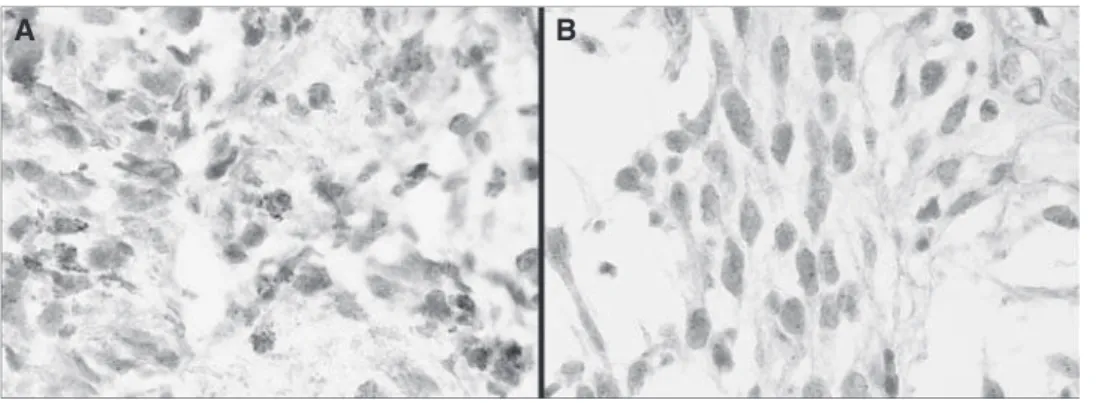
PDGFA and PDGFRA expression was found in 81.2% (130 out of 160) and 29.6% (48 out of 160) of gliomas, respectively. Immunohistochemical analysis showed that tumour cells express PDGFA in the cytoplasm (Figure 1A), whereas PDGFRA was preferentially observed in the cytoplasmic compartment and rarely on membranes (Figure 1B). PDGFA expression was also observed in tumour-associated endothelial cells and in basal membrane of
Figure 1 Immunohistochemistry analysis of PDGFA and PDGFRA in gliomas; (A) Glioblastoma with (þ þ þ) score for PDGFA expression (200);
(B) Glioblastoma with (þ þ þ) score for PDGFRA expression (200). (C) Glioblastoma with () score for PDGFA expression in tumour cells and positive in endothelial cells (200). (D) Glioblastoma with (þ) score for PDGFRA expression (200).
976
Molecular
Diagnostic
blood vessels in approximately 12% (19 out of 160) of the cases (Figure 1C). Representative PDGFRA-negative staining is shown in Figure 1D. Twenty five percent (40 out of 160) of gliomas coexpressed PDGFA and their receptor PDGFRA; however, no statistically significant association between the expression of the ligand and its receptor was observed (P40.05). When the presence or absence of PDGFA/PDGFRA overexpression (þ þ þ) was defined according to histological type, a trend for PDGFA and PDGFRA coexpression in glioblastomas was found (P¼0.069).
PDGFA and PDGFRA expression was significantly correlated with histological type (P¼0.004 and 0.0001, respectively) (Table 1). When tumours were classified into the three major groups according to their histogenesis (astrocytic, oligodendroglial and mixed), only PDGFRA expression was statistically significantly correlated with astrocytic lineage (Po0.001) (Table 1).
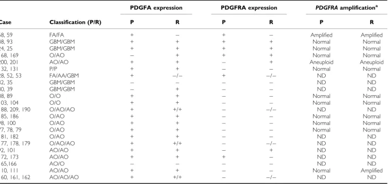
In 22 cases, it was possible to analyse PDGFA and PDGFRA expression in both primary and recurrent tumours (Table 2). Overall, the results were concordant; however, we observed loss or gain of PDGFA and PDGFRA in recurrent tumours of some patients (Table 2).
No statistically significant correlations were found between PDGFRA expression and clinical – pathological parameters includ-ing age, gender, WHO histological grade and prognosis (Table 1). However, the absence of PDGFA expression was significantly associated (P¼0.023) with age (445 years) and with a poor prognosis in glioma patients (P¼0.026) (Table 1 and Figure 2).
PDGFRAgene mutations
PCR – SSCP analysis forPDGFRAgene mutations in exons 12, 18 and 23 produced optimal results in 86 cases, 30 of which were PDGFRA-positive tumours. No activating mutations were found. However, four silent mutations and an intronic insertion were identified in 45.3% (39 out of 86) of glioma patients (Table 3). Five patients showed the simultaneous presence of two different mutations. No association was found between the presence of PDGFRAgene mutations and PDGFRA expression (P40.05).
PDGFRAgene amplification
Analysis ofPDGFRAgene copy number status as defined by QPCR was successfully performed in 57 gliomas.PDGFRAcopy number changes (ratio 42) were observed in 52.6% (30 out of 57) of glioma patients: 18 displayed ratios o5 and were considered representative of aneuploidy/aneusomy and 12 (21.1%) harboured
Table 2 PDGFA /PDGFRA expression andPDGFRAamplification in glioma patients with recurrences
PDGFA expression PDGFRA expression PDGFRAamplificationa
Case Classification (P/R) P R P R P R
58, 59 FA/FA + + Amplified Amplified
48, 93 GBM/GBM + + + + Normal Normal
24, 25 GBM/GBM + + + + Normal Normal
168, 169 O/AO + + + Normal Normal
200, 201 AO/AO + + + Aneuploid Aneuploid
132, 131 P/P + + Normal Normal
28, 52, 53 FA/AA/GBM + / + / ND ND
32, 35 GBM/GBM ND ND
30, 39 GBM/GBM + ND ND
88, 89 O/O + + Normal Normal
103, 104 O/O + + Normal Normal
188, 209, 190 O/AO/AO + +/+ / ND ND
185, 186 O/AO + + Normal Normal
98, 100 O/AO + + Normal Normal
77, 78, 79 O/AO + + Normal Normal
181, 182 O/AO + + ND ND
177, 178, 179 O/AO/AO + +/+ / ND ND
92, 101 AO/AO + + + ND ND
172, 173 AO/AO + + + ND ND
165,166 AO/O ND ND
110, 111 AO/AO + + Normal Amplified
160, 161, 162 AO/AO/AO + +/+ / ND ND
aAssessed by QPCR; P¼Primary tumour; R¼Recurrent tumour; P¼Pilocytic astrocytoma (grade I); FA¼fibrilar astrocytoma (grade II); AA¼anaplastic astrocytoma (grade III);
GBM¼glioblastoma (grade IV); O¼oligodendroglioma (grade II); AO¼anaplastic oligodendroglioma (grade III); ()¼Negative expression (score 0 and +); (+)¼Positive expression (score ++ and +++); ND¼not done.
Cum
ulativ
e sur
viv
al
1.0
0.8
0.6
0.4
0.2
0.0
0.00 50.00 100.00 150.00 200.00 250.00
P= 0.026
Follow-up (months) PDGFA-negative gliomas
PDGFA-positive gliomas
Figure 2 Kaplan – Meier curve illustrating the impact of PDGFA
expression on overall survival (months) of glioma patients.
977
Molecular
ratiosX5 and were considered amplified (Table 1). No statistically significant associations were found between thePDGFRA ampli-fication and PDGFRA expression. However, a borderline associa-tion (P¼0.058) was observed between amplification and overexpression (þ þ þ). In addition, a statistically significant association (P¼0.038) was found betweenPDGFRAamplification and overexpression in grade II diffuse astrocytomas (Table 4). After statistical analysis, no significant correlations were found between thePDGFRAgene amplification and clinical – pathological features (P40.05) (Table 1).
In 12 cases, data on PDGFRAamplification status in both the primary and recurrent tumours were available (Table 2). In all but one case (tumours 110 and 111) primary and recurrent tumours displayed identical PDGFRA copy number status. Interestingly acquisition of PDGFRA amplification was observed in the recurrent tumour 111; however, this was not associated with expression of PDGFRA.
To validate QPCR results we performed CISH in six cases, three with and three without PDGFRA amplification as defined by QPCR. CISH analysis of cases defined as harbouringPDGFRAgene amplification by QPCR revealed clusters ofPDGFRAsignals in the nuclei of neoplastic cells (Figure 3A), confirming gene amplifica-tion. In normal cases, only one-to-two PDGFRAgene signals per nucleus were found in neoplastic cells (Figure 3B). To further validate the results of the QPCR analysis, we performed micro-array-based comparative genomic hybridisation in two cases, one with and one withoutPDGFRAamplification as defined by QPCR. The case withoutPDGFRAgene amplification by QPCR showed no changes on chromosome 4 (Figure 4A). In the case with amplification, the peak of the amplicon on 4q was restricted to 4q12, encompassing a genomic region of 3.7 Mb (Figure 4B), flanked by the BACs RP11-654K2 (54025,329 kb) and RP11-284L3
(57933,681 kb), including the genes: LNX1, CHIC2, GSH2, PDGFRA, KIT, KDR, SRD5A2 L, TMEM165, CLOCK, PDCL2, NMU, EXOC1, CEP135, AASDH, PPAT, PAICS, SRP72, ARL9, HOP, SPINK2, REST, C4orf14, POLR2B, and IGFBP7. Taken together, the above CISH and aCGH findings provide a robust validation for the results obtained with QPCR.
DISCUSSION
The PDGF pathway is one of the most consistently altered cellular signalling system in glial tumourigenesis (Shih and Holland, 2006; Fomchenko and Holland, 2007). PDGF and PDGFRs have both been found to be overexpressed in glial tumour cell lines and tumour surgical samples (Nisteret al, 1988, 1991; Hermansonet al, 1992; Black et al, 1996; Di Roccoet al, 1998; Varelaet al, 2004). PDGFRA and PDGFA have been shown to be expressed in tumour cells, whereas PDGFB and PDGFRB have been found in glioma-associated endothelial cells (Hermanssonet al, 1988; Hermanson et al, 1992; Plateet al, 1992). Furthermore, studies on the PDGFC and D ligands, also demonstrate their expression in gliomas; however, the clinical and biological significance of their expression remain to be determined (Lokker et al, 2002). Importantly, the function of PDGF signalling in gliomagenesis has been enlightened by its potential role in cancer stem cell hypothesis of gliomagen-esis. It has been suggested that activation of PDGFRA signalling, directly or indirectly through creating a favourable microenviron-ment niche, can contribute to the transformation of neural stem/ progenitor cells into glioma tumours (Jackson et al, 2006; Fomchenko and Holland, 2007; Siebzehnrublet al, 2008).
Clinical trials evaluating the efficacy of anti-PDGFRA drugs in patients with glioblastomas are ongoing (Reardon et al, 2005;
Table 3 Sequence variants ofPDGFRAgene in glioma patients
Exon Nucleotide change Amino-acid substitution No of cases dbSNP
Exon 12
1686 T4C I562I 1 Not yet described
1701 G4A P567P 6 rs1873778
1777 C4T L593 L 1 Not yet described
Exon 18
2472 C4T V824V 21 rs2228230
2449_50insA IVS18-50insA 15 rs3830355
dbSNP¼single nucleotide polymorphism database (http://www.ncbi.nlm.nih.gov/SNP/).
Table 4 Correlation between PDGFRA overexpression andPDGFRAamplification in gliomas
PDGFRAamplificationa
Histological type (WHO grade) N PDGFRA overexpression Not amplified (%) Amplified (%) P-value
Pilocytic astrocytoma (I) 1 Negative 1 (100) 0 (0) NP
overexpression 0 (0) 0 (0)
Diffuse astrocytoma (II) 10 Negative 5 (100) 2 (40.0) 0.038*
overexpression 0 (0) 3 (60.0)
Anaplastic astrocytoma (III) 2 Negative 2 (100) 0 (0) NP
overexpression 0 (0) 0 (0)
Glioblastoma (IV) 19 Negative 4 (25.0) 1 (33.3) 0.624
overexpression 12 (75.0) 2 (66.7)
Oligodendroglioma (II) 10 Negative 8 (88.9) 1 (100) 0.900
overexpression 1 (11.1) 0 (0)
Anaplastic oligodendroglioma (III) 13 Negative 11 (100) 1 (50) 0.154
overexpression 0 (0) 1 (50)
Anaplastic oligoastrocytoma (III) 2 Negative 1 (100) 0 (0) 0.500
overexpression 0 (0) 1 (100)
aAssessed by QPCR; Negative expression¼scores 0, + and ++; Overexpression¼score +++; (*) Statistically significant values (Po0.05); NP¼not possible. 978
Molecular
Diagnostic
Wenet al, 2006; Desjardinset al, 2007; Newton, 2007; Raymond et al, 2008). Despite the positive response, none of the new targeted therapies has shown significant clinical activity as a single agent in phase II studies (Brandsma and van den Bent, 2007; Raymond et al, 2008). Currently, combination of anti-PDGFRA drugs with chemotherapy agents is being evaluated (Reardon et al, 2008). However, the molecular alterations underlying glioma patients’ response to PDGFRA antagonists are unknown. One study analysed the prevalence ofPDGFRAmutations in patients enrolled in a phase I/II study of imatinib mesylate in recurrent malignant gliomas; however, no activating mutations were observed
(Wen et al, 2006). The in vitro studies using PDGFR-targeted drugs (e.g., imatinib) have provided conflicting information about the molecular underpinning of sensitivity to those agents. Although some suggested that sensitivity to targeted agents is associated with overall PDGFR activation; others point to the putative role of PDGFRB or failed to show any association between PDGFR status and response to imatinib (Gross et al, 2006; Ha¨gerstrandet al, 2006; Servideiet al, 2006).
Here, we observed PDGFA expression in 81.2% of gliomas. Overall, PDGFA was highly expressed in all histological types of gliomas. Previous studies on PDGFA mRNA expression reported
Figure 3 CISH analysis ofPDGFRAin a glioblastoma (A) withPDGFRAamplification (600, no HE counterstaining) and other (B) withoutPDFRA
amplification (600, no HE counterstaining). The colour reproduction of this figure is available on the html full text version of the article.
Log2 ratio
p16.3 p16.1 p15.33 p15.31 p15.2 p15.1 p14 p13 p12
q12 PDGFRA q13.1
q13.2 q13.3
q21.21
q22.1 q22.3
q24 q25 q26
q28.1
q28.3
q31.21
q31.3 q32.1
q32.3 q34.1 q34.3 q35.1 q35.2 – 2.0 – 1.5 – 1.0 – 0.5 0.0 0.5 1.0 1.5 2.0
Log2 ratio
– 2.0 – 1.5 – 1.0 – 0.5 0.0 0.5 1.0 1.5 2.0
Chromosome 4 (Mb)
0
20
40
60
80
100
120
140
160
180
Chromosome 4 (Mb)
0
20
40
60
80
100
120
140
160
180
A B
Figure 4 Ideogram and microarray CGH chromosome plots of chromosome 4, in whichPDGFRAgene is located, for a case without (A) and other with
(B) PDGFRA amplification as defined by QPCR. Log2ratios are plotted on thexaxis against each clone according to genomic location on theyaxis. The centromere is represented by a horizontal dotted line. Vertical dashed lines correspond to log2ratios of 0.12 (green) and0.12 (red). Grey dots: Log2 ratios; Blue dots: aws-smoothed Log2ratios. The colour reproduction of this figure is available on the html full text version of the article.
979
Molecular
high levels of PDGFA in gliomas (Maxwellet al, 1990; Mapstone, 1991; Hermansonet al, 1992; Di Roccoet al, 1998). It should be noted, however, that there is a paucity of data on prevalence of PDGFA protein expression in primary glioma specimens. We have previously shown that 100% of gliosarcomas express PDGFA (Reis et al, 2005). PDGFRA expression was detected in 29.6% of gliomas, and more frequently expressed in 45 – 60% of malignant astrocytic tumours. These frequencies are in agreement with previous studies, where approximately 50% for malignant astrocytomas were reported to express this receptor (Nister et al, 1988, 1991; Hermanson et al, 1992; Black et al, 1996; Di Rocco et al, 1998; Ribomet al, 2002; Varelaet al, 2004; Lianget al, 2008; Takeiet al, 2008; Thorarinsdottir et al, 2008). A previous study showed PDGFRA expression in approximately 50% of gliomas and a correlation with poor prognosis in low-grade gliomas (Varelaet al, 2004). However, in a report of 40 patients with grade II astrocytomas and oligoastrocytomas, there was an association between high PDGFRA expression and a favourable patient outcome (Ribom et al, 2002). Recently, Liang et al, (2008) in a paediatric high-grade glioma series failed to find any significant impact of PDGFRA expression on survival. In our series, PDGFRA expression was not correlated with patients’ survival. Interestingly, we found that the absence of PDGFA expression is significantly associated with age and poor prognosis in patients with glioma. Given the retrospective nature of our study, further analysis of the prognostic impact of PDGFA and PDGFRA expression in gliomas is warranted.
Overexpression of RTKs in cancer has been shown to be driven by underlying genetic events in a substantial proportion of cases (Gschwindet al, 2004). For instance, KIT overexpression in GISTs is driven by activatingKITmutations (Gomeset al, 2008), whereas HER2 overexpression in breast cancer is driven by HER2 gene amplification (Arriola et al, 2008). Here, we investigated the prevalence of PDGFRA-activating mutations and gene amplifica-tion in gliomas. In agreement with previous studies (Hartmann et al, 2004; Randet al, 2005; Reiset al, 2005; Sihtoet al, 2005; Wen et al, 2006; McLendon et al, 2008; Parsons et al, 2008), no PDGFRA-activating mutations were found. However, four silent mutations and an intronic insertion were identified. Apart from two silent mutations in PDGFRA exon 12, the other mutations have been previously described and considered to be genetic poly-morphisms (http://www.ncbi.nlm.nih.gov/projects/SNP/; Carvalho et al, 2005; Reiset al, 2005; Wenet al, 2006). The impact of these genetic polymorphisms in PDGFRA function remains to be elucidated.
PDGFRAgene amplification analysis revealed PDGFRA ampli-fication in 21.1% (12 out of 57) of gliomas, a frequency similar to that described in previous studies (Fleming et al, 1992; Kumabe
et al, 1992; Smithet al, 2000; Alonsoet al, 2005; Arjonaet al, 2005; Puputtiet al, 2006; Beroukhimet al, 2007; McLendonet al, 2008). We have further shown by aCGH that the amplicon encompasses a region of 3.6 Mb, which, in addition toPDGFRA, also includesKIT and KDR oncogenes. Co-amplification of these three oncogenes has already been detected with other methodologies in gliomas (Joensuuet al, 2005; Puputtiet al, 2006; Holtkampet al, 2007). A statistically significant association betweenPDGFRAamplification and overexpression was found only in diffuse astrocytomas (grade II). Given that PDGFRA overexpression appears to be an early event in gliomagenesis (Hermanson et al, 1996), our results provide support to the contention that gene amplification may be one of the underlying mechanisms at this stage. In a way akin to other oncogenes, such as EGFR, overexpression of PDGFRA was more pervasive than gene amplification. It should be noted, however, that there wereB42% of cases withPDGFRA amplifica-tion that lacked PDGFRA protein expression, suggesting that in some cases the target of 4q12 amplification may be a gene other thanPDGFRA.
In conclusion, here we show that PDGFA is expressed in different types of gliomas and its absence is associated with a poor prognosis. PDGFRA is significantly highly expressed in malignant astrocytic tumours. Based on the concurrent expression of PDGFA and PDGFRA in glioblastomas, it could be hypothesised that autocrine/paracrine loops may be present in these tumours, corroborating the importance of this signalling pathway in gliomas (Nister et al, 1991; Guha et al, 1995; Fomchenko and Holland, 2007).PDGFRAgene amplification may be the underlying genetic mechanism driving PDGFRA overexpression in gliomas. However, B42% of cases with amplification of PDGFRA did not display PDGFRA protein expression, suggesting that a gene other than PDGFRAmay be the driver of this amplicon. Further studies are needed to correlate these molecular alterations and response to anti-PDGFRA drugs and to investigate the alternative drivers of the 4q12 amplicon. However, our results provide a step forward in the identification of a molecular basis for tailoring the therapies for specific subgroups of glioma patients.
ACKNOWLEDGEMENTS
OM is a recipient of a PhD fellowship (SFRH/BD/36463/2007) from Fundac¸a˜o para a Cieˆncia e a Tecnologia (FCT), Portugal. JSRF, MBKL, AM, NT, KF and AA are funded by Breakthrough Breast Cancer. This study was supported in part by Pfizer/Sociedade de Cieˆncias Me´dicas de Lisboa with the prize: ‘Research in oncology diseases, Professor Francisco Gentil’ and Breakthrough Breast Cancer.
REFERENCES
Alonso ME, Bello MJ, Arjona D, Martinez-Glez V, de Campos JM, Isla A, Kusak E, Vaquero J, Gutierrez M, Sarasa JL, Rey JA (2005) Real-time quantitative PCR analysis of gene dosages reveals gene
amplification in low-grade oligodendrogliomas.Am J Clin Pathol123:
900 – 906
Arjona D, Bello MJ, Alonso ME, Isla A, de Campos JM, Vaquero J, Sarasa JL, Gutierrez M, Rey JA (2005) Real-time quantitative PCR analysis of regions involved in gene amplification reveals gene overdose in
low-grade astrocytic gliomas.Diagn Mol Pathol14:224 – 229
Arriola E, Lambros MBK, Jones C, Dexter T, Mackay A, Tan DSP, Tamber N, Fenwick K, Ashworth A, Dowsett M, Reis JS (2007) Evaluation of Phi29-based whole-genome amplification for microarray-based
com-parative genomic hybridisation.Lab Invest87:75 – 83
Arriola E, Marchio` C, Tan DS, Drury SC, Lambros MB, Natrajan R, Rodriguez-Pinilla SM, Mackay A, Tamber N, Fenwick K, Jones C, Dowsett M, Ashworth A, Reis-Filho JS (2008) Genomic analysis of the
HER2/TOP2A amplicon in breast cancer and breast cancer cell lines.Lab
Invest88:491 – 503
Basto D, Trovisco V, Lopes JM, Martins A, Pardal F, Soares P, Reis RM
(2005) Mutation analysis of B-RAF gene in human gliomas. Acta
Neuropathol (Berlin)109:207 – 210
Beroukhim R, Getz G, Nghiemphu L, Barretina J, Hsueh T, Linhart D, Vivanco I, Lee JC, Huang JH, Alexander S, Du J, Kau T, Thomas RK, Shah K, Soto H, Perner S, Prensner J, DeBiasi RM, Demichelis F, Hatton C, Rubin MA, Garraway LA, Nelson SF, Liau L, Mischel PS, Cloughesy TF, Meyerson M, Golub TA, Lander ES, Mellinghoff IK, Sellers WR (2007) Assessing the significance of chromosomal aberrations in cancer:
methodology and application to glioma.Proc Natl Acad Sci USA104:
20007 – 20012
Black P, Carroll R, Glowacka D (1996) Expression of platelet-derived growth factor transcripts in medulloblastomas and ependymomas.
Pediatr Neurosurg24(2):74 – 78
980
Molecular
Diagnostic
Blume-Jensen P, Hunter T (2001) Oncogenic kinase signalling.Nature411: 355 – 365
Brandsma D, van den Bent MJ (2007) Molecular targeted therapies and
chemotherapy in malignant gliomas.Curr Opin Oncol19:598 – 605
Capdeville R, Buchdunger E, Zimmermann J, Matter A (2002) Glivec
(STI571, imatinib), a rationally developed, targeted anticancer drug.Nat
Rev Drug Discov1:493 – 502
Carvalho I, Milanezi F, Martins A, Reis RM, Schmitt F (2005) Over-expression of platelet-derived growth factor receptor alpha in breast
cancer is associated with tumour progression. Breast Cancer Res 7:
R788 – R795
Coe BP, Ylstra B, Carvalho B, Meijer GA, Macaulay C, Lam WL (2007)
Resolving the resolution of array CGH.Genomics89:647 – 653
Demetri GD, von Mehren M, Blanke CD, Van den Abbeele AD, Eisenberg B, Roberts PJ, Heinrich MC, Tuveson DA, Singer S, Janicek M, Fletcher JA, Silverman SG, Silberman SL, Capdeville R, Kiese B, Peng B, Dimitrijevic S, Druker BJ, Corless C, Fletcher CD, Joensuu H (2002) Efficacy and safety of imatinib mesylate in advanced gastrointestinal stromal tumors.
N Engl J Med347:472 – 480
Desjardins A, Quinn JA, Vredenburgh JJ, Sathornsumetee S, Friedman AH, Herndon JE, McLendon RE, Provenzale JM, Rich JN, Sampson JH, Gururangan S, Dowell JM, Salvado A, Friedman HS, Reardon DA (2007) Phase II study of imatinib mesylate and hydroxyurea for recurrent grade
III malignant gliomas.J Neuro Oncol83:53 – 60
Di Rocco F, Carroll RS, Zhang J, Black PM (1998) Platelet-derived growth factor and its receptor expression in human oligodendrogliomas.
Neurosurgery42:341 – 346
Druker BJ, Sawyers CL, Kantarjian H, Resta DJ, Reese SF, Ford JM, Capdeville R, Talpaz M (2001) Activity of a specific inhibitor of the BCR-ABL tyrosine kinase in the blast crisis of chronic myeloid leukemia and
acute lymphoblastic leukemia with the Philadelphia chromosome.N Engl
J Med344:1038 – 1042
Fleming TP, Saxena A, Clark WC, Robertson JT, Oldfield EH, Aaronson SA, Ali IU (1992) Amplification and/or overexpression of platelet-derived growth factor receptors and epidermal growth factor receptor in human
glial tumors.Cancer Res52:4550 – 4553
Fomchenko EI, Holland EC (2007) Platelet-derived growth factor-mediated
gliomagenesis and brain tumor recruitment.Neurosurg Clin N Am18:
39 – 58
Gomes AL, Gouveia A, Capelinha AF, de la CD, Silva P, Reis RM, Pimenta A, Lopes JM (2008) Molecular alterations of KIT and PDGFRA in GISTs:
evaluation of a Portuguese series.J Clin Pathol61:203 – 208
Gomes AL, Reis-Filho JS, Lopes JM, Martinho O, Lambros MB, Martins A, Schmitt F, Pardal F, Reis RM (2007) Molecular alterations of KIT
oncogene in gliomas.Cell Oncol29:399 – 408
Gross D, Bernhardt G, Buschauer A (2006) Platelet-derived growth factor receptor independent proliferation of human glioblastoma cells: selective
tyrosine kinase inhibitors lack antiproliferative activity.J Cancer Res Clin
Oncol132:589 – 599
Gschwind A, Fischer OM, Ullrich A (2004) Timeline—The discovery of
receptor tyrosine kinases: targets for cancer therapy.Nat Rev Cancer4:
361 – 370
Guha A, Dashner K, Black PM, Wagner JA, Stiles CD (1995) Expression of
PDGF and PDGF receptors in human astrocytoma operation
specimens supports the existence of an autocrine loop.Int J Cancer60:
168 – 173
Gunnarsson R, Staaf J, Jansson M, Ottesen AM, Goransson H, Liljedahl U, Ralfkiaer U, Mansouri M, Buhl AM, Smedby KE, Hjalgrim H, Syvanen AC, Borg A, Isaksson A, Jurlander J, Juliusson G, Rosenquist R (2008) Screening for copy-number alterations and loss of heterozygosity in chronic lymphocytic leukemia–a comparative study of four differently
designed, high resolution microarray platforms. Genes Chromosomes
Cancer47:697 – 711
Ha¨gerstrand D, Hesselager G, Achterberg S, Wickenberg Bolin U, Kowanetz M, Kastemar M, Heldin CH, Isaksson A, Niste´r M, Ostman A (2006) Characterization of an imatinib-sensitive subset of high-grade human
glioma cultures.Oncogene25:4913 – 4922
Hartmann C, Xu X, Bartels G, Holtkamp N, Gonzales IA, Tallen G, von
Deimling A (2004) Pdgfr-alpha in 1p/19q LOH oligodendrogliomas.Int J
Cancer112:1081 – 1082
Hermanson M, Funa K, Hartman M, Claesson Welsh L, Heldin CH, Westermark B, Nister M (1992) Platelet-derived growth factor and its receptors in human glioma tissue: expression of messenger RNA and
protein suggests the presence of autocrine and paracrine loops.Cancer
Res52:3213 – 3219
Hermanson M, Funa K, Koopmann J, Maintz D, Waha A, Westermark B, Heldin CH, Wiestler OD, Louis DN, von Deimling A, Nister M (1996) Association of loss of heterozygosity on chromosome 17p with high platelet-derived growth factor alpha receptor expression in human
malignant gliomas.Cancer Res56:164 – 171
Hermansson M, Nister M, Betsholtz C, Heldin CH, Westermark B, Funa K (1988) Endothelial cell hyperplasia in human glioblastoma: coexpression of mRNA for platelet-derived growth factor (PDGF) B chain and PDGF
receptor suggests autocrine growth stimulation.Proc Natl Acad Sci USA
85:7748 – 7752
Holtkamp N, Ziegenhagen N, Malzer E, Hartmann C, Giese A, von Deimling A (2007) Characterization of the amplicon on chromosomal segment
4q12 in glioblastoma multiforme.Neuro Oncol9:291 – 297
Huang J, Gusnanto A, O0
Sullivan K, Staaf J, Borg A, Pawitan Y (2007) Robust smooth segmentation approach for array CGH data analysis.
Bioinformatics23:2463 – 2469
Jackson EL, Garcia-Verdugo JM, Gil-Perotin S, Roy M,
Quinones-Hinojosa A, VandenBerg S, Alvarez-Buylla A (2006) PDGFR alpha-positive B cells are neural stem cells in the adult SVZ that form
glioma-like growths in response to increased PDGF signaling.Neuron51:
187 – 199
Joensuu H, Puputti M, Sihto H, Tynninen O, Nupponen NN (2005) Amplification of genes encoding KIT, PDGFRalpha and VEGFR2 receptor tyrosine kinases is frequent in glioblastoma multiforme.
J Pathol207:224 – 231
Kumabe T, Sohma Y, Kayama T, Yoshimoto T, Yamamoto T (1992) Amplification of alpha-platelet-derived growth factor receptor gene lacking an exon coding for a portion of the extracellular region in a
primary brain tumor of glial origin.Oncogene7:627 – 633
Lacroix M, Abi-Said D, Fourney DR, Gokaslan ZL, Shi W, DeMonte F, Lang FF, McCutcheon IE, Hassenbusch SJ, Holland E, Hess K, Michael C, Miller D, Sawaya R (2001) A multivariate analysis of 416 patients with glioblastoma multiforme: prognosis, extent of resection, and survival.
J Neurosurg95:190 – 198
Lambros MB, Simpson PT, Jones C, Natrajan R, Westbury C, Steele D, Savage K, Mackay A, Schmitt FC, Ashworth A, Reis-Filho JS (2006) Unlocking pathology archives for molecular genetic studies: a reliable
method to generate probes for chromogenic and fluorescent in situ
hybridization.Lab Invest86:398 – 408
Liang ML, Ma J, Ho M, Solomon L, Bouffet E, Rutka JT, Hawkins C (2008)
Tyrosine kinase expression in pediatric high grade astrocytoma.J Neuro
Oncol87:247 – 253
Lokker NA, Sullivan CM, Hollenbach SJ, Israel MA, Giese NA (2002) Platelet-derived growth factor (PDGF) autocrine signaling regulates survival and mitogenic pathways in glioblastoma cells: evidence that the novel PDGF-C and PDGF-D ligands may play a role in the development
of brain tumors.Cancer Res62:3729 – 3735
Louis DN (2006) Molecular pathology of malignant gliomas. Annu Rev
Pathol1:97 – 117
Louis DN, Ohgaki H, Wiestler OD, Cavenee WK, Burger PC, Jouvet A, Scheithauer BW, Kleihues P (2007) The 2007 WHO classification
of tumours of the central nervous system.Acta Neuropathologica114:
97 – 109
Mapstone TB (1991) Expression of platelet-derived growth factor and transforming growth factor and their correlation with cellular
morphol-ogy in glial tumors.J Neurosurg75:447 – 451
Marchio` C, Iravani M, Natrajan R, Lambros MB, Savage K, Tamber N, Fenwick K, Mackay A, Senetta R, Di PS, Schmitt FC, Bussolati G, Ellis LO, Ashworth A, Sapino A, Reis-Filho JS (2008a) Genomic and immuno-phenotypical characterization of pure micropapillary carcinomas of the
breast.J Pathol215:398 – 410
Marchio` C, Natrajan R, Shiu KK, Lambros MB, Rodriguez-Pinilla SM, Tan DS, Lord CJ, Hungermann D, Fenwick K, Tamber N, Mackay A, Palacios J, Sapino A, Buerger H, Ashworth A, Reis-Filho JS (2008b) The genomic profile of HER2-amplified breast cancers: the influence of ER status.
J Pathol216:399 – 407
Mackay A, Tamber N, Fenwick K, Iravani M, Grigoriadis A, Dexter T, Lord CJ, Reis-Filho JS, Ashworth A (2009) A high-resolution integrated analysis of genetic and expression profiles of breast cancer cell lines.
Breast Cancer Res Treat(e-pub ahead of print 24 January 2009)
Maxwell M, Naber SP, Wolfe HJ, Galanopoulos T, Hedley-Whyte ET, Black PM, Antoniades HN (1990) Coexpression of platelet-derived growth factor (PDGF) and PDGF-receptor genes by primary human
astro-cytomas may contribute to their development and maintenance.J Clin
Invest86:131 – 140
981
Molecular
McLendon R, Friedman A, Bigner D, Van Meir EG, Brat DJ, Mastrogianakis GM, Olson JJ, Mikkelsen T, Lehman N, Aldape K, Yung WK, Bogler O, Weinstein JN, VandenBerg S, Berger M, Prados M, Muzny D, Morgan M, Scherer S, Sabo A, Nazareth L, Lewis L, Hall O, Zhu Y, Ren Y, Alvi O, Yao J, Hawes A, Jhangiani S, Fowler G, San Lucas A, Kovar C, Cree A, Dinh H, Santibanez J, Joshi V, Gonzalez-Garay ML, Miller CA, Milosavljevic A, Donehower L, Wheeler DA, Gibbs RA, Cibulskis K, Sougnez C, Fennell T, Mahan S, Wilkinson J, Ziaugra L, Onofrio R, Bloom T, Nicol R, Ardlie K, Baldwin J, Gabriel S, Lander ES, Ding L, Fulton RS, McLellan MD, Wallis J, Larson DE, Shi X, Abbott R, Fulton L, Chen K, Koboldt DC, Wendl MC, Meyer R, Tang Y, Lin L, Osborne JR, Dunford-Shore BH, Miner TL, Delehaunty K, Markovic C, Swift G, Courtney W, Pohl C, Abbott S, Hawkins A, Leong S, Haipek C, Schmidt H, Wiechert M, Vickery T, Scott S, Dooling DJ, Chinwalla A, Weinstock GM, Mardis ER, Wilson RK, Getz
G, Winckler W, Verhaak RG, Lawrence MS, O0
Kelly M, Robinson J, Alexe G, Beroukhim R, Carter S, Chiang D, Gould J, Gupta S, Korn J, Mermel C, Mesirov J, Monti S, Nguyen H, Parkin M, Reich M, Stransky N, Weir BA, Garraway L, Golub T, Meyerson M, Chin L, Protopopov A, Zhang J, Perna I, Aronson S, Sathiamoorthy N, Ren G, Yao J, Wiedemeyer WR, Kim H, Kong SW, Xiao Y, Kohane IS, Seidman J, Park P, Kucherlapati R, Laird PW, Cope L, Herman JG, Weisenberger DJ, Pan F, Van den Berg D, Van Neste L, Yi JM, Schuebel KE, Baylin SB, Absher DM, Li JZ, Southwick A, Brady S, Aggarwal A, Chung T, Sherlock G, Brooks JD, Myers RM, Spellman PT, Purdom E, Jakkula LR, Lapuk AV, Marr H, Dorton S, Choi YG, Han J, Ray A, Wang V, Durinck S, Robinson M, Wang NJ, Vranizan K, Peng V, Van Name E, Fontenay GV, Ngai J, Conboy JG, Parvin B, Feiler HS, Speed TP, Gray JW, Brennan C, Socci ND, Olshen A, Taylor BS, Lash A, Schultz N, Reva B, Antipin Y, Stukalov A, Gross B, Cerami E, Wang WQ, Qin LX, Seshan VE, Villafania L, Cavatore M, Borsu L, Viale A, Gerald W, Sander C, Ladanyi M, Perou CM, Hayes DN, Topal MD, Hoadley KA, Qi Y, Balu S, Shi Y, Wu J, Penny R, Bittner M, Shelton T, Lenkiewicz E, Morris S, Beasley D, Sanders S, Kahn A, Sfeir R, Chen J, Nassau D, Feng L, Hickey E, Barker A, Gerhard DS, Vockley J, Compton C, Vaught J, Fielding P, Ferguson ML, Schaefer C, Zhang J, Madhavan S, Buetow KH, Collins F, Good P, Guyer M, Ozenberger B, Peterson J, Thomson E (2008) Comprehensive genomic characterization defines human glioblastoma genes and core pathways.
Nature455(7216):1061 – 1068
Newton HB (2007) Small-molecule and antibody approaches to
molecular chemotherapy of primary brain tumors. Curr Opin Invest
Drugs8:1009 – 1021
Nister M, Claesson-Welsh L, Eriksson A, Heldin CH, Westermark B (1991) Differential expression of platelet-derived growth factor receptors in
human malignant glioma cell lines.J Biol Chem266:16755 – 16763
Nister M, Libermann TA, Betsholtz C, Pettersson M, Claesson Welsh L, Heldin CH, Schlessinger J, Westermark B (1988) Expression of messenger RNAs for platelet-derived growth factor and transforming growth factor-alpha and their receptors in human malignant glioma cell
lines.Cancer Res48:3910 – 3918
Parsons DW, Jones S, Zhang X, Lin JC, Leary RJ, Angenendt P, Mankoo P, Carter H, Siu IM, Gallia GL, Olivi A, McLendon R, Rasheed BA, Keir S, Nikolskaya T, Nikolsky Y, Busam DA, Tekleab H, Diaz Jr LA, Hartigan J, Smith DR, Strausberg RL, Marie SK, Shinjo SM, Yan H, Riggins GJ, Bigner DD, Karchin R, Papadopoulos N, Parmigiani G, Vogelstein B, Velculescu VE, Kinzler KW (2008) An integrated genomic analysis of
human glioblastoma multiforme.Science321(5897):1807 – 1812
Pietras K, Sjoblom T, Rubin K, Heldin CH, Ostman A (2003) PDGF
receptors as cancer drug targets.Cancer Cell3:439 – 443
Plate KH, Breier G, Farrell CL, Risau W (1992) Platelet-derived growth factor receptor-beta is induced during tumor development and upregulated during tumor progression in endothelial cells in human
gliomas.Lab Invest67:529 – 534
Puputti M, Tynninen O, Sihto H, Blom T, Maenpaa H, Isola J, Paetau A, Joensuu H, Nupponen NN (2006) Amplification of KIT, PDGFRA,
VEGFR2, and EGFR in gliomas.Mol Cancer Res4:927 – 934
Rand V, Huang J, Stockwell T, Ferriera S, Buzko O, Levy S, Busam D, Li K, Edwards JB, Eberhart C, Murphy KM, Tsiamouri A, Beeson K, Simpson AJ, Venter JC, Riggins GJ, Strausberg RL (2005) Sequence survey of
receptor tyrosine kinases reveals mutations in glioblastomas.Proc Natl
Acad Sci USA102:14344 – 14349
Raymond E, Brandes AA, Dittrich C, Fumoleau P, Coudert B, Clement PM, Frenay M, Rampling R, Stupp R, Kros JM, Heinrich MC, Gorlia T, Lacombe D, van den Bent MJ, European Organisation for Research and Treatment of Cancer Brain Tumor Group Study (2008) Phase II study of imatinib in patients with recurrent gliomas of various histologies: a
European Organisation for Research and Treatment of Cancer Brain
Tumor Group Study.J Clin Oncol26:4659 – 4665
Reardon DA, Desjardins A, Vredenburgh JJ, Sathornsumetee S, Rich JN, Quinn JA, Lagattuta TF, Egorin MJ, Gururangan S, McLendon R, Herndon JE, Friedman AH, Salvado AJ, Friedman HS (2008) Safety and pharmacokinetics of dose-intensive imatinib mesylate plus
temozolo-mide: Phase 1 trial in adults with malignant glioma.Neuro Oncol10:
330 – 340
Reardon DA, Egorin MJ, Quinn JA, Rich JN, Gururangan S, Vredenburgh JJ, Desjardins A, Sathornsumetee S, Provenzale JM, Herndon JE, Dowell JM, Badruddoja MA, McLendon RE, Lagattuta TF, Kicielinski KP, Dresemann G, Sampson JH, Friedman AH, Salvado AJ, Friedman HS (2005) Phase II study of imatinib mesylate plus hydroxyurea in adults with recurrent
glioblastoma multiforme.J Clin Oncol23:9359 – 9368
Reis-Filho JS, Drury S, Lambros MB, Marchio C, Johnson N, Natrajan R, Salter J, Levey P, Fletcher O, Peto J, Ashworth A, Dowsett M (2008) ESR1
gene amplification in breast cancer: a common phenomenon?Nat Genet
40:809 – 810
Reis RM, Martins A, Ribeiro SA, Basto D, Longatto-Filho A, Schmitt FC, Lopes JM (2005) Molecular characterization of PDGFR-alpha/PDGF-A
and c-KIT/SCF in gliosarcomas.Cell Oncol27:319 – 326
Ribom D, Andrae J, Frielingsdorf M, Hartman M, Nister M, Smits A (2002) Prognostic value of platelet derived growth factor alpha receptor
expression in grade 2 astrocytomas and oligoastrocytomas. J Neurol
Neurosurg Psychiatry72:782 – 787
Richardson WD, Pringle N, Mosley MJ, Westermark B, Dubois Dalcq M (1988) A role for platelet-derived growth factor in normal gliogenesis in
the central nervous system.Cell53:309 – 319
Servidei T, Riccardi A, Sanguinetti M, Dominici C, Riccardi R (2006) Increased sensitivity to the platelet-derived growth factor (PDGF) receptor inhibitor STI571 in chemoresistant glioma cells is associated with enhanced PDGF-BB-mediated signaling and STI571-induced Akt
inactivation.J Cell Physiol208:220 – 228
Shih AH, Holland EC (2006) Platelet-derived growth factor (PDGF) and
glial tumorigenesis.Cancer Lett232:139 – 147
Siebzehnrubl FA, Jeske I, Mu¨ller D, Buslei R, Coras R, Hahnen E, Huttner HB, Corbeil D, Kaesbauer J, Appl T, von Ho¨rsten S, Blu¨mcke I (2008)
Spontaneous in vitrotransformation of adult neural precursors into
stem-like cancer cells.Brain Pathol19: 399–408
Sihto H, Sarlomo-Rikala M, Tynninen O, Tanner M, Andersson LC, Franssila K, Nupponen NN, Joensuu H (2005) KIT and platelet-derived growth factor receptor alpha tyrosine kinase gene mutations and KIT
amplifications in human solid tumors.J Clin Oncol23:49 – 57
Smith JS, Wang XY, Qian J, Hosek SM, Scheithauer BW, Jenkins RB, James CD (2000) Amplification of the platelet-derived growth factor receptor-A (PDGFRA) gene occurs in oligodendrogliomas with grade IV anaplastic
features.J Neuropathol Exp Neurol59:495 – 503
Takei H, Yogeswaren ST, Wong KK, Mehta V, Chintagumpala M, Dauser RC, Lau CC, Adesina AM (2008) Expression of oligodendroglial differentiation markers in pilocytic astrocytomas identifies two clinical subsets and shows a significant correlation with proliferation index and
progression free survival.J Neuro Oncol86:183 – 190
Thorarinsdottir HK, Santi M, McCarter R, Rushing EJ, Cornelison R, Jales A, MacDonald TJ (2008) Protein expression of platelet-derived growth factor receptor correlates with malignant histology and PTEN with
survival in childhood gliomas.Clin Cancer Res14:3386 – 3394
Uhrbom L, Hesselager G, Nister M, Westermark B (1998) Induction of brain tumors in mice using a recombinant platelet-derived growth factor
B-chain retrovirus.Cancer Res58:5275 – 5279
Varela M, Ranuncolo SM, Morand A, Lastiri J, De Kier Joffe EB, Puricelli LI, Pallotta MG (2004) EGF-R and PDGF-R, but not bcl-2, overexpression
predict overall survival in patients with low-grade astrocytomas.J Surg
Oncol86:34 – 40
Wen PY, Kesari S (2008) Malignant gliomas in adults.N Engl J Med359:
492 – 507
Wen PY, Yung WKA, Lamborn KR, Dahia PL, Wang YF, Peng B, Abrey LE, Raizer J, Cloughesy TF, Fink K, Gilbert M, Chang S, Junck L, Schiff D, Lieberman F, Fine HA, Mehta M, Robins HI, DeAngelis LM, Groves MD, Puduvalli VK, Levin V, Conrad C, Maher EA, Aldape K, Hayes M, Letvak L, Egorin MJ, Capdeville R, Kaplan R, Murgo AJ, Stiles C, Prados MD (2006) Phase I/II study of imatinib mesylate for recurrent malignant
gliomas: North American Brain Tumor Consortium Study 99-08. Clin
Cancer Res12:4899 – 4907
Westermark B, Heldin CH, Nister M (1995) Platelet-derived growth factor
in human glioma.Glia15:257 – 263
982