Noninvasive Ventilation During Immediate
Postoperative Period in Cardiac Surgery
Patients: Systematic Review and Meta-Analysis
Suzimara Monteiro Pieczkoski
1, PT; Ane Glauce Freitas Margarites
2, PT; Graciele Sbruzzi
1,3, PT; ScD
Abstract
Objective: To verify the effectiveness of noninvasive ventilation compared to conventional physiotherapy or oxygen therapy in the mortality rate and prevention of pulmonary complications in patients during the immediate postoperative period of cardiac surgery.
Methods: Systematic review and meta-analysis recorded in the International Prospective Register of Ongoing Systematic Reviews (number CRD42016036441). The research included the following databases: MEDLINE, Cochrane Central, PEDro, LILACS and manual search of the references of studies published until March 2016. The review included randomized controlled trials with patients during the immediate postoperative period of cardiac surgery, which compared the use of noninvasive ventilation, BiLevel modes, continuous positive airway pressure, intermittent positive pressure breathing and positive pressure ventilation with conventional physiotherapy or oxygen therapy, and assessed the mortality rate, occurrence of pulmonary complications (atelectasis,
pneumonia, acute respiratory failure, hypoxemia), reintubation rate, ventilation time, time spent in the intensive care unit (ICU), length of hospital stay and partial pressure of oxygen.
Results: Among the 479 selected articles, ten were included in the systematic review (n=1050 patients) and six in the meta-analysis. The use of noninvasive ventilation did not significantly reduce the risk for atelectasis (RR: 0.60; CI95% 0.28-1.28); pneumonia (RR: 0.20; CI95% 0.04-1.16), reintubation rate (RR: 0.51; CI95%: 0.15-1.66), and time spent in the ICU (-0.04 days; CI95%: -0.13; 0.05).
Conclusion: Prophylactic noninvasive ventilation did not significantly reduce the occurrence of pulmonary complications such as atelectasis, pneumonia, reintubation rate and time spent in the ICU. The use is still unproven and new randomized controlled trials should be carried out.
Keywords: Thoracic Surgery. Cardiovascular Surgical Procedures. Noninvasive Ventilation. Meta-Analysis.
1Multi-Professional Integrated Residency in Health and Cardiovascular Care of the
Hospital de Clínicas of Porto Alegre (HCPA), Porto Alegre, RS, Brazil.
2Hospital de Clínicas of Porto Alegre (HCPA), Porto Alegre, RS, Brazil. 3Universidade Federal do Rio Grande do Sul (UFGRS), Porto Alegre, RS, Brazil.
This study was carried out at the Multi-professional Integrated Residency in Health and Cardiovascular Care of the Hospital de Clínicas of Porto Alegre (HCPA), Porto Alegre, RS, Brazil.
No financial support. No conflict of interest.
Correspondence Address: Graciele Sbruzzi
Rua Felizardo, 750 - Bairro Jardim Botânico - Porto Alegre, RS, Brazil - Zip code: 90690-200 Email: [email protected]
Article received on February 20th, 2017.
Article accepted on March 12th, 2017. DOI: 10.21470/1678-9741-2017-0032
Abbreviations, acronyms & symbols
ARF CABG CP CPAP CPB ICU IPPB
= Acute respiratory failure = Coronary artery bypass grafting = Conventional physiotherapy = Continuous positive airway pressure = Cardiopulmonary bypass
= Intensive care unit
= Intermittent positive pressure breathing
IS NIV PaO2
PO PSV RCTs
= Incentive spirometer = Noninvasive ventilation = Partial pressure of oxygen = Postoperative
INTRODUCTION
Patients in the postoperative (PO) period of cardiac surgery have greater risk to develop pulmonary complications. These complications can increase hospitalization time, morbidity,
mortality, and costs for the health system[1]. Among the most
frequent pulmonary complications are atelectasis, pneumonia, pulmonary edema and acute respiratory failure (ARF). Atelectasis
is one of the most common[1,2].
The etiology of pulmonary complications results from a multifactorial process. Surgical factors such as the use of cardiopulmonary bypass (CPB), anesthesia, surgery time, mechanical ventilation time, pleural opening, phrenic nerve alteration, use of the mammary artery in myocardial revascularization surgery, pain in the sternal surgical wound and in the surgical drains lead to a decrease in the functional residual
capacity and increase of intrapulmonary shunt[3-6]. In addition,
preoperative factors regarding the patient, such as previously existing lung diseases, smoking, old age, poor nutritional health,
among others, are a predisposition to complications[7].
Certain measures are used during the PO of cardiac surgeries, in an attempt to minimize pulmonary complications, such as adequate analgesia, oxygen therapy and physiotherapy. The physiotherapist uses the resources and chest physiotherapy techniques, such as deep breathing stimulation, cough stimulation, use of incentive spirometers, early patient
mobilization and ambulation[1,8]. However, sometimes these
features and techniques are not enough, and additional measures, such as the use of noninvasive ventilation (NIV), are necessary.
NIV is a support for spontaneous ventilation with portable ventilators. Its use as a prophylactic measure aims to reduce the incidence of endotracheal intubation, length of hospital stay
and prevent pulmonary complications[9,10]. However, even with
randomized controlled trials (RCTs) and a systematic review, there is no consensus in the literature regarding its use as a prophylactic measure after cardiac surgery.
Zarbock et al.[10] carried out a study with 468 elective heart
surgery patients during the period of postoperative care and showed that the use of prophylactic continuous positive airway pressure (CPAP) reduced the incidence of pulmonary complications such as hypoxemia, pneumonia, reintubation rate, and reduced the readmission rate in intensive care, compared to the control group. However, another study with 30 patients undergoing coronary artery bypass grafting (CABG) showed that CPAP therapy minimized the decrease in partial pressure of
oxygen (PaO2) after extubation, however, it was unable to prevent
the decrease of oxygenation on the second PO day[11]. In 2011,
a systematic review investigated the use of NIV as a preventive measure in patients undergoing heart surgery, including four studies. The authors found that NIV, compared to standard treatment with oxygen therapy and chest physiotherapy, significantly improved gas exchange without any significant
difference in the rate of atelectasis[1].
Thus, the existence of new RCTs related to the prophylactic use of NIV in patients during the immediate PO period of cardiac surgery, and the absence of meta-analysis, justify a systematic
review with a recent meta-analysis on the subject. Thus, the objective of this review is to verify the effectiveness of the use of NIV compared to conventional physiotherapy (CP) or oxygen therapy in the mortality rate and prevention of pulmonary
complications in patients in the immediate PO of cardiac surgery.
METHODS
This is a systematic review and meta-analysis of RCTs, registered with the International Prospective Register of Ongoing Systematic Reviews (PROSPERO) under the number CRD42016036441, and following the recommendations of the Preferred Reporting Items for Systematic Reviews and
Meta-Analyses: The PRISMA Statement[12] and the Cochrane
Collaboration[13].
Eligibility Criteria
The review included RCTs with patients during the immediate PO period of heart surgery (CABG, valve replacement, among others) that compared the use of NIV, BiLevel, CPAP, intermittent positive pressure breathing (IPPB) and positive pressure ventilation (PSV) with CP or oxygen therapy. And also, that assessed mortality rate and incidence of pulmonary complications (atelectasis, pneumonia, ARF, hypoxemia) as primary outcomes, and reintubation rate, ventilation time, time spent in the intensive care unit (ICU), length of hospital stay and
PaO2 as secondary outcomes. Studies that included patients in
the PO period of other types of surgery, and patients who were heart transplant recipients, were excluded from the review.
Search Strategy
The studies were found using a systematic search in the databases MEDLINE (via PubMed), Cochrane Central, PEDro, LILACS, in addition to a manual search of the references of published studies on the subject. There was no restriction of date and language for this research. The search included studies published from the start of the databases until March, 2016, and comprised the key descriptors and synonyms terms referring to "cardiac surgery", "coronary artery bypass", "tricuspid valve replacement", "mitral valve replacement", "aortic valve replacement", "noninvasive ventilation", "continuous positive airway pressure", and "positive-pressure respiration", combined with a sensitive list of ECR search terms developed by Robinson
& Dickersin[14]. The complete search strategy used for PubMed is
shown on Table 1. Other strategies will be available upon request.
Study Selection
Table 1. Research strategy used on PubMed.
#1 “Cardiac Surgery”[MeSH] OR “Cardiac Surgery” OR “Surgery, Thoracic” OR “Surgery, Cardiac” OR “Surgery, Heart” OR “Heart
Surgery” OR “Procedure, Cardiac Surgical” OR “Procedures, Cardiac Surgical” OR “Surgical Procedure, Cardiac” OR “Surgical
Procedures, Cardiac” OR “Surgical Procedures, Heart” OR “Cardiac Surgical Procedure” OR “Heart Surgical Procedures” OR “Procedure, Heart Surgical” OR “Procedures, Heart Surgical” OR “Surgical Procedure, Heart” OR “Heart Surgical Procedure”
#2 “Coronary Artery Bypass”[Mesh] OR “Coronary Artery Bypass” OR “Artery Bypass, Coronary” OR “Artery Bypasses, Coronary”
OR “Bypasses, Coronary Artery” OR “Coronary Artery Bypasses” OR “Coronary Artery Bypass Surgery” OR “Bypass, Coronary Artery” OR “Aortocoronary Bypass” OR “Aortocoronary Bypasses” OR “Bypass, Aortocoronary” OR “Bypasses, Aortocoronary”
OR “Bypass Surgery, Coronary Artery” OR “Coronary Artery Bypass Grafting”
#3 “Tricuspid Valve Replacement” OR “Tricuspid Valve Surgery” OR “Valve Replacement” OR “Valve Surgery” OR “Mitral Valve
Replacement” OR “Mitral Valve Surgery” OR “Aortic Valve Replacement” OR “Aortic Valve Surgery”
#4 #1 OR #2 OR #3
#5 “Noninvasive ventilation”[MeSH] OR “Noninvasive Ventilation” OR “Noninvasive Ventilations” OR “Ventilation, Noninvasive”
OR “Ventilations, Noninvasive” OR “Non-Invasive Ventilation” OR “Non-Invasive Ventilations” OR “Ventilation, Non-Invasive” OR “Ventilations, Non-Invasive” OR “Non Invasive Ventilation” OR “Non Invasive Ventilations” OR “Ventilation, Non Invasive” OR “Ventilations, Non Invasive” OR “bilevel ventilation” OR “Noninvasive Positive Pressure Ventilation” OR “CPAP” OR “BIPAP”
#6 “Continuous Positive Airway Pressure”[Mesh] OR “Continuous Positive Airway Pressure” OR “CPAP Ventilation” OR “Ventilation,
CPAP” OR “Biphasic Continuous Positive Airway Pressure” OR “Bilevel Continuous Positive Airway Pressure” OR “Nasal Continuous Positive Airway Pressure” OR “Ncpap Ventilation” OR “Ventilation, ncpap”
#7 “Positive-Pressure Respiration”[Mesh] OR “Positive-Pressure Respiration
#8 #5 OR #6 OR #7
#9 (randomized controlled trial[pt] OR controlled clinical trial[pt] OR randomized controlled trials[mh] OR random allocation[mh]
OR double-blind method[mh] OR single-blind method[mh] OR clinical trial[pt] OR clinical trials[mh] OR (“clinical trial”[tw])
OR ((single*[tw] OR double*[tw] OR OR triple*[tw]) AND (mask*[tw] OR blind*[tw])) OR ("latin square"[tw]) OR placebos[mh] OR placebo*[tw] OR random*[tw] OR research design[mh:noexp] OR follow-up studies[mh] OR prospective studies[mh] OR cross-over studies[mh] OR control*[tw] OR prospective*[tw] OR volunteer*[tw]) NOT (animal[mh] NOT
human[mh])
#10 #4 AND #8 AND #9
Data Extraction
The data extraction was performed independently by the same two assessors, using a standardized form. Information on patient characteristics, intervention, outcomes, and methodological quality were extracted. Disagreements were resolved by consensus. The main outcomes were mortality rate and incidence of pulmonary complications (atelectasis, pneumonia, ARF, hypoxemia) and the secondary outcomes were reintubation rate, ventilation time (hours), time spent in the ICU,
length of hospital stay (days), and PaO2.
Risk of Bias Assessment
The methodological quality was evaluated independently by the same two assessors, in a descriptive manner, based on
the recommendations of the Cochrane Collaboration[13]. The
following items were evaluated: random sequence generation, allocation concealment, patient blinding, blinding of therapists
and outcome assessors, intention-to-treat analysis, and description of losses and exclusions. These characteristics were considered as “not informed” in studies without clear description of them.
Data Analysis
and high heterogeneity, respectively. All analyses were carried out using the Review Manager software, version 5.3 (Cochrane Collaboration).
RESULTS
Description of the Studies
Four hundred and seventy-nine articles were identified with the search strategy, of which 21 studies were considered for detailed analysis. After the analysis, ten articles met the eligibility criteria and were included in this review, with a total of 1050 patients. Among these studies, three used the BiLevel mode[8,15,16], four the CPAP[10,11,17,18], one the BiLevel mode and
CPAP in the same study[3], one the IPPB[19] and one the PSV[20].
Regarding the control groups, seven studies[3,10,15-19] performed
CP. Of these, one performed only CP[16]; the other used CP
associated with incentive spirometer (IS)[3,19], standard treatment
(oxygen therapy and CPAP for some patients, pharmacological
treatment)[10], usual care (pharmacological measures and IS)
[15] and oxygen therapy[17,18]. Two studies[8,11] received oxygen
therapy exclusively and one of the studies[20] did not clearly
describe the comparison. Figure 1 shows the flowchart of the included studies and Table 2 the characteristics of these studies.
Risk of Bias
Of all the studies included in the systematic review, 30%
described the random sequence generation[8,15,17], 10% described
allocation concealment[15]; none of the studies described blinding
of the therapist and the patient, or presented this information; 40% of the studies described blindness of the outcome
assessors, but for only one outcome in each study[3,15,17,19]. All
studies described the losses and exclusions[3,8,10,11,15-20], and 60%
described the intention-to-treat analysis[8,10,11,16,18,19] (Table 3).
Intervention Effect
Mortality rate
Only one study (n=126) assessed the mortality rate. The authors compared the use of NIV associated with usual care (chest physiotherapy, bronchodilator and saline nebulization,
cough exercises, mobilization and IS) vs. usual care. The mortality
rate was the same in both groups (1.6%)[15].
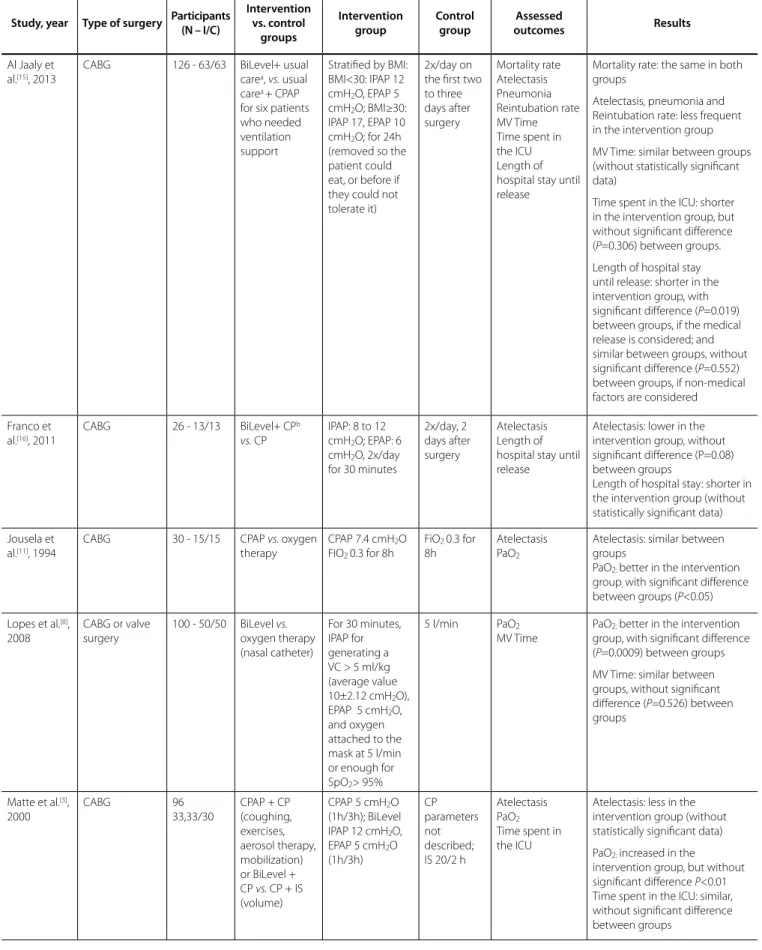
Table 2. Characteristics of the studies included in the review.
Study, year Type of surgery Participants (N – I/C)
Intervention vs. control groups Intervention group Control group Assessed outcomes Results
Al Jaaly et al.[15], 2013
CABG 126 - 63/63 BiLevel+ usual
carea, vs. usual
carea + CPAP
for six patients who needed ventilation support
Stratified by BMI: BMI<30: IPAP 12 cmH2O, EPAP 5
cmH2O; BMI≥30:
IPAP 17, EPAP 10 cmH2O; for 24h
(removed so the patient could eat, or before if they could not tolerate it)
2x/day on the first two to three days after surgery Mortality rate Atelectasis Pneumonia Reintubation rate MV Time Time spent in the ICU Length of hospital stay until release
Mortality rate: the same in both groups
Atelectasis, pneumonia and Reintubation rate: less frequent in the intervention group
MV Time: similar between groups (without statistically significant data)
Time spent in the ICU: shorter in the intervention group, but without significant difference (P=0.306) between groups.
Length of hospital stay until release: shorter in the intervention group, with significant difference (P=0.019) between groups, if the medical release is considered; and similar between groups, without significant difference (P=0.552) between groups, if non-medical factors are considered
Franco et al.[16], 2011
CABG 26 - 13/13 BiLevel+ CPb
vs. CP
IPAP: 8 to 12 cmH2O; EPAP: 6
cmH2O, 2x/day
for 30 minutes
2x/day, 2 days after surgery
Atelectasis Length of hospital stay until release
Atelectasis: lower in the intervention group, without significant difference (P=0.08) between groups
Length of hospital stay: shorter in the intervention group (without statistically significant data)
Jousela et al.[11], 1994
CABG 30 - 15/15 CPAP vs. oxygen
therapy
CPAP 7.4 cmH2O
FiO2 0.3 for 8h
FiO2 0.3 for
8h
Atelectasis PaO2
Atelectasis: similar between groups
PaO2: better in the intervention
group, with significant difference
between groups (P<0.05)
Lopes et al.[8],
2008
CABG or valve surgery
100 - 50/50 BiLevel vs.
oxygen therapy (nasal catheter)
For 30 minutes, IPAP for generating a VC > 5 ml/kg (average value 10±2.12 cmH2O),
EPAP 5 cmH2O,
and oxygen attached to the mask at 5 l/min or enough for SpO2> 95%
5 l/min PaO2
MV Time
PaO2: better in the intervention
group, with significant difference
(P=0.0009) between groups
MV Time: similar between groups, without significant difference (P=0.526) between groups
Matte et al.[3],
2000
CABG 96
33,33/30
CPAP + CP (coughing, exercises, aerosol therapy, mobilization) or BiLevel + CP vs. CP + IS (volume)
CPAP 5 cmH2O
(1h/3h); BiLevel IPAP 12 cmH2O,
EPAP 5 cmH2O
(1h/3h)
CP parameters not described; IS 20/2 h
Atelectasis PaO2
Time spent in the ICU
Atelectasis: less in the intervention group (without statistically significant data)
PaO2: increased in the
Pulmonary Complications
Atelectasis
Six studies assessed the incidence of atelectasis[3,11,15-17,19].
Among them, four[3,11,15,16] were included in the meta-analysis
(n=407). One study compared NIV associated with usual care vs.
usual care[15]; one compared NIV associated with conventional
physiotherapy (CP) vs. CP[16]; another compared NIV vs. oxygen
therapy, exclusively[11]; another study, with two intervention
groups, compared NIV/CPAP mode associated with CP vs. CP; and
NIV/BiLevel mode associated with CP vs. CP[3]. The use of NIV in
the postoperative period of cardiac surgery did not significantly reduce the risk of atelectasis (RR: 0.60, CI95% 0.28, 1.28, I-square: 69%) (Figure 2).
This high statistical heterogeneity can be explained by a
study by Al Jaaly et al.[15], which presented a more favorable result
Mazullo, et al.[20], 2010
CABG, valve replacement, Combined surgeries, Interatrial communication, aneurysm repair
32 - 14/18 NIV (PSV) vs. not
described
PSV PEEP 5 cmH2O; levels of
PSV adjusted to reach a current volume of 5 to 8 ml/kg; FiO2 40%,
for 2h
Not described
ARF after extubation
ARF: control group presented higher incidence. (without statistically significant data)
Oikkonenet al.[19], 1991
CABG 52 - 26/26 IPPB+CP (chest
physiotherapy techniques) vs.
IS (volume) + CP
Airway peak pressure 10 to 15 cmH2O at
least four times/ day, minimum of 10 satisfactory inspirations, five to 10 minutes each session 1x/day, more frequently, if necessary CP Atelectasis PaO2
Atelectasis: less in the intervention group; without significant difference between groups (P>0.1)
PaO2: similar values between
groups on the first three days
Pinilla et al.[17],
1990
CABG 58 - 32/26 CPAP+ CP (chest
physiotherapy)
vs. oxygen therapy + CP
Between 5 and 7.5 cmH2O, for
12h
Not described
Atelectasis Hypoxemia (PaO2/FiO2 )
Time spent in the ICU
Atelectasis: not different between groups
Hypoxemia (PaO2/FiO2):
significantly improved ratio in the intervention group, (P<0.05), half an hour until 24h after extubation; after that, a decrease could be noted in both groups Time spent in the ICU: similar between groups, without significant difference between groups
Thomas et al.[18], 1992
CABG 28 - 14/14 CPAP+CP vs.
oxygen therapy + CP
5 cmH2O, for 1h Not
described
Hypoxemia Hypoxemia: significantly reduced
the pulmonary shunt in the intervention group (P=0.016)
Zarbock et al.[10], 2009
CABG or heart valve replacement
468 - 232/236
CPAP vs.
standard
treatmentc
10 cmH2O, for at
least 6h
Intermittent CPAP for 10 min every 4h at 10 cm H2O; other
information was not described
Hypoxemia (PaO2/
FiO2 <100)
Nosocomial Pneumonia Reintubation rate MV Time Time spent in the ICU and at the hospital
Hypoxemia (PaO2/FiO2<100),
pneumonia, reintubation rate: lower in the intervention group, with significant difference
between groups (P=0.03)
MV Time late extubation group: similar between groups, without significant difference (P>0.05) Time spent in the ICU and at the
hospital:similar between groups;
without significant difference (P>0.05) between groups
to the use of NIV. One of the factors that may justify this has to do with the time of application of the therapy. The study applied the NIV for a longer period of time, and it was withdrawn when the patient had to eat, drink, or before if the patient could not tolerate the ventilation support, with average application time of 16 hours. In the other studies, the time was shorter (8 hours after
extubation[11], 1h every 3h, totaling 8h[3], and 2 times a day for 30
minutes, totaling 1h[16]).
Other factors that could influence the effects of the NIV on the results are the ventilation parameters and type of intervention performed in the control group.
The other two studies[17,19], which were not included in the
meta-analysis due to lack of data, showed that the incidence of atelectasis decreased at the end of the intervention with NIV, but it did not differ from the control groups.
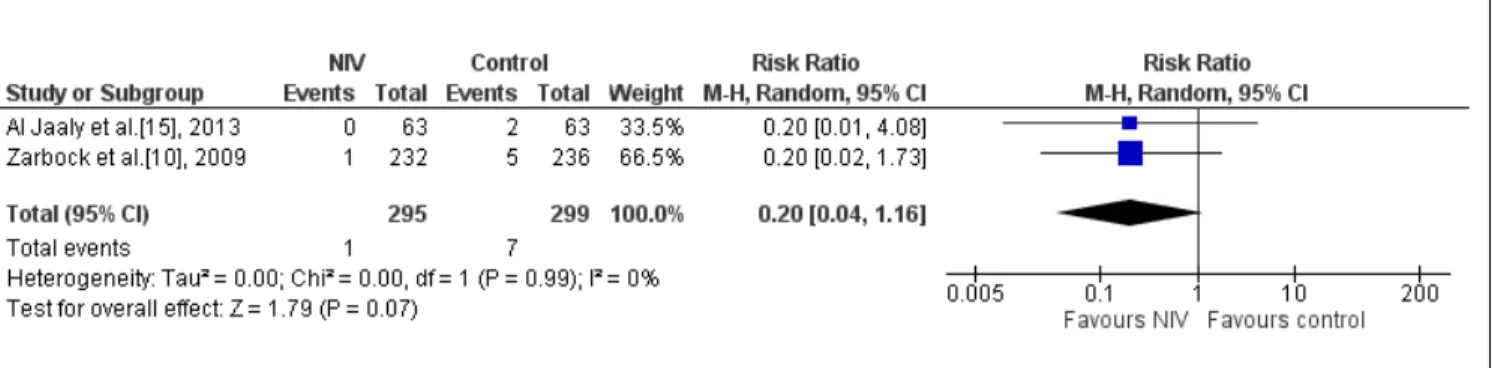
Pneumonia
Two studies[10,15] assessed this outcome (n=594). One
study compared NIV associated to usual care vs. usual care[15];
and the other, NIV vs. standard treatment (oxygen therapy, CP,
nasal intermittent CPAP for 10 min every 4h at 10 cmH2O and
pharmacological treatment)[10].From the analysis, we observed
that the use of NIV did not significantly reduce the probability of
pneumonia (RR: 0.20; CI95% 0.04; 1.16; I-square: 0%) (Figure 3).
Acute Respiratory Failure
Only one study assessed this outcome. Mazzulo Filho et
al.[20] carried out a study with 32 patients during the immediate
postoperative period of cardiac surgery. The patients were randomly divided into two groups: control (n=18) and
Table 3. Risk of bias assessment.
Study, year
Random Sequence Generation
Allocation Concealment
Blinding Therapist
Blinding Patient
Blinding of the Outcome
Assessment
Description of Losses and
Exclusions
Intention-to-treat Analysis
Al Jaaly et al.[15], 2013 Yes Yes No Not informed Yes* Yes No
Franco et al.[16], 2011 Not informed Not informed Not informed Not informed Not informed Yes Yes
Jousela et al.[11], 1994 Not informed Not informed Not informed Not informed Not informed Yes Yes
Lopes et al.[8], 2008 Yes Not informed Not informed Not informed Not informed Yes Yes
Matte et al.[3], 2000 Not informed Not informed Not informed Not informed Yes* Yes No
Mazullo et al.[20], 2010 Not informed Not informed Not informed Not informed Not informed Yes No
Oikkonen et al.[19], 1991 Not informed Not informed Not informed Not informed Yes** Yes Yes
Pinilla et al.[17], 1990 Yes Not informed Not informed Not informed Yes** Yes No
Thomas et al.[18], 1992 Not informed Not informed Not informed Not informed Not informed Yes Yes
Zarbock et al.[10], 2009 Not informed Not informed Not informed Not informed Not informed Yes Yes
Yes*=for the Atelectasis outcome; Yes**= for the chest X-ray
intervention (n=14), which received NIV/PSV mode during 2 hours, after extubation. As to the pulmonary complication, none of the patients from the intervention group presented ARF; on the other hand, nine patients from the control group did.
Hypoxemia
Three studies[10,17,18] assessed this outcome. It was impossible
to perform the meta-analysis of this outcome, because the studies did not present sufficient data for the analysis.
Zarbock et al.[10] performed a study with 468 patients
that underwent elective heart surgery, and compared the
NIV/CPAP mode vs. standard treatment (oxygen therapy, CP,
nasal intermittent CPAP for 10 min every 4h at 10 cmH2O
and pharmacological treatment). The study showed that the
incidence of hypoxemia (PaO2/FiO2< 100) was lower in the
intervention group, when compared with the control group (1 of 232; 5 of 236 patients, respectively).
Pinilla et al.[17] carried out a study with 58 patients, and
compared NIV/CPAP mode associated to CP vs. CP and oxygen
therapy. They found that the PaO2/FiO2 ratiowas significantly
better in the intervention group (P<0.05), half an hour until
24h after extubation, when compared with the control group; after that, it decreased in both groups (325±62, 320±37, in the intervention and control groups, respectively, without difference between groups).
Thomas et al.[18] compared two groups with 14 patients
after CABG, applying NIV/CPAP mode associated to CP vs. CP
and oxygen therapy. The fraction of the pulmonary shunt was of 16.3% before, 12.6% during and 15.7% after the CPAP; in the control group, the shunt was reduced from 17.3% to 16.8%. This reduction was significantly higher in the CPAP group when
compared with the control group (P=0.016).
PaO2
Four studies[3,8,11,19] assessed this outcome. It was not possible
to perform the meta-analysis of this outcome, because the studies did not present sufficient data for the analysis.
Matte et al.[3] performed a study with 96 patients, randomly
divided into three groups. The study assessed two intervention groups: one group compared NIV/CPAP mode associated to CP
(coughing, aerosol therapy, exercises, mobilization) vs. CP, and
the other compared NIV/BiLevel mode associated to CP vs. CP.
In the three groups, the PaO2 (mmHg) significantly decreased
Fig. 3 –Analysis of the pneumonia regarding the studies that compared the prophylactic NIV to the control group. NIV=noninvasive ventilation
in the 1st day of postoperative care (preoperative: control group
78±10, CPAP 76±12, BiLevel 81±10; 1st day before treatment:
control group 65±12, CPAP 63±9, BiLevel 66 ±11; P<0.001). For
the patients of the control group, this decrease was still present
on the 2nd day; however, the patients in the intervention group
presented a slightly improved PaO2 (P<0.01).
Lopes et al.[8] developed a study with 100 patients that
underwent CABG or heart valve surgery, randomly divided into two groups. The study applied NIV/BiLevel mode for 30 minutes
after extubation vs. oxygen therapy. The NIV improved the PaO2,
with significant difference (P=0.0009) between groups; the same
happened with time, comparing the moment before extubation
with 30, 120 and 360 minutes after the procedure (P=0.00008)[8].
Conversely, two studies[11,19] found different results. In a
study performed with 30 patients that underwent CABG, who
were randomly divided into two groups, NIV/CPAP mode vs.
oxygen therapy, the PaO2 decreased significantly in the control
group after extubation (from 19.2±5.3 kPa to 12.4±2.7 kPa), but it decreased less in the CPAP group (from 16.4±3.3 kPa to
14.0±2.1kPa). In the 2nd PO, the PaO
2 was equally low in both
groups (control: 8.4±1.5 kPa, CPAP: 8.9±1.9 kPa)[11].
Oikkonen et al.[19] performed a study with 52 patients who
were randomly divided into two groups: IPPB associated to CP
vs. CP and IS. On the first three days of PO care, the values of
PaO2 (kPa) were similar in both groups (1st PO: control 14±1, IPPB
15±1; 2nd PO: control 12±1, IPPB 11 ± 1; 3rd PO: control 10±1, IPPB
11±1), without statistically significant differences. Based on this, both resources are equally efficient.
Reintubation Rate
Two studies assessed this outcome (n= 594)[10,15].It was observed
that using NIV does not significantly reduce the probability of
reintubation (RR: 0.51; CI95%: 0.15; 1.66; I-square: 0%) (Figure 4).
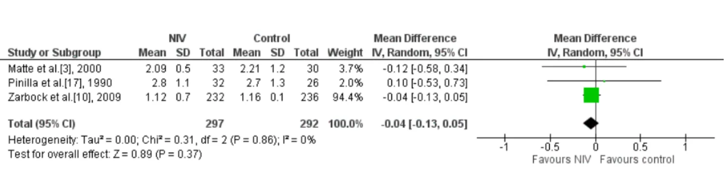
Time spent in the ICU
Three studies assessed this outcome (n=641)[3,10,17].One study
assessed two intervention groups: one compared NIV/CPAP mode
associated to CP vs. CP, and the other compared NIV/BiLevel mode
associated to CP vs. CP[3]. Another study compared NIV associated
to CP vs. oxygen therapy associated to CP[17]; and another study
assessed NIV vs. standard treatment[10]. It was observed that using
Fig. 5 –Analysis of the time spent in the intensive care unit regarding the studies that compared the prophylactic NIV to the control group. NIV=noninvasive ventilation
Fig. 4 –Analysis of the reintubation rate regarding the studies that compared the prophylactic NIV to the control group. NIV=noninvasive ventilation
Length of Hospital Stay
Only two studies assessed length of hospital stay (n=594)
[10,15],however, it was not possible to perform the meta-analysis
due to lack of data. According to Al Jaaly et al.[15],the length of
hospital stay until release from hospital care, in days, was shorter in the intervention group, when compared with the control
group (5±1.5; 6±1.5, respectively, P=0.019). Considering only
the doctors’ opinion, the patients could be released. However, because of non-medical reasons (such as social factors), the patients could not be released and the total length of hospital stay until the release was similar, without significant difference
between groups (P=0.552).
A study by Zarbock et al.[10] showed that the total length of
hospital stay was similar between groups, without significant difference (intervention group 13±0.5 days, control group 14±0.6 days; P>0.05).
Mechanical Ventilation Time
Three studies assessed this outcome (n=694)[8,10,15]; however,
it was not possible to perform the meta-analysis due to lack of data.
In the study by Lopes et al.[8], comparing NIV vs. oxygen
therapy, the average time of mechanical ventilation was 3.77±0.94h, with no significant difference between the control
and intervention groups (P=0.526). Zarbock et al.[10] assessed NIV
vs. standard treatment after admission to the ICU. The patients
were divided into two groups: the late extubated group and the
extubated group; after that, each group was subdivided into intervention and control groups. Patients in the late extubated group (intervention and control) were ventilated for the same
length of time (6.2±0.5 h, 6.±0.7 h, respectively, P>0.05); data
from the extubated group were not described. Finally, the third
study assessed NIV associated with usual care vs. usual care, and
the average time was 6 hours for the intervention and control groups[15].
DISCUSSION
Evidence Summary
The objective of this study was to search for the best available scientific evidence regarding the prophylactic use of NIV in patients during immediate postoperative care for cardiac surgery. Regarding the mortality rate, only one study assessed this outcome, and the result was similar between the intervention and control groups. We observed that prophylactic NIV, when compared to conventional physiotherapy or oxygen therapy, did not significantly reduce the probability of pulmonary complications such as atelectasis, pneumonia, reintubation rate and time spent in the ICU. As to the outcome of ARF, the incidence was higher in the control group. Regarding the outcome of hypoxemia, in most studies, the use of NIV
improved oxygenation. Regarding PaO2, only half of the studies
For the outcome mortality rate, study showed low incidence between the intervention and control groups. This can be explained by the fact that the patients included in the studies had preoperative comorbidities, and similar surgical characteristics,
among others, without the need for emergency surgery[15].
However, more studies should be carried out to expand this information.
Another outcome assessed by our study was pulmonary complications. These are frequent in the PO of cardiac surgery, and the etiology of the dysfunction results from a multifactorial process, which involves surgical and preoperative factors, well
reported in the literature[4-7]. Thus, early therapy is necessary to
avoid further degradation[3].
From the review, it was observed that using NIV during the postoperative care of heart surgery did not significantly reduce the risk of atelectasis, but it improved oxygenation. This complication is often caused by a compromised ventilation lung perfusion ratio due to atelectasis. Even after cardiac surgery without complications, atelectatic areas decrease functional residual capacity and increase pulmonary shunt. These unventilated areas can account for up to 20% of the total lung
volume, thus causing hypoxemia in the PO[10]. In this sense, NIV
avoids alveolar collapse and enables better alveolar recruitment, reducing the formation of atelectasis and increasing functional
residual capacity[10,16].
Even though NIV presents positive effects in other populations, such as patients with ARF in the postoperative period of abdominal
surgery[21], in our study we have not yet found a beneficial effect of
this intervention in relation to atelectasis. One thing that may justify these unfavorable outcomes are the NIV pressure parameters, with
CPAP values between 5 and 7.5 cmH2O[3,11]. These pressures low
in the airways have transitory effects on gas exchange[10,17]. It is
believed that, for a prolonged effect, high pressure values are
required to keep airways open[10]. A previous study performed
with patients after thoracic surgery demonstrated that pressures
of at least 9 to 10 cmH2O should be used to maintain positive
tracheal pressure throughout the respiratory cycle[22].
Another pulmonary complication is pneumonia. From the analysis, it was observed that using NIV did not significantly
reduce the probability of pneumonia[10,15]. We observed that
there is a favorable tendency to the use of NIV; however, more RCTs, with larger sample sizes, are necessary to corroborate this information.
Regarding the outcomes of reintubation rate and time spent in the ICU, it was observed that using NIV does not significantly reduce the probability of reintubation and the time spent in the ICU. This may have occurred because of the small number of included studies (two and three, respectively). It is possible that with more RCTs, with larger sample sizes, this result could change; therefore, there is no evidence on the effectiveness of NIV on the reintubation rate and time spent in the ICU.
Another assessed outcome was the length of hospital
stay, but only two studies assessed it[10,15]. The time was similar
between groups in both studies, with no significant difference between groups. This similarity can be justified by the low incidence of pulmonary complications in both studies, which could prolong this time if an increase occurred.
The Strengths and Limitations of the Study
Regarding the strong methodological points of this study, it is important to point out the systematic and sensitive bibliographic search, with explicit and reproducible eligibility criteria, without restriction of language and date, independently performed by two assessors; as well as the selection of studies, data extraction and analysis of methodological quality of included articles, also performed independently by two assessors. In addition to that, a meta-analysis was performed with the results of the studies, provided they allowed such analysis, since the meta-analysis can give more reliable estimates as to the efficacy of the treatment.
As to the limitations, the low methodological quality of the included studies stands out, since the indispensable items for assessing the risk of bias were presented incompletely or not informed. In addition, the included studies were quite different regarding the physiotherapy techniques, resources and exercises used, time of intervention, time of application of NIV and frequency of examinations. All this may compromise the results found in the meta-analyses.
In addition to the methodological differences, we highlight the small number of studies found in the literature, and the sample size, with small number of patients, which suggest the need for new RCTs with more patients and more methodological rigor.
CONCLUSION
Our study showed that no difference between the use of prophylactic NIV and conventional physiotherapy or oxygen therapy could be found in patients during the postoperative period of cardiac surgery, in relation to mortality rate and pulmonary complications such as atelectasis, pneumonia, reintubation rate, time spent in the ICU, length of hospital stay and mechanical ventilation time, with an improvement in oxygenation. Therefore, due to the low methodological rigor of the included articles and small sample size, new RCTs should be carried out to corroborate this information.
Authors’ roles & responsibilities
SMP
AGFM
GS
REFERENCES
1. Chiumello D, Chevallard G, Gregoretti C. Non-invasive ventilation in postoperative patients: a systematic review. Intensive Care Med. 2011;37(6):918-29.
2. Niyayeh Saffari NH, Nasiri E, Mousavinasab SN, Ghafari R, Soleimani A, Esmaeili R. Frequency rate of atelectasis in patients following coronary artery bypass graft and its associated factors at Mazandaran Heart Center in 2013-2014. Glob J Health Sci. 2015;7(7 Spec No):97-105.
3. Matte P, Jacquet L, Van Dyck M, Goenen M. Effects of conventional physiotherapy, continuous positive airway pressure and non-invasive ventilatory support with bilevel positive airway pressure after coronary artery bypass grafting. Acta Anaesthesiol Scand. 2000;44(1):75-81. 4. Weissman C. Pulmonary complications after cardiac surgery. Semin
Cardiothorac Vasc Anesth. 2004;8(3):185-211.
5. Luchesa CA, Greca FH, Guarita-Souza LC, Santos JLV, Aquim EE. Papel da eletroanalgesia na função respiratória de pacientes submetidos à operação de revascularização do miocárdio. Rev Bras Cir Cardiovasc. 2009,24(3):391-6.
6. Huffmyer JL, Groves DS. Pulmonary complications of cardiopulmonary bypass. Best Pract Res Clin Anaesthesiol. 2015;29(2):163-75.
7. Laizo A, Delgado FEF, Rocha GM. Complicações que aumentam o tempo de permanência na unidade de terapia intensiva na cirurgia cardíaca. Rev Bras Cir Cardiovasc. 2010 25(2):166-71.
8. Lopes CR, Brandão CMA, Nozawa E, Auler Junior JOC. Benefícios da ventilação não-invasiva após extubação no pós-operatório de cirurgia cardíaca. Rev Bras Cir Cardiovasc. 2008;23(3):344-50.
9. Jaber S, Chanques G, Jung B. Postoperative noninvasive ventilation. Anesthesiology. 2010;112(2):453-61.
10. Zarbock A, Mueller E, Netzer S, Gabriel A, Feindt P, Kindgen-Milles D. Prophylactic nasal continuous positive airway pressure following cardiac surgery protects from postoperative pulmonary complications: a prospective, randomized, controlled trial in 500 patients. Chest. 2009;135(5):1252-9.
11. Jousela I, Räsänen J, Verkkala K, Lamminen A, Mäkeläinen A, Nikki P. Continuous positive airway pressure by mask in patients after coronary surgery. Acta Anaesthesiol Scand. 1994;38(4):311-6.
12. Moher D, Liberati A, Tetzlaff J, Altman DG; PRISMA Group. Preferred reporting items for systematic reviews and meta-analyses: the PRISMA statement. BMJ. 2009;339:b2535.
13. Higgins JPT, Green S. Cochrane handbook for systematic reviews of interventions. Version 5.3.0 [updated October 2015]. The Cochrane Collaboration; 2015.
14. Robinson KA, Dickersin K. Development of a highly sensitive search strategy for the retrieval of reports of controlled trials using PubMed. Int J Epidemiol. 2002;31(1):150-3.
15. Al Jaaly E, Fiorentino F, Reeves BC, Ind PW, Angelini GD, Kemp S, et al. Effect of adding postoperative noninvasive ventilation to usual care to prevent pulmonary complications in patients undergoing coronary artery bypass grafting: a randomized controlled trial. J Thorac Cardiovasc Surg. 2013;146(4):912-8.
16. Franco AM, Torres FC, Simon IS, Morales D, Rodrigues AJ. Assessment of noninvasive ventilation with two levels of positive airway pressure in patients after cardiac surgery. Rev Bras Cir Cardiovasc. 2011;26(4):582-90. 17. Pinilla JC, Oleniuk FH, Tan L, Rebeyka I, Tanna N, Wilkinson A, et al. Use
of a nasal continuous positive airway pressure mask in the treatment of postoperative atelectasis in aortocoronary bypass surgery. Crit Care Med. 1990;18(8):836-40.
18. Thomas AN, Ryan JP, Doran BR, Pollard BJ. Nasal CPAP after coronary artery surgery. Anaesthesia. 1992;47(4):316-9.
19. Oikkonen M, Karjalainen K, Kähärä V, Kuosa R, Schavikin L. Comparison of incentive spirometry and intermittent positive pressure breathing after coronary artery bypass graft. Chest. 1991;99(1):60-5.
20. Mazzulo Filho JBR, Bonfim VJG, Aquim EE. Ventilação mecânica não invasiva no pós-operatório imediato de cirurgia cardíaca. Rev Bras Ter Intensiva. 2010;22(4):363-8.
21. Associação de Medicina Intensiva Brasileira (AMIB), Sociedade Brasileira de Pneumologia e Tisiologia (SBPT). Diretrizes brasileiras de ventilação mecânica - 2013. Disponível em: http://itarget.com.br/newclients/sbpt. org.br/2011/downloads/arquivos/Dir_VM_2013/Diretrizes_VM2013_ SBPT_AMIB.pdf