Effectiveness of prophylactic non-invasive ventilation
on respiratory function in the postoperative phase of
pediatric cardiac surgery: a randomized controlled trial
Camilla R. S. Silva1, Lívia B. Andrade1, Danielle A. S. X. Maux1,
Andreza L. Bezerra1, Maria do Carmo M. B. Duarte1
ABSTRACT | Objective: To evaluate the effectiveness of prophylactic, non-invasive ventilation (NIV) on respiratory function in seven- to 16-year-old children in the post-operative phase of cardiac surgery. Method: A randomized, controlled trial with 50 children who had undergone cardiac surgery with median sternotomy. After extubation, patients were randomly assigned to one of two groups: control group (n=26), which received instructions regarding posture, early ambulation, and cough stimulation, and CPAP group (continuous positive airway pressure; n=24), which received the same instructions as the control group and CPAP=10 cmH20 twice daily for 30 minutes from the 1st to the 5th post-operative day (POD).
As a primary outcome, lung function was evaluated before and on the 1st, 3rd, and 5th PODs with measures of respiratory
rate (RR), tidal volume (TV), slow vital capacity (SVC), inspiratory capacity (IC), minute volume (MV), peak expiratory low (PEF), and maximal inspiratory pressure (MIP). As secondary outcomes, the time of hospitalization and intensive care were recorded. A mixed, linear regression model and z-test were used to analyze respiratory function, considering
p<0.05. Results: All variables, except RR and MV, showed a signiicant drop on the 1st POD, with gradual recovery;
however, only MIP had returned to pre-operative values on the 5th POD in both groups. The RR showed a signiicant
increase on the 1st POD, with a gradual reduction but without returning to baseline. In the intergroup analysis, signiicant
improvement (p=0.04) was observed only in PEF in the CPAP group on the 1st DPO. The length of hospitalization and
intensive care showed no signiicant differences. Conclusion: NIV was safe and well accepted in this group of patients, and the protocol used was effective in improving PEF on the 1st DPO in the CPAP group.
Keywords: cardiac surgical procedures; pediatrics; continuous positive airway pressure; non-invasive ventilation; physical therapy.
Clinical Trials Identiier: RBR-9j4thm. http://www.ensaiosclinicos.gov.br/rg/RBR-9j4thm/
BULLET POINTS
• NIV may be used to prevent or minimize the deterioration of respiratory function in the post-operative period of pediatric cardiac surgery.
• The prophylactic use of NIV in the form of CPAP was effective in improving peak expiratory low in the post-operative period of pediatric cardiac surgery.
• New protocols and new ways of offering prophylactic, non-invasive ventilation in the post-operative period of pediatric cardiac surgery must be evaluated.
HOW TO CITE THIS ARTICLE
Silva CRS, Andrade LB, Maux DASX, Bezerra AL, Duarte MCMB. Effectiveness of prophylactic non-invasive ventilation on respiratory function in the postoperative phase of pediatric cardiac surgery: a randomized controlled trial. Braz J Phys Ther. 2016 Nov-Dec; 20(6):494-501. http://dx.doi.org/10.1590/bjpt-rbf.2014.0191
1 Instituto de Medicina Integral Prof. Fernando Figueira (IMIP), Recife, PE, Brazil
Received: Apr. 09, 2015 Revised: Nov. 10, 2015 Accepted: Mar. 07, 2016
Introduction
Pulmonary complications are the most frequent causes of morbidity in patients undergoing cardiac surgery. Complications range from 6% to 76% of cases1, depending on the severity of the disease, and are responsible for prolonging the period of
hospitalization with increased hospital costs2, as well
as being a major cause of mortality3.
from bed, ambulation, deep breathing stimulation, use of incentive spirometers, and cough stimulation. However, these methods are often not effective, resulting in the need to employ other measures, such as using positive airway pressure4.
Currently in clinical practice, the use of non-invasive ventilation (NIV) has been shown to be a method capable of offering positive pressure, as it is easy
to use and does not require the presence of artiicial
airways5. NIV can be provided at two pressure levels,
bilevel (BiPAP) or continuous positive airway pressure
(CPAP), and is an alternative proposal to prevent pulmonary complications, thereby reducing muscle fatigue, improving functional residual capacity and gas exchanges6.
Despite numerous studies on the prophylactic use of NIV in adults in the post-operative period of cardiac surgery7, the literature is scarce in pediatrics. Most
studies performed in this population show the beneits
of NIV in treating pulmonary complications8-10, but only one retrospective study analyzed this feature in a prophylactic way11. Thus, further investigation is required to clarify the use of prophylactic NIV and its relationship to respiratory function in the post-operative period of cardiac surgery in pediatric
patients. Therefore, the objective of this clinical
trial is to evaluate the effectiveness of prophylactic, non-invasive ventilation on the respiratory function of patients in the post-operative period of pediatric cardiac surgery.
Method
Study type
A randomized, controlled clinical trial was conducted.
Participants
The study included patients aged seven to 16 years, who had undergone elective cardiac surgery with median sternotomy at the Instituto de Medicina
Integral Prof. Fernando Figueira (IMIP), Recife, PE, Brazil, from June 2010 to March 2013. The guardians
of all of the participants signed a consent form after receiving information regarding the proposed protocol.
This study was approved by the Research Ethics
Committee of IMIP (protocol 1489-09).
Patients were excluded who were pre-operative and who presented with hemodynamic instability, contraindications to the use of NIV, chronic lung disease, or inability to perform the evaluation techniques.
Randomization and allocation
After surgery, the patients had ventilation tubes removed within 24 hours and were randomly assigned to one of two groups: control (n=26) and CPAP (n=24). The randomization for the use of NIV or not was performed according to a list of sequential numbers from one to 62 (number of patients to be randomized) generated by the software Random Allocation version 1.0, using the words CONTROL and CPAP.
The blinding of allocation (concealed allocation) was obtained by opaque, sealed envelopes, which were numbered consecutively and contained the name of each group. A person not involved in the research received the list of random numbers and prepared sequentially numbered, opaque envelopes from one to 62 containing the name of the group to which each patient would be allocated.
Interventions
The control group received instructions on posture, early ambulation, and cough stimulus. In terms of posture, patients were advised to avoid antalgic positions (increased thoracic kyphosis, protraction of the shoulders, and bending of the head) due to sternotomy, as these antalgic positions could
compromise lung function. Early ambulation was
encouraged when the patient presented clinical and hemodynamic stability, and after removal of drains. Patients were instructed to cough while protecting the incision with their hands resting on the surgical site, providing greater security and therefore a more effective cough.
The intervention group, in addition to the above-mentioned guidelines, was submitted to non-invasive ventilation with continuous positive airway pressure (CPAP) twice a day for 30 minutes, from the 1st to the 5th post-operative day (POD) through
a low-generating system (Boussignac system, Vygon SA, Écouen, France) coupled to a medium-sized, Vygon
pneumatic face mask attached to the face by a system
of silicon strips of the same brand. The low rate was
adjusted to reach the pressure of 10 cmH2O, measured
by a Vygon manometer connected to the system via
a circuit between the outlet of the low-generating
device and the manometer.
Outcomes
slow vital capacity (SVC), inspiratory capacity
(IC), peak expiratory low (PEF), and maximal
inspiratory pressure (MIP). These variables were assessed pre-operatively, and re-evaluated on the 1st, 3rd, and 5th PODs. As secondary outcomes, the length of hospitalization and in the intensive care unit (ICU) were recorded.
Respiratory function was assessed with patients in
their beds in the Fowler position at 45º. Ventilatory
variables were measured using an analog spirometer (nSpire Health Inc., Longmont, CO, USA) coupled
to a face mask. PEF was measured using a portable peak-low device (Mini-Wright Standard, Clement
Clarke International, Harlow, UK) coupled to a mouthpiece and using a nose clip on the patient after a forced maximal inhalation and exhalation with the glottis open. MIP was measured by means of a maximum inspiration from functional residual
capacity (FRC) using an analog manometer with a
scale to -120 cmH20 (Comercial Médica) coupled to a mouthpiece and using a nose clip on the patient. To ensure the reliability of measurements for each parameter evaluated, three attempts were performed and the highest values were recorded.
Sample size
The sample size calculation was performed using
the Statcalc feature of the Epi Info software, version 3.5.3. For the calculation, a pilot study was made
involving 26 patients (12 in the control group and 14 in the intervention group), using as a basis the maximal inspiratory pressure (MIP). Groups of a
size equal to 26 would be suficient to identify a
difference in the mean of MIP of at least 25% with a power of 80% and type 1 error of 5%, assuming a mean ± standard deviation of 87.7 (±34) cmH2O for the Control Group, and 100 (±30) cmH2O for the Intervention Group. A rate of loss of 20% rate was forecast, therefore 62 patients should be included in the study, randomly assigned to one of two groups, and distributed equally.
Statistical analysis
Statistical analysis was performed using Stata/SE 12.1 (StataCorp LP, College Station, TX, USA). Data
were summarized in terms of mean and standard
deviation and standard error. For the analysis of the
respiratory function variables, given that the study design involved two groups of patients observed on four different occasions, data analysis was based on
the adjustment of mixed linear regression models.
After adjustment of each model for the variables of
respiratory function, comparisons were performed between groups on each occasion and among the times in each group, using the z test in these comparisons.
For all tests, a signiicance level of 0.05 was used.
Results
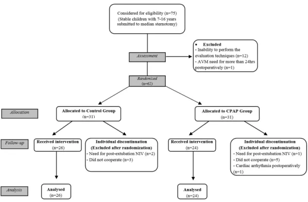
Of the 75 patients initially considered eligible to perform median sternotomy with cardiopulmonary bypass, 62 were randomized and 50 (19% loss of follow-up) completed the study as presented in the CONSORT12 lowchart (Figure 1). The most frequent diagnoses were secondary valvular disease related to rheumatic heart disease (46%), followed by intra-atrial communication (14%).
Clinical and anthropometric characteristics of the sample are presented in Table 1. There were no extubation failures in the patients studied.
In the analysis of the variables of respiratory function between groups, it was observed that the
PEF on the 1st POD was greater in the CPAP group
compared to the control group (p=0.04), but there
were no signiicant differences in this variable on
the 3rd and 5th PODs between groups. The remaining respiratory-function variables evaluated showed no
signiicant difference between groups at any of the
assessed moments (Table 2 and Figure 2).
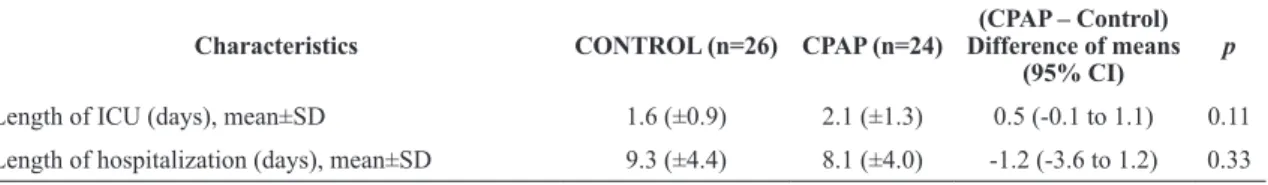
Regarding the time spent in the hospital and the
ICU, there were no signiicant differences between
the groups (Table 3).
Discussion
NIV administered continuously or intermittently has been used alone or in combination with physical therapeutic techniques to prevent atelectasis and hypoxemia during the post-operative period of cardiac surgery in adults8. However, to the authors’ knowledge,
this study represents the irst randomized, controlled
trial to evaluate prophylactic NIV in improving respiratory function in children who have undergone cardiac surgery with cardiopulmonary bypass.
The comparison of respiratory function variables
intragroup over time in this study conirms the indings
of the study by Caséca et al.13, a prospective study that evaluated children who underwent mitral valve replacement or reconstruction. The authors demonstrated
that PEF and lung volume and capacity values evaluated
in the post-operative period, except MV, remained
compared to pre-operative values. In the present study, only MIP returned to pre-operative values on the 5th POD, a variable that was not analyzed in the study by Caséca et al.13 cited above.
In the intergroup comparison, only PEF showed
a signiicant difference on the 1st POD in relation to
the pre-operative values. The variable PEF is related
to the effectiveness of coughing. The higher this variable is, the better the elimination of secretions14 and consequently fewer pulmonary complications will
be seen in the post-operative period. This inding can possibly be explained by an increased FRC provided
by the use of CPAP, thus generating a higher lung
volume and consequent increase in expiratory low.
The study of Franco et al.15, which also assessed
respiratory function, showed no signiicant difference
in these variables between groups.
In our study, there were also no signiicant differences
in the days spent in the hospital and ICU between the groups. Studies in the adult population in which NIV was used prophylactically in the post-operative period of cardiac surgery also showed no reduction in these times16-18.
In contrast, a retrospective, observational study in children with heart disease, which evaluated the prophylactic and non-prophylactic use of NIV in
preventing extubation failure, observed a signiicant
reduction in length of stays in the hospital and ICU in the group using prophylactic NIV. However, although
the majority of children were in the post-operative
phase of cardiac surgery, there were some who were only undergoing drug treatment, and in these patients
NIV was used by means of CPAP or BiPAP11.
Hemodynamic changes were not observed, nor any kind of complications related to the application of NIV in our patients, showing that its preventive use in the cardiac post-operative period was safe and well accepted in the pediatric population. This aspect has also been reported by Gupta et al.11, who concluded that NIV is a well-tolerated and safe therapy that can be successfully applied in critically ill children with heart disease to avoid extubation failure.
Despite the importance of this study, it is necessary
to highlight some limitations. Firstly, because it is
a study of children and adolescents from seven to
16 years old, it was dificult to standardize assessments
of respiratory function. This is because there are no
age-speciic reference patterns in the literature for these
variables; however, we used the mean values for each group (control and CPAP). Secondly, the short period of follow-up of patients, until the 5th post-operative
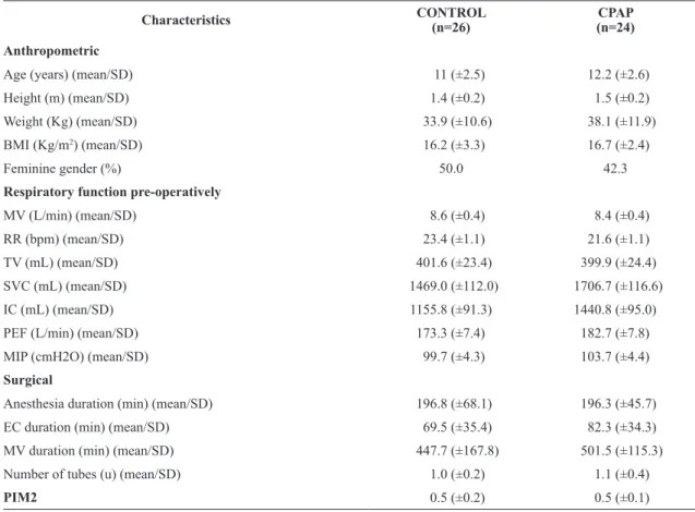
Table 1. Clinical and anthropometric characteristics of the sample.
Characteristics CONTROL
(n=26)
CPAP (n=24)
Anthropometric
Age (years) (mean/SD) 11 (±2.5) 12.2 (±2.6)
Height (m) (mean/SD) 1.4 (±0.2) 1.5 (±0.2)
Weight (Kg) (mean/SD) 33.9 (±10.6) 38.1 (±11.9)
BMI (Kg/m2) (mean/SD) 16.2 (±3.3) 16.7 (±2.4)
Feminine gender (%) 50.0 42.3
Respiratory function pre-operatively
MV (L/min) (mean/SD) 8.6 (±0.4) 8.4 (±0.4)
RR (bpm) (mean/SD) 23.4 (±1.1) 21.6 (±1.1)
TV (mL) (mean/SD) 401.6 (±23.4) 399.9 (±24.4)
SVC (mL) (mean/SD) 1469.0 (±112.0) 1706.7 (±116.6)
IC (mL) (mean/SD) 1155.8 (±91.3) 1440.8 (±95.0)
PEF (L/min) (mean/SD) 173.3 (±7.4) 182.7 (±7.8)
MIP (cmH2O) (mean/SD) 99.7 (±4.3) 103.7 (±4.4)
Surgical
Anesthesia duration (min) (mean/SD) 196.8 (±68.1) 196.3 (±45.7)
EC duration (min) (mean/SD) 69.5 (±35.4) 82.3 (±34.3)
MV duration (min) (mean/SD) 447.7 (±167.8) 501.5 (±115.3)
Number of tubes (u) (mean/SD) 1.0 (±0.2) 1.1 (±0.4)
PIM2 0.5 (±0.2) 0.5 (±0.1)
m: meters; Kg: kilograms; L: liters; min: minute; mL: milliliters; cmH2O: centimeters of water; bpm: breath per minute; BMI: body mass index; MV: minute volume; RR: respiratory rate; TV: tidal volume; SVC: slow vital capacity; IC: inspiratory capacity; PEF: peak expiratory low; MIP: maximal inspiratory pressure; EC: extracorporeal circulation; MV: mechanical ventilation; u: unit; PIM2: Pediatric Index of Mortality 2; SD: standard deviation.
Figure 2. Monitoring of inspiratory capacity (IC), slow vital capacity (SVC), peak expiratory low (PEF), and maximal inspiratory
pressure (MIP) over time in the control and CPAP groups. (A) Distribution of the changes in IC preoperatively (pre-op) and on the 1st,
3rd, and 5th postoperative days (PODs) (p<0.05 intragroup; p<0.05 between groups, except for pre-op: p = 0.03); (B) Distribution of the
changes in SVC pre-op and on the 1st, 3rd, and 5th PODs (p <0.05 intragroup; p<0.05 between groups); (C) Distribution of the changes in PEF pre-op and on the 1st, 3rd, and 5th PODs (p <0.05 intragroup,; p<0.05 between groups, except on 1st POD: p=0.042); (D) Distribution
Table 2. Comparison of intra- and inter-group variables for respiratory function of the assessed moments.
Group
Moment
Pre-operative 1st POD 3rd POD 5th POD
Mean (SD†) Mean (SD†) Mean (SD†) Mean (SD†)
RR
Control (G1) 23.4 (5.6) 35.1* (7.9) 29.3* (5.9) 27.4* (5.8)
CPAP (G2) 21.6 (5.2) 35.0* (9.9) 29.0* (6.6) 27.1* (6.6)
G1vs G2: p value 0.23 0.95 0.86 0.88
G2 - G1, mean (95%CI) -1.8 (-4.8 to 1.2) -0.2 (-5.1 to 4.7) -0.3 (-3.7 to 3.1) -0.3 (-3.7 to 3.1)
TV
Control (G1) 401.6 (153.2) 228.7* (49.1) 273.0* (262.3) 299.3* (104.5)
CPAP (G2) 399.9 (114.0) 256.6* (65.1) 294.7* (79.6) 333.9* (119.4)
G1 vs G2: p value 0.96 0.09 0.35 0.18
G2 - G1, mean (95% CI) -1.7 (-68.0 to 64.6) 27.9 (-4.9 to 60.7) 21.7 (-23.8 to 67.3) 34.6 (-16.1 to 85.4)
MV
Control (G1) 8.6 (2.0) 7.9 (2.0) 7.7* (1.9) 7.9* (1.5)
CPAP (G2) 8.4 (2.2) 8.7 (2.6) 8.2 (1.9) 8.7 (2.1)
G1 vs G2: p value 0.66 0.24 0.36 0.14
G2 - G1, mean (95% CI) -0.3 (-1.4 to 0.9) 0.8 (-0.5 to 2.0) 0.5 (-0.6 to 1.5) 0.8 (-0.3 to 1.8)
IC
Control (G1) 1155.8 (383.7) 413.1* (225.8) 579.2* (309.0) 720.2* (369.6)
CPAP (G2) 1440.8 (557.3) 473.7* (181.7) 661.7* (202.8) 842.5* (282.1)
G1vs G2: p value 0.03 0.28 0.25 0.18
G2 - G1, mean (95% CI) 285.0 (26.9 to 543.2) 60.6 (-51.3 to 172.6) 82.5 (-60.8 to 225.7) 122.3 (-57.4 to 302.0)
SVC
Control (G1) 1469.0 (592.5) 445.8* (241.7) 646.9* (394.2) 813.1* (465.5)
CPAP (G2) 1706.7 (572.0) 512.1* (212.9) 733.1* (304.0) 960.4* (402.5)
G1vs G2: p value 0.14 0.29 0.38 0.22
G2 - G1, mean (95% CI) 237.7 (-79.2 to 554.4) 66.3 (-57.9 to 190.5) 86.2(-106.2 to 278.6) 147.3 (-89.9 to 384.6)
PEF
Control (G1) 173.3 (39.1) 56.2* (33.2) 89.4* (42.2) 133.7* (51.6)
CPAP (G2) 182.7 (38.3) 76.3* (37.9) 109.2* (38.0) 152.7* (55.1)
G1vs G2: p value 0.38 0.04 0.07 0.19
G2 - G1, mean (95% CI) 9.4 (-11.6 to 30.5) 20.1 (0.8 to 39.4) 19.8 (-2.2 to 41.7) 19.0 (-10.0 to 48.1)
MIP
Control (G1) 99.7 (24.7) 70.2* (35.9) 85.3* (31.7) 98.5 (27.9)
CPAP (G2) 103.7 (18.9) 77.9* (36.6) 95.2* (33.2) 108.3 (24.7)
G1vs G2: p value 0.50 0.44 0.27 0.18
G2 - G1, mean (95% CI) 4.0 (-8.0 to 16.1) 7.7 (-12.1 to 27.4) 9.9 (-7.8 to 27.6) 9.8 (-4.6 to 24.2)
† SD: standard deviation; Intragroup comparisons between times: preoperative was chosen as a reference. In each group, occasions marked
* were statistically signiicant compared with the preoperative to a p value <0.05. POD: postoperative day; RR: respiratory rate; TV: tidal volume; MV: minute volume; IC: inspiratory capacity; SVC: slow vital capacity; PEF: peak expiratory low; MIP: maximal inspiratory capacity.
results in respiratory function. Finally, the 19% loss
of follow-up should be considered a limitation of the study.
Table 3. Comparison of length of hospitalization and ICU between CPAP and Control groups.
Characteristics CONTROL (n=26) CPAP (n=24)
(CPAP – Control) Difference of means
(95% CI)
p
Length of ICU (days), mean±SD 1.6 (±0.9) 2.1 (±1.3) 0.5 (-0.1 to 1.1) 0.11
Length of hospitalization (days), mean±SD 9.3 (±4.4) 8.1 (±4.0) -1.2 (-3.6 to 1.2) 0.33
ICU: Intensive care unit, SD: standard deviation.
provide greater increases in lung volumes and be able to demonstrate greater gains in respiratory function
for these patients. Furthermore, a longer follow-up
period of these children could show effective results of this therapeutic resource in the post-operative period of pediatric cardiac surgery.
Conclusion
It was found that pediatric patients who had undergone cardiac surgery by median sternotomy
with CPB showed signiicant losses in respiratory
function, which were perpetuated to the 5th POD, by which time only inspiratory pressure had returned to pre-operative values.
The post-operative use of CPAP was safe and well accepted by patients, but the protocol used was
effective only in the improvement of PEF on the
1st POD. There was no reduction in hospitalization and ICU times when compared to the control group.
Further studies are suggested in the pediatric
population to assess new protocols and new ways of offering non-invasive ventilation in the post-operative period.
References
1. Boisseau N, Rabary O, Padovani B, Staccini P, Mouroux J, Grimaud D, et al. Improvement of “dynamic analgesia” does not decrease atelectasis after thoracotomy. Br J Anaesth. 2001;87(4):564-9. http://dx.doi.org/10.1093/bja/87.4.564. PMid:11878725.
2. Barbosa RAG, Carmona MJC. Avaliação da função pulmonar em pacientes submetidos à cirurgia cardíaca com circulação extracorpórea. Rev Bras Anestesiol. 2002;52(6):689-99.
http://dx.doi.org/10.1590/S0034-70942002000600005. PMid:19475240.
3. Higgins TL, EstafanousFG, Loop FD, BeckGJ, BlumJM, Paranandi L. Stratification of morbidity and mortality outcome by preoperative risk factors in coronary artery bypass patients: a clinical severity score. JAMA. 1992;267(17):2344-8. http://dx.doi.org/10.1001/jama.1992.03480170070031. PMid:1564774.
4. Altschuler E. A breathing tape: a non-invasive prophylaxis/ preventative measure for post-surgical atelectasis which
supplies, rather than requires, patient motivation. Med Hypotheses. 1999;53(1):78-9. http://dx.doi.org/10.1054/ mehy.1998.0721. PMid:10499832.
5. Meduri GU, Cook TR, Turner RE, Cohen M, Leeper KV. Noninvasive positive pressure ventilation in status asthmaticus. Chest. 1996;110(3):767-74. http://dx.doi.
org/10.1378/chest.110.3.767. PMid:8797425.
6. Silva DCB, ForondaFAK, Troster EJ. Ventilação não invasiva em pediatria. J Pediatr. 2003;79(8):161-8. http://
dx.doi.org/10.2223/JPED.1092.
7. Olper L, Redaelli V, Corbetta D. Efficacy of non-invasive ventilation for cardiothoracic surgical patients: a systematic review. It J Physiother. 2011;1(1):17-26.
8. Essouri S, Chevret L, Durand P, Haas V, FaurouxB, Devictor D. Noninvasive positive pressure ventilation: five years of experience in a pediatric intensive care unit. Pediatr Crit Care Med. 2006;7(4):329-34. http://dx.doi.org/10.1097/01.
PCC.0000225089.21176.0B. PMid:16738493.
9. Kovacikova L, Dobos D, Zahorec M. Non-invasive positive pressure ventilation for bilateral diaphragm paralysis after pediatric cardiac surgery. Interact Cardiovasc Thorac Surg. 2009;8(1):171-2. http://dx.doi.org/10.1510/icvts.2008.187096. PMid:18835855.
10. Najaf-Zadeh A, Leclerc F. Noninvasive positive pressure ventilation for acute respiratory failure in children: a concise review. Ann Intensive Care. 2011;1(1):15. http://
dx.doi.org/10.1186/2110-5820-1-15. PMid:21906346. 11. Gupta P, Kuperstock JE, Hashmi S, Arnolde V, Gossett
JM, Prodhan P, et al. Efficacy and predictors of success of noninvasive ventilation for prevention of extubation failure in critically ill children with heart disease. Pediatr Cardiol. 2013;34(4):964-77. http://dx.doi.org/10.1007/s00246-012-0590-3. PMid:23196891.
12. Costa LOP, Maher CG, Lopes AD, Noronha MA, Costa LCM. Transparent reporting of studies relevant to physical therapy practice. Rev Bras Fisioter. 2011;15(4):267-71. http://dx.doi.
org/10.1590/S1413-35552011005000009. PMid:21975681. 13. Caséca MB, Andrade LB, Britto MCA. Pulmonary function assessment in children and teenagers before and after surgical treatment for rheumatic valve disease. J Pediatr. 2006;82(2):144-50. http://dx.doi.org/10.2223/JPED.1462. PMid:16614770.
14. FreitasFS, Parreira VF, Ibiapina CC. Aplicação clínica do pico de fluxo da tosse: uma revisão de literatura. Fisioter Mov. 2010;23(3):495-502. http://dx.doi.org/10.1590/ S0103-51502010000300016.
15. Franco AM, Torres FCC, Simon ISL, Morales D, Rodrigues
Rev Bras Cir Cardiovasc. 2011;26(4):582-90. http://dx.doi.
org/10.5935/1678-9741.20110048. PMid:22358273. 16. Zarbock A, Mueller E, Netzer S, Gabriel A, Feindt P,
Kindgen-Milles D. Prophilactic nasal continuous positive arway pressure following cardiac surgery protects from postoperative pulmonare complications. Chest. 2009;135(5):1252-9. http://
dx.doi.org/10.1378/chest.08-1602. PMid:19017864. 17. Jousela I, Räsänen J, Verkkala K, Lamminen A, Mäkeläinen
A, Nikki P. Continuous positive airway pressure by mask in patients after coronary surgery. Acta Anaesthesiol Scand. 1994;38(4):311-6. http://dx.doi.org/10.1111/j.1399-6576.1994. tb03899.x. PMid:8067215.
18. Pinilla JC, Oleniuk FH, Tan L, Rebeyka I, Tanna N, Wilkinson A, et al. Use of a nasal continuous positive airway pressure mask in the treatment postoperative atelectasias in aortocoronary bypass surgery. Crit Care Med. 1990;18(8):836-40. http://dx.doi.org/10.1097/00003246-199008000-00008. PMid:2199148.
Correspondence
Maria do Carmo Menezes Bezerra Duarte
Instituto de Medicina Integral Prof. Fernando Figueira Rua Visconde de Jequitinhonha, 1140, Apto. 302, Setúbal CEP 51030-020, Recife, PE, Brazil