Luci Mara Fachardo Jaqueira(a) Mônica Costa Armond(a) Luciano José Pereira(a) Carlos Eduardo Pinto de Alcântara(b)
Leandro Silva Marques(a)
(a) Department of Orthodontics, School of Dentistry, Vale do Rio Verde University, Três Corações, MG, Brazil.
(b) School of Dentistry, Federal University of Vales do Jequitinhonha e Mucuri, Diamantina, MG, Brazil.
Corresponding author: Leandro Silva Marques Rua Arraial dos Forros, 215 Diamantina - MG - Brazil CEP: 39100-000
E-mail: [email protected]
Received for publication on Jun 07, 2010 Accepted for publication on Jul 07, 2010
Determining skeletal maturation stage
using cervical vertebrae: evaluation of
three diagnostic methods
Abstract: The aim of the present study was to compare the use of three cervical vertebral evaluation methods (Hassel-Farman, Baccetti et al., and Seedat-Forsberg) for determinating skeletal maturation stage in orth-odontic patients. Twenty-three radiographs were randomly selected from a private orthodontic practice. Each radiograph was analyzed on three separate occasions by four evaluators (one radiologist and three ortho-dontists), who determined the skeletal maturation stage using the refer-ences established by each of the three methods. Intraevaluator and in-terevaluator comparisons were performed, and the degree of agreement was established using the weighted Kappa coeficient (95% CI). Good agreement (Kappa between 0.61 and 0.80) was observed between the de-terminations of most of the evaluators. The three methods demonstrated clinical applicability. However, the method proposed by Baccetti et al. achieved the best results, followed by the Hassel-Farman and the Seedat-Forsberg methods.
Descriptors: Growth and Development; Cervical Vertebrae; Orthodontics.
Introduction
The most frequently employed methods for determining skeletal mat-uration stage in orthodontics are the use of radiographs of the hand and wrist, also the cervical vertebrae. A number of authors state that such methods are reliable and that they can be routinely employed in orth-odontic treatment planning.1-7
Recent studies have shown that patients might beneit more by assess-ing x-rays of the cervical vertebrae than the hand and wrist.6,7 The main argument is that evaluation of the vertebrae can be made from routine orthodontic documentation, thereby avoiding the second dose of radia-tion necessary for radiography of the hand and wrist.
The chief methods for determining skeletal maturation in orthodontic patients through the assessment of cervical vertebrae are those proposed by Hassel and Farman8 (1995), Baccetti et al.9 (2002), and Seedat and Forsberg10 (2005). Although these methods have been systematically em-ployed in studies and clinical practice, the literature does not offer any consistent evidence regarding their advantages and disadvantages or the limitations of one method compared with another.
evaluation methods (Hassel-Farman,8 Baccetti et
al.,9 and Seedat-Forsberg10) for determining skeletal maturation stage in orthodontic patients.
Materials and Methods
Twenty-three radiographs of good quality and no overlapping of the cervical vertebrae were ran-domly selected. These were diagnostic radiographs of patients never having had any type of orthodontic or orthopedic treatment, had no congenital or ac-quired malformation of the cervical vertebrae, nor having any developmental abnormalities. The pa-tients were between 7 and 49 years of age and there-fore in different stages in relation to the adolescent growth spurt.
The study was based on identifying the skeletal maturation stage of the cervical vertebrae by four evaluators (three orthodontists and one radiologist) who were duly instructed in performing the evalua-tion using the three proposed methods. Each exam-iner received a folder containing the 23 radiographs with patient data blacked out as well as a pencil sharpener, eraser, ruler, and an explanatory text with drawings illustrating the three cervical verte-bral maturation identiication methods. Each evalu-ator was instructed to irst perform all evaluations using the Hassel-Farman method,8 followed by the Baccetti et al. method,9 and inally the Seedat-Fors-berg method.10 There was a 15-day interval between evaluations with the aim of eliminating possible in-luences of one method over the other. Radiograph inspection was performed using an x-ray ilm viewer with standard light intensity and the use of a mask to isolate the radiograph and limit excess light.
In the Hassel-Farman method,8 there are six cervical vertebral maturation stages (CVMSs): ini-tiation, acceleration, transition, deceleration, matu-ration, and inalization. The method proposed by Baccetti et al.9 has ive CVMSs, and it allows two variations for each stage. In the method proposed by Seedat and Forsberg,10 the authors use the six stag-es proposed by Hassel and Farman,8 but evaluate changes occurring only in the body of the third cer-vical vertebra (C3). The authors justify the option for the C3 vertebra for its easier visualization in the cephalogram. The S-shape on the upper edge of C3
is indicative of active adolescent growth, occurring in the initiation and acceleration stages. The lip- shape on the lower edge of C3 occurs in the transi-tion and deceleratransi-tion stages, whereas the rounding of the lower corners of the vertebra suggests little growth or the end of growth, corresponding to the maturation and inalization stages.
For comparison purposes, an adjustment was made to allow for the Hassel-Farman method hav-ing six CVMSs and the method proposed by Bac-cetti et al.9 having ive (two variants for each stage, with the exception of Stage III). Thus, all patients determined to be in Stage VI of the Hassel-Farman8 and Seedat-Forsberg10 methods were considered to be in Stage V. We deemed this adjustment to be val-id, as there is a form of Hassel-Farman8 Stage VI in Stage V of the Baccetti et al. method.9
There is no “gold standard” for estimating skele-tal maturation through the use of cervical vertebrae. Therefore, a radiologist was used in this study as a
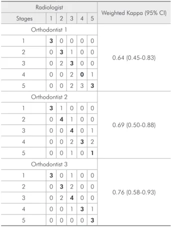
Table 1 - Interexaminer agreement (radiologist vs. ortho-dontists) for the Hassel-Farman method,8 expressed as
skel-etal maturation stage.
Radiologist
Weighted Kappa (95% CI) Stages 1 2 3 4 5
Orthodontist 1
0.64 (0.45-0.83)
1 3 0 0 0 0
2 0 3 1 0 0
3 0 2 3 0 0
4 0 0 2 0 1
5 0 0 2 3 3
Orthodontist 2
0.69 (0.50-0.88)
1 3 1 0 0 0
2 0 4 1 0 0
3 0 0 4 0 1
4 0 0 2 3 2
5 0 0 1 0 1
Orthodontist 3
0.76 (0.58-0.93)
1 3 0 1 0 0
2 0 3 2 0 0
3 0 2 4 0 0
4 0 0 1 3 1
reference point, being the most experienced among the evaluators as well as a researcher in the ield of imaging diagnostics. After we collected the data, the weighted Kappa test was performed to assess the de-gree of intrareference examiner (radiologist) ade-gree- agree-ment and interexaminer agreeagree-ment between the ra-diologist and the three orthodontists. Orthodontist 1 had 4 years of clinical experience, whereas ortho-dontists 2 and 3 had 9 and 15 years of experience, respectively.
The data were analyzed using the Stata com-puter program (Stata, Version 9.0, Stata Corp., College Station, TX, USA) and the degree of agree-ment was obtained based on the Altman classi-ication:11 0.00 – 0.20 = very low; 0.21 – 0.40 = low; 0.41 – 0.60 = fair; 0.61 – 0.80 = good; and 0.81 – 1.00 = very good. The conidence inter-val (CI) was 95%. The study was approved by the Ethics Committee of the University of Vale do Rio Verde, Faculty of Dentistry.
Results
Agreement comparing the three methods with one another, as determined by the principal examin-er (radiologist) was considexamin-ered good (> 0.64). Table 1 shows good agreement between the principal aminer (radiologist) and the three orthodontist ex-aminers (0.64, 0.69, and 0.76, respectively) for the Hassel-Farman method.8
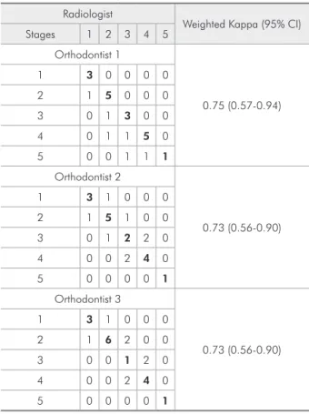
There was also good agreement between the ra-diologist and the three orthodontist examiners con-cerning the Bacceti et al. method9 (0.75, 0.73, and 0.73, respectively) (Table 2) and the Seedat-Forsberg method10 (0.63, 0.66, and 0.54, respectively) (Table 3).
Discussion
Results of our study indicate that determination of skeletal maturation stage via cervical vertebrae had satisfactory clinical applicability, within the limits of this study, using any of the three principal
Table 2 - Interexaminer agreement (radiologist vs. ortho-dontists) for the Baccetti et al. method9 expressed as skeletal
maturation stage.
Radiologist
Weighted Kappa (95% CI) Stages 1 2 3 4 5
Orthodontist 1
0.75 (0.57-0.94)
1 3 0 0 0 0
2 1 5 0 0 0
3 0 1 3 0 0
4 0 1 1 5 0
5 0 0 1 1 1
Orthodontist 2
0.73 (0.56-0.90)
1 3 1 0 0 0
2 1 5 1 0 0
3 0 1 2 2 0
4 0 0 2 4 0
5 0 0 0 0 1
Orthodontist 3
0.73 (0.56-0.90)
1 3 1 0 0 0
2 1 6 2 0 0
3 0 0 1 2 0
4 0 0 2 4 0
5 0 0 0 0 1
Table 3 - Interexaminer agreement (radiologist vs. ortho-dontists) for the Seedat-Forsberg method10 expressed as
skeletal maturation stage.
Radiologist
Weighted Kappa (95% CI) Stages 1 2 3 4 5
Orthodontist 1
0.63 (0.43-0.82)
1 3 0 0 0 0
2 2 3 0 0 0
3 0 4 3 1 0
4 0 0 0 1 0
5 0 0 2 1 3
Orthodontist 2
0.66 (0.43-0.89)
1 4 0 0 0 0
2 0 4 0 0 0
3 1 3 5 0 0
4 0 0 0 1 0
5 0 0 1 1 3
Orthodontist 3
0.54 (0.33-0.74)
1 3 1 0 0 0
2 1 3 0 0 0
3 1 3 0 4 1
4 0 0 2 1 0
methods referenced in the literature. A number of studies state that cervical vertebrae can be used as a reliable parameter for estimating skeletal age, but stress that this method should not be used exclusive-ly and that further information should be acquired in order to reach a precise determination.4-7,12
In both the principal intraexaminer (radiologist) comparison and comparisons between the princi-pal examiner and the orthodontists, the best results were obtained using the Baccetti et al. method.9 The authors propose a new classiication, in which there are ive maturation stages and variations in each stage, thereby allowing improved radiograph clas-siication. As one maturation stage does not abrupt-ly evolve into another, the Baccetti et al. method9 eliminates many of the doubts as to which stage an individual belongs. In this method, denominated CVMS, only Stage III does not have a variation. The evaluators also agreed that the Baccetti et al. method9 was easier to administer than the others, as the two anatomical variables in each stage simplify the classiication. Although new, the validity of this method has been proven, and it has been used in a study aimed at developing a new formula for assess-ing the potential of mandibular growth.9,12-15
On the other hand, the Seedat-Forsberg
meth-od10 has a more simpliied form, as it only assess-es the C3 vertebra (its shape, the concavity on the lower edge, the S-shape on the upper edge, the lip-shape on the lower corners, and the rounding of the lower corners of the vertebral body) and uses the six stages proposed by Hassel and Farman.8 However, this method was the one that generated the great-est amount of doubt regarding the identiication of the maturation stage. All evaluators stated that the method proposed by Seedat and Forsberg10 was the most dificult for identifying the skeletal maturation stage of orthodontic patients, reporting that the an-atomical details viewed in this method are very sub-tle, thereby hindering their visualization. One alter-native would be a combination of this method with another, such as that of Baccetti et al.9 Although this method achieved the least degree of agreement between evaluators, the results do not contraindi-cate its use.
Conclusion
The three methods demonstrated clinical appli-cability. However, the method proposed by Baccetti
et al.9 achieved the best results, followed by the Has-sel-Farman method8 and the Seedat-Forsberg meth-od.10
References
1. San Román P, Palma JC, Oteo MD, Nevado E. Skeletal matu-ration determined by cervical vertebrae development. Eur J Orthod. 2002 Jun;24(3):303-11.
2. Baccetti T, Franchi L, McNamara JA Jr. The cervical vertebral maturation (CVM) method for the assessment of optimal treatment timing in dentofacial orthopedics. Semin Orthod. 2005 Sep;11(3):119-29.
3. Generoso R, Sadoco EC, Armond MC, Gameiro GH. Evalu-ation of mandibular length in subjects with Class I and Class II skeletal patterns using the cervical vertebrae maturation. Braz Oral Res. 2010 Jan-Mar;24(1):46-51.
4. Flores-Mir C, Burgess CA, Champney M, Jensen RJ, Pitcher MR, Major PW. Correlation to skeletal maturation stages determined by cervical vertebrae and hand-wrist evaluations. Angle Orthod. 2006 Jan;76(1):1-5.
5. Gandini P, Mancini M, Andreani F. A comparison of hand-wrist bone and cervical vertebral analyses in measuring skel-etal maturation. Angle Orthod. 2006 Nov;76(6):984-9.
6. Soegiharto BM, Moles DR, Cunningham SJ. Discriminatory ability of the skeletal maturation index and the cervical ver-tebrae maturation index in detecting peak pubertal growth in Indonesian and white subjects with receiver operating char-acteristics analysis. Am J Orthod Dentofacial Orthop. 2008 Aug;134(2):227-37.
7. Lai EH, Liu JP, Chang JZ, Tsai SJ, Yao CC, Chen MH, et al. Radiographic assessment of skeletal maturation stages for orthodontic patients: hand-wrist bones or cervical vertebrae? J Formos Med Assoc. 2008 Apr;107(4):316-25.
8. Hassel B, Farman AG. Skeletal maturation evaluation us-ing cervical vertebrae. Am J Orthod Dentofacial Orthop. 1995 Jan;107(1):58-66.
10. Seedat AK, Forsberg CD. An evaluation of the third cervical vertebra (C3) as a growth indicator in Black subjects. SADJ. 2005 May;60(4):156,158-60.
11. Altman DG. Practical statistics for medical research. London: Chapman and Hall, 1991. 611 p.
12. Grave K, Townsend G. Cervical vertebral maturation as a predictor of the adolescent growth spurt. Aust Orthod J. 2003 Apr;19(1):25-32.
13. Santos EC, Bertoz FA, Arantes FM, Reis PM, Bertoz AP. Skel-etal maturation analysis by morphological evaluation of the
c er v ic a l ver tebrae. J C l i n Ped iat r D ent . 20 0 6 Spring;30(3):265-70.
14. Franchi L, Baccetti T, McNamara JA Jr. Mandibular growth as related to cervical vertebral maturation and body height. Am J Orthod Dentofacial Orthop. 2000 Sep;118(3):335-340.