Oral Radiology
Otavio Shoiti Umetsubo Bruno Felipe Gaia Felipe Ferreira Costa Marcelo Gusmão Paraiso Cavalcanti
Departmentof Stomatology, School of Dentistry, University of São Paulo, São Paulo, SP, Brazil.
Corresponding Author:
Marcelo Gusmão Paraiso Cavalcanti E-mail: [email protected]
Received for publication on Mar 06, 2012 Accepted for publication on Apr 29, 2012
Detection of simulated incipient
furcation involvement by CBCT: an
in
vitro
study using pig mandibles
Abstract: The aim of the present study was to test the reproducibility, sensitivity, and speciicity of cone-beam computed tomography (CBCT) in detecting incipient furcation involvement. Fifteen macerated pig man-dibles, with intact second molar teeth and preserved adjacent cortical ar-eas, were used. Simulated lesions were created in the furcation region of these teeth by applying 70% perchloric acid in up to four possible buc-cal/lingual sites in the right/left sides of each mandible. The mandibles were then submitted to a CBCT scan. Two blinded and calibrated expe-rienced oral and maxillofacial radiologists interpreted the exams. Furca-tion involvement was also assessed in the regions without simulated le-sions. CBCT showed high levels of accuracy, ranging from 78% to 88%. The variations in Kappa values for intra- and inter-observer agreement (0.41–0.59) were considered moderate. CBCT can be considered a reli-able and accurate method for detecting incipient furcation involvement.
Descriptors: Cone-beam Computed Tomography; Periodontics; Furcation Defects.
Introduction
Furcation involvement is one of the major problems in the treatment of periodontal disease, and is directly associated with tooth loss. There-fore, furcation lesions must be detected with accuracy at an early stage of the disease.1,2 Periapical and interproximal intraoral radiographs are
widely accepted as valuable tools for establishing the diagnosis and guid-ing the treatment of periodontal disease, mainly to assess the pattern and extent of bone resorption.3,4 These characteristics can be clearly viewed
in the interproximal alveolar bone. However, intraoral radiographs have the inherent inaccuracy of a two-dimensional exam. In the free faces, structural overlapping usually impairs the demarcation of bone loss ex-tension in the vestibular and lingual cortical areas.5 Studies have shown
a signiicant advantage of CBCT over intraoral radiographs owing to its three-dimensional capability to assess buccal and lingual surfaces.6,7
Recently, cone-beam computed tomography (CBCT) has been used to evaluate dental structures, providing volumetric and 3D information of submillimeter resolution and without any image distortion of the ana-tomic structures.8-10 Several studies have evaluated the potential of this
imaging technique both in vivo (in advanced disease conditions, such as chronic periodontitis)5,11,12 and in vitro (using simulated lesions created
with high-speed spherical drills).6,13-17 These
stud-ies have shown high levels of accuracy for CBCT in detecting lesions owing to lesion size (in cases of chronic periodontal disease) or to well-deined le-sion boundaries resulting from a methodology that uses drills (in vitro studies).
In previous studies, the performance of periapi-cal radiography in detecting larger periodontal le-sions proved inferior to that of CBCT.5-7,11-17 In
clini-cal studies,5,11,12 the primary purpose of radiographic
examination was not as much to perform an early detection of periodontitis, as it was to characterize the disease and support the treatment follow-up.
In vitro studies6,13-17 simulate lesions by
pro-ducing them with high-speed spherical drills. This methodology makes it possible to create defects with deined contours18 and to simulate an advanced
stage of the disease.
The aim of the present study was thus to test the reproducibility, sensitivity, and speciicity of cone-beam computed tomography (CBCT) in detecting incipient furcation involvement.
Methodology
Selection of specimens
The present study was submitted to the Ethics and Research Committee of our institution for ap-proval under protocol no. 104/2010.
Fifteen mandibles of young adult pigs (previous-ly slaughtered for human consumption) were used. The mandibular second molars were intact and the adjacent cortex was preserved.19 The mandibles
were boiled in water (for 8 hours, twice) and their soft tissues (gingivae, mucosa, and muscles) were removed. The mandibles were then washed with neutral soap to remove lipid remnants. Before the simulated lesions were created, the specimens were assessed clinically and tomographically in order to assess whether they had any original bone defect that could be misinterpreted in the ensuing diagnos-tic imaging. Any specimen that had a bone defect was excluded from the study.
Simulating lesions in the alveolar process in the furcation region
Simulated lesions were created in the
furca-tion region of the second molars in up to four pos-sible sites (buccally and lingually, and in the right and left sides) of each mandible, for a total of 60 possible sites. These sites were divided into a con-trol group (with no lesion) and a test group (with a lesion), which were then analyzed simultaneously. Twenty of these sites were selected randomly, so that a mandible could have a variable number (0–4) of lesions in the furcation region of the molar teeth, bilaterally. One of the authors, whose job was not to evaluate the images (O.S.U.), created the lesions. The lesions were made to simulate incipient bone loss caused by periodontal disease at an early stage. Two-millimeter cotton pledgets soaked in 70% per-chloric acid were kept in contact with the bone for 2 hours (Figure 1); after each application, the mandi-bles were washed for 1 min under tap water, accord-ing to the methodology used in previous studies.19,20
Image acquisition
CBCT images of the mandibles were acquired with a single apparatus (i-CAT New Generation, Imaging Sciences International, Hatield, USA), us-ing the followus-ing acquisition protocol:
• voxel size: 0.2 mm,
• acquisition time: 26.9 s,
• tube voltage: 120 kV,
• ilament current: 5 mA,
• ield of view (FOV): 6 × 16 cm.
mandibular molar region and identify any possible furcation involvement, even in the regions where no simulated lesion was created. They were blinded to any information on the procedures performed and on the conditions of the teeth. Each observer ana-lyzed the images independently using multiplanar reconstructed images (axial, coronal and sagittal, si-multaneously), in an independent workstation (Fig-ure 2). They were free to use the software visual-ization tools (contrast, magniication, and window width and level). When interpreting the images, the room lights were turned off.
The interpretation time was not restricted, and the second assessment of the same images was re-peated within a 14-day interval by the same observ-ers to produce Observation 1’ and Observation 2’. The absence or presence of a bone defect was as-sessed in each of the four sites on the 15 mandibles
(n = 60) to compare the imaging diagnosis of the lesions with that of the gold standard (macerated mandible).
Statistical analysis was carried out using the validity and Kappa tests (SPSS software, v. 17.0.0, SPSS Inc., Chicago, USA). The values for the Kappa (κ) coeficient were calculated to assess the intra- and inter-observer agreement, which were classiied according to the following criteria:
• low (κ = 0.20–0.39),
• moderate (κ = 0.40–0.59), and
• substantial (κ = 0.60–0.79).
The values for sensitivity and speciicity were es-timated in order to check the agreement of the as-sessments against the gold standard, and to distin-guish the false positives and false negatives observed in each group.
Figure 1 - Positioning of the cotton fragment (A), and chemical lesion simulation. (B) Final aspect, two hours after acid ap-plication.
Figure 2 - CBCT axial (A) and coronal (B) images. The lesion on the right vestibular surface (arrows) was detected by both observers.
B
B A
Results
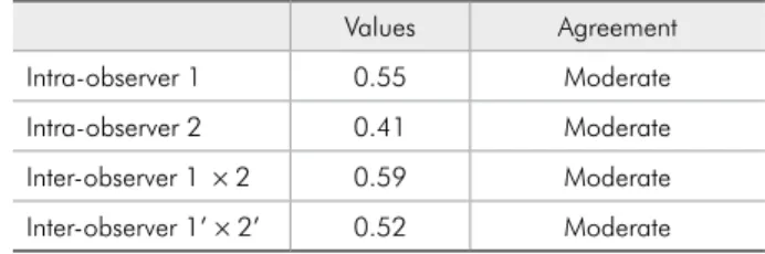
The kappa values for the intra-observer agree-ment in detecting incipient furcation involveagree-ment ranged from 0.41 to 0.55. The kappa values for in-ter-observer agreement, ranged from 0.52 to 0.59. All of these values were considered as representing moderate levels of agreement (Table 1).
Table 2 shows the results for overall sensitivity, speciicity, and accuracy in detecting incipient furca-tion involvement. These data represent all observa-tions for both observers. CBCT showed moderate sensitivity (ranging from 50% to 75%), represented by the true positive results of detecting incipient fur-cation involvement. However, high speciicity (rang-ing from 93% to 100%) was observed, represented by the correlation between the true negative results and the sites where there was actually no lesion. The accuracy of CBCT (ranging from 78% to 88%) was represented by the proportion between true positive and true negative results, considered simultaneously in relation to all results
Discussion
According to Mol,4 an imaging examination
(ei-ther intraoral radiography or CBCT) in periodontol-ogy will beneit the patient only if it can inluence the treatment. Therefore, an exam should be indi-cated only if the additional information it provides will make a difference in patient management and if this difference will have an impact on the outcomes that are important to him/her. The same author em-phasizes that three-dimensional (3D) information may have advantages over information obtained from two-dimensional (2D) examinations. Howev-er, it is necessary to evaluate whether an increase in diagnostic eficacy has an impact on both the treat-ment and the overall results for the patient. Corbet et al.3 reported that neither helical nor multislice CT
offer any favorable cost-beneit ratio or therapeutic advantage to periodontal practice. These authors disagree with Mol4 and state that CBCT should be
used more often because it involves a lower radiation dose. We agree with the statements made by Mol,4
Vandenberghe et al.16 and Walter et al.21 that a
diag-nostic method must produce a clinical outcome, and we emphasize that the biological effects of ionizing
radiation must always be taken into account, even though the radiation dose from CBCT is reduced.
Naitoh et al.5 presented a case of upper molars
with pocket depth measurements of 4 mm (mesio-buccal), 6 mm (disto-(mesio-buccal), 6 mm (mesio-palatal), and 5 mm (disto-palatal). They also used 3D recon-structed images from CBCT to characterize alveolar bone loss. However, this procedure is controversial, since several factors involved in 3D image acquisi-tion may interfere with the results, namely:
• the operator’s subjective analysis,
• the acquisition parameters (kV, mA, voxel size and FOV size), and
• the software parameters (threshold, reconstruc-tion algorithms).22
Walter et al.11,12 presented clinical cases with
se-vere chronic periodontitis, in which a furcation in-volvement was clinically detected. CBCT was used for lesion assessment and treatment planning, not for the early detection and prevention of lesions. Grimard et al.8 evaluated the outcomes of a
regen-erative treatment using intraoral radiographs and CBCT images. According to Naitoh et al.5 and
Wal-ter et al.,11,12 the subjects included in these studies
presented advanced stages of periodontitis. In the present study, our goal was to analyze CBCT im-ages of incipient furcation lesions using a rigorous Table 2 - Sensitivity, specificity and accuracy values for both observers in both analyses.
Sensitivity Specificity Accuracy First
assessment
Observation 1 70% 98% 88%
Observation 2 75% 90% 85%
Second assessment
Observation 1’ 50% 100% 83%
Observation 2’ 50% 93% 78% Table 1 - Intra- and inter-observer agreement.
Values Agreement
Intra-observer 1 0.55 Moderate
Intra-observer 2 0.41 Moderate
experimental control. Lesions of this size were not analyzed in previous studies.
Misch et al.,6 Grimard et al.8 and Noujeim et al.15
addressed periodontal lesions on the proximal sur-faces of teeth. Interproximal lesions render CBCT a less decisive tool for the prognosis of affected teeth (access to and hygiene of a tooth’s mesial and distal faces are easier than access/hygiene in the furcation region). Periodontal resorption begins with immu-nological and inlammatory events, leading to bone demineralization. Therefore, it is possible that bone loss is not related to the degree of inlammation or the qualitative nature of the inlammation, but rath-er to whrath-ere the inlammation is located in the bone and to how long it persists.
In the in vitro studies of Misch et al.,6 Mengel et
al.,13 Vandenberghe et al.16,17 and Noujeim et al.,15
drills were used to simulate lesions. In these studies, CBCT images demonstrated good reliability in both detecting the lesions and measuring their dimen-sions. However, lesions simulated with a drill would represent a more advanced stage of the disease, which could be detected by either periodontal prob-ing or intraoral radiography. Fuhrmann et al.23 also
used drills to simulate lesions, and reported that he-lical CT is a reliable technique to classify furcation involvement both vertically and horizontally.
In previous studies, Mol & Balasundaram14
con-cluded that CBCT is better than intraoral radiogra-phy in terms of both aiding in establishing the di-agnosis and providing quantitative information on periodontal bone levels. However, the authors ad-mitted that CBCT failed to provide suficient spatial resolution in cases where the diagnostic decisions depended on small details. Periodontal resorption starts with inlammatory and immune events, re-sulting in bone demineralization. At an initial stage, the cavities are small and have irregular borders.24,25
We believe that lesions are more dificult to detect with either intraoral radiography or CBCT at this stage. In the simulations shown in the present study, we intended to explore the extent to which addi-tional information obtained by CBCT images can impact the periodontal treatment. We simulated the bone loss caused by inlammation occurring more deeply, or otherwise not easily detected clinically.
We also simulated a situation in which CBCT could detect a bone resorption process in time to allow re-moving the cause of inlammation and resuming the physiological cycle, thus allowing bone formation and restoration of the lost bone.
The lesions created in the present study were smaller than those produced in previous stud-ies,6,13-17 with the aim of simulating an initial stage
of bone resorption. Studies validating the use of CBCT to detect lesions, simulating an early stage of periodontitis, with a rigorous experimental control, were not found in the related literature.
The research published by Mol & Balasunda-ram14 demonstrated low levels of intra-observer
agreement. In our study, moderate levels of intra- and inter-observer agreement were obtained. The intra-observer agreement may have increased owing to the advanced technology of the equipment. A high amount of false-negative diagnoses were reached, which may be explained by both the reduced lesion dimensions and the changes in the technique used to simulate the lesions.
It is clear that CBCT is still not the irst choice for periodontal bone support imaging. This func-tion continues to be well performed by periapical ra-diographs. However, CBCT can help in cases where periapical radiographs are not suficiently illustra-tive owing to overlapping structures. Nevertheless, CBCT imaging can be indicated for other applica-tions, such as assessments for implant placement and periodontal furcation surgery. Even though incipient furcation lesions are not considered major indications for CBCT, any changes detected in the furcation region of molar teeth in patients scanned for other reasons should nevertheless be reported.
Following the guidelines of imaging interpreta-tion criteria, the whole volume of data obtained by a CBCT examination should be carefully assessed, regardless of the original diagnostic question under-lying the examination.
Conclusion
re-liable tool for detecting incipient furcation involve-ment, thus expanding the applicability reported by other authors, who tested the CBCT technique as applied to larger furcation lesions, which were not created chemically using acid.
Acknowledgements
We wish to acknowledge the National Coun-cil for Scientiic and Technological Development (CNPq), Brasília, DF, Brazil, for inancially sup-porting the work of Dr. Marcelo Cavalcanti,
Uni-versal Research Project, grant no. 472895/2009-5, and Research Productivity Scholarship, grant no. 303847/2009-3. We also wish to thank the Coor-dination for the Improvement of Higher Education Personnel (CAPES), Brasília, DF, Brazil, for provid-ing PhD scholarships to Felipe Costa and Bruno Gaia, and a Master’s degree scholarship to Otavio Umetsubo. Additionally, we would like to thank the Kavo Research Institute for providing the CBCT equipment support.
References
1. Hishikawa T, Izumi M, Naitoh M, Furukawa M, Yoshinari N, Kawase H, et al. The effect of horizontal X-ray beam an-gulation on the detection of furcation defects of mandibular first molars in intraoral radiography. Dentomaxillofac Radiol. 2010 Feb;39(2):85-90.
2. Walter C, Weiger R, Zitzmann NU. Periodontal surgery in furcation-involved maxillary molars revisited—an introduc-tion of guidelines for comprehensive treatment. Clin Oral Investig. 2011 Feb;15(1):9-20.
3. Corbet EF, Ho DKL, Lai SML. Radiographs in periodontal disease diagnosis and management. Aust Dent J. 2009 Sep;54 Suppl 1:S27-43.
4. Mol A. Imaging methods in periodontology. Periodontol 2000. 2004 Feb;34(1):34-48.
5. Naitoh M, Yamada S, Noguchi T, Ariji E, Nagao J, Mori K, et al. Three-dimensional display with quantitative analysis in alveolar bone resorption using cone-beam computerized tomography for dental use: a preliminary study. Int J Peri-odontics Restorative Dent. 2006 Dec;26(6):607-12. 6. Misch KA, Yi ES, Sarment DP. Accuracy of cone beam
com-puted tomography for periodontal defect measurements. J Periodontol. 2006 Jul;77(7):1261-6.
7. De Faria Vasconcelos K, Evangelista K, Rodrigues C, Estrela C, de Sousa T, Silva M. Detection of periodontal bone loss using cone beam CT and intraoral radiography. Dentomaxil-lofac Radiol. 2012 Jan;41(1):64-9.
8. Grimard BA, Hoidal MJ, Mills MP, Mellonig JT, Nummikoski PV, Mealey BL. Comparison of clinical, periapical radiograph, and cone-beam volume tomography measurement techniques for assessing bone level changes following regenerative peri-odontal therapy. J Periodontol. 2009 Jan;80(1):48-55. 9. Tetradis S, Anstey P, Graff-Radford S. Cone beam computed
tomography in the diagnosis of dental disease. J Calif Dent Assoc. 2010 Jan;38(1):27-32.
10. Cavalcanti MG. Cone beam computed tomographic imaging: perspective, challenges, and the impact of near-trend future applications. J Craniofac Surg. 2012 Jan;23(1):279-82.
11. Walter C, Kaner D, Berndt DC, Weiger R, Zitzmann NU. Three-dimensional imaging as a pre-operative tool in deci-sion making for furcation surgery. J Clin Periodontol. 2009 Mar;36(3):250-7.
12. Walter C, Weiger R, Zitzmann NU. Accuracy of three-dimen-sional imaging in assessing maxillary furcation involvement. J Clin Periodontol. 2010 May;37(5):436-41.
13. Mengel R, Candir M, Shiratori K, Flores-de-Jacoby L. Digital volume tomography in the diagnosis of periodontal defects: an in vitro study on native pig and human mandibles. J Peri-odontol. 2005 May;76(5):665-73.
14. Mol A, Balasundaram A. In vitro cone beam computed tomog-raphy imaging of periodontal bone. Dentomaxillofac Radiol. 2008 Sep;37(6):319-24.
15. Noujeim M, Prihoda T, Langlais R, Nummikoski P. Evalua-tion of high-resoluEvalua-tion cone beam computed tomography in the detection of simulated interradicular bone lesions. Den-tomaxillofac Radiol. 2009 Mar;38(3):156-62.
16. Vandenberghe B, Jacobs R, Yang J. Diagnostic validity (or acuity) of 2D CCD versus 3D CBCT – images for assessing periodontal breakdown. Oral Surg Oral Med Oral Pathol Oral Radiol Endod. 2007 Sep;104(3):395-401.
17. Vandenberghe B, Jacobs R, Yang J. Detection of periodontal bone loss using digital intraoral and cone beam computed tomography images: an in vitro assessment of bony and/or in-frabony defects. Dentomaxillofac Radiol. 2008 Jul;37(5):252-60.
18. Tirrell BC, Miles DA, Brown CE, Legan JJ. Interpretation of chemically created lesions using direct digital imaging. J Endod. 1996 Feb;22(2):74-8.
19. Stravopoulos A, Wenzel A. Accuracy of cone beam dental CT, intraoral digital and conventional film radiography for the detection of periapical lesions. An ex vivo study in pig jaws. Clin Oral Investig. 2007 Mar;11(1):101-6.
con-ventional film radiography. Dentomaxillofac Radiol. 2009 Oct;38(7):458-64.
21. Walter C, Weiger R, Dietrich T, Lang NP, Zitzmann NU. Does three-dimensional imaging offer a financial benefit for treating maxillary molars with furcation involvement? – A pilot clini-cal case series. Clin Oral Implants Res. 2012 Mar;23(3):351-8. 22. Hassan B, Couto Souza P, Jacobs R, de Azambuja Berti S, van
der Stelt P. Influence of scanning and reconstruction param-eters on quality of three-dimensional surface models of the dental arches from cone beam computed tomography. Clin Oral Investig. 2010 Jun;14(3):303-10.
23. Fuhrmann RAW, Bücker A, Diedrich PR. Furcation involve-ment: comparison of dental radiographs and HR-CT-slices in human specimens. J Periodontal Res. 1997 Jul;32(5):409-18. 24. Graves DT, Li J, Cochran DL. Inflammation and uncoupling
as mechanisms of periodontal bone loss. J Dent Res. 2011 Feb;90(2):143-53.