This study used micro-computed tomography (micro-CT) to evaluate the fit of the master gutta-percha cone at time of cone fit, gutta-percha volume in the filling material, and the filling material volume in relation to the canal at the apical limit of the working length. Root canals of 20 maxillary central incisors were prepared with rotary instruments and distributed into two groups (n=10). The gutta-percha cone tip was either plasticized (apical thermal impression technique - ATI) or not (conventional technique - CT), and its apical fit was checked. The apical 1 mm of working length was examined with a micro-CT, canals were filled with gutta-percha and sealer, and new micro-CT scans were obtained. In CT, gutta-percha filled 35.83 ± 15.05% of the canal at cone selection and 38.72 ± 11.64% after filling. In ATI, these values were 23.14 ± 7.74% and 26.98 ± 20.40%, respectively. Gutta-percha volume in the filling material, and filling material volume in relation to the canal were, respectively, 61.28 ± 11.64% and 87.76 ± 9.98% for CT, and 73.00 ± 20.41% and 89.96 ± 9.08% for ATI. No significant difference was found between cone selection and after canal filling, for either CT (p=0.593) or ATI (p=0.4975). The techniques did not differ significantly with respect to gutta-percha volume in the filling material (p=0.132) and filling material volume in relation to the canal (p=0.612). An ideal fit of the master gutta-percha cone at working length was not achieved regardless of the cone selection technique, and the material-filled area was similar for both techniques.
Comparison of Two Techniques for
Selection of Master Gutta-Percha Cone
Using Micro-Computed Tomography
João Manoel Silva-Filho1, Aline Evangelista Souza-Gabriel2, Graziela Bianchi Leoni2, Samuel Henrique Câmara De-Bem2, Edson Alfredo1, Ricardo Gariba Silva1
1School of Dentistry, University of Ribeirão Preto, Ribeirão Preto, SP, Brazil
2Department of Restorative Dentistry, Ribeirão Preto School of Dentistry, USP - University of São Paulo, Ribeirão Preto, SP, Brazil
Correspondence: Profa. Dra. Aline Evangelista Souza-Gabriel, Avenida Portugal 1690, Apto 84, 14020-380 Ribeirão Preto, SP, Brasil. Tel: +55-16-3602-4078. e-mail: [email protected]
Key Words: Endodontics, gutta-p e r c h a , m i c r o - c o m gutta-p u t e d tomography, root canal.
Introduction
The combination of gutta-percha cones with a sealer is still the most common method for obturation (1,2). During selection of the gutta-percha master cone, retention or tug-back of the cone at the apical canal end does not ensure an ideal apical fit, which occurs only when the cross-section of the cone coincides with the cross-section of the canal. The lack of accuracy in the cone manufacturing process allied to an incorrect choice of cone size might permit a tug-back sensation without an actual fit of the master cone to root canal, which leaves gaps and endangers the apical seal of the filling (3,4).
Although it is not routinely performed in the clinical practice, the gutta-percha cone can be dipped for two or three seconds in heated water or solvent for plasticization, inserted into the apical stop and then removed a few times in order to take an "impression" of the apical portion of the canal. Plasticized gutta-percha can fill the canals more efficiently, especially those with a more complex anatomy (5-7).
The correct cone selection can be confirmed by periapical radiography or computed tomography (8,9). High-resolution micro-computed tomography (micro-CT) can be used for the evaluation of 3D shapes and volumes
of root canals (10,11). This method does not requires tooth preparation (11,12), producing images with good geometric resolution, relatively short acquisition times, simple operational procedures and fast scanning protocols (13,14).
In this study, micro-CT was used to evaluate the fit of the master percha cone at time of cone fit, gutta-percha volume in the filling material, and also check the filling material volume of in relation to the canal at the apical limit of the working length. For illustration purposes, the cases in which sealer extrusion occurred beyond the apical limit of the master gutta-percha were recorded.
Material and Methods
Twenty human maxillary central incisors were used in the study. Access to the pulp chamber was done and a size 15 K-file (Dentsply Maillefer, Ballaigues, Switzerland) was introduced into each canal until its tip was visible at the apical foramen. The working length was established by subtracting 1 mm from this length. The coronal third of the canals was preflared with Gates Glidden drills (Dentsply Maillefer) and then the canals were prepared with K3® rotary instruments (SybronEndo, Glendora, CA, USA) in the following sequence: 20/.02, 25/.02, 30/.02, 30/.04, 35/.02, 40/.02, 45/.02, 50/.02. The apical stop was prepared with ISSN 0103-6440
Braz Dent J 24(4) 2013
368
J. M. Silva-Filho et al.
the last instrument. The canals were irrigated with 2 mL of 1.0% NaOCl at each change of file. The canals were flooded with 17% EDTA for 5 min followed by flushing with distilled water and drying.
The roots were randomly distributed into two groups (n=10), according to the technique used for selection of the master percha cone. In both groups, a gutta-percha cone of the same size as the master apical file (50/.02) was chosen after confirming its diameter with a calibrating ruler. The tip of the cone was either softened with a heated #24 spatula (apical impression technique) or not (conventional technique), and the gutta-percha cone was tried into the canal to check apical fit according to three tests: visual - the cone tip reached the full working length; tactile - tug-back was felt when the cone tip was at the apical length indicating its tight fit in the apical end of the working length; and radiographic - the cone tip reached the radiographic working length. In both techniques, marks were made on the cone and tooth crown to ensure that the cone could always be placed in the same position.
The specimens were subjected to an initial tomographic analysis using a micro-CT scanner (SkyScan 1174v2; SkyScan NV, Kontich, Belgium) set at an isotropic resolution of 22.6 mm, max 50 kV, max 800 mA, 40 W maximum power, 30 µm spatial resolution, and projections from 360-degree acquisition rotation. The volume of gutta-percha and the volume of the root canal were estimated in the apical 1 mm of the working length, which correspond to the first 35 µCT slices (35 slices x 30 µm = 1050 µm ± 1 mm) and was considered as the region of interest for analysis. The coincidence/discrepancy between the gutta-percha cone volume and the canal volume was considered as fit/non-fit of the master gutta-percha cone at the apical canal, expressed as the percentage of voids relative to the canal volume, and was obtained using the following formula: (canal volume–gutta-percha volume ÷ canal volume) X 100.
After the initial micro-CT imaging, the root canals were filled with the master gutta-percha cone, AH Plus sealer (De Trey-Dentsply, Konstanz, Germany) and R8 accessory cones (Dentsply Ind. e Com. Ltda., Petrópolis, RJ, Brazil),
according to the lateral compaction technique. The sealer was taken to the canal with a #20 finger spreader up to 3 mm short of the working length and then the master gutta-percha cone was coated with sealer and positioned in the canal according to marks made on the cone and tooth crown. Accessory cones coated with sealer were inserted. Quality and apical extension of the filling was assessed radiographically and the teeth were stored at 37 °C for 72 h.
A new micro-CT imaging was performed to determine the volume of gutta-percha in the filling material and the volume of filling material in relation to the canal volume in the region of interest (apical 1 mm of the working length), using the following formulas, respectively: (gutta-percha volume ÷ canal volume) X 100 and (filling material volume ÷ canal volume) X 100. For illustration purposes, the cases in which there was sealer extrusion beyond the apical limit of the master gutta-percha cone were recorded.
Data were analyzed statistically by the paired t-test at 5% significance level, using the software SPSS version 19 (SPSS Inc., Chicago, IL, USA).
Results
No significant difference was found between the values obtained at the moment of cone selection and after root canal filling for either the conventional technique (p=0.593) or the apical impression technique (p=0.4975) (Table 1).
There was no significant difference between the conventional and apical thermal impression techniques with respect to gutta-percha volume in the filling material (p= 0.132) and filling material volume in relation to the canal volume (p=0.612) (Table 2).
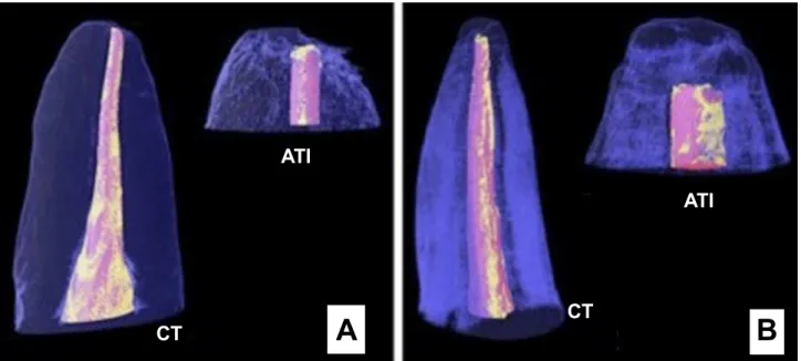
The micro-CT imaging revealed extravasation of sealer in 90% of the cases, regardless of the master gutta-percha cone selection technique. Figure 1 is a micro-CT scan illustrating sealer extrusion beyond the apical limit of the master gutta-percha cone in one specimen. Figure 2 shows the partial three-dimensional reconstruction of root-filled teeth in which the master gutta-percha cone was selected by the conventional and by the apical thermal impression technique. Part of the mineralized dental structure,
Table 1. Mean values (SD) of the difference between the gutta-percha volume and the canal volume at apical 1 mm of the working length at time of cone fit and after root canal filling for both cone selection techniques
Cone selection Conventional technique
Apical thermal impression
At time of cone fit 35.83 ± 15.05A 23.14 ± 7.74A
After root canal filling 38.72 ± 11.64B 26.98 ± 20.40B
Different letters indicate statistically significant difference (p<0.05).
Table 2. Mean values (SD) of gutta-percha volume in the filling material and volume of filling material in relation to the canal at the apical 1 mm of the working length for the conventional and apical thermal impression techniques
Cone selection Conventional technique
Apical thermal impression
Gutta-percha volume 61.28 ± 11.64A 73.00 ± 20.41A
Filling material volume 87.76 ± 9.98B 89.96 ± 9.08B
Braz Dent J 24(4) 2013
369
T
echniques of cone selection and micro-CT
gutta-percha and sealer were observed throughout canal extension and at the apical 1 mm of the working length.
Discussion
The selection of the master gutta-percha cone and sealer are very important factors in obtaining a tight seal of root canal filling (3,4). In the present study, a ‘thermal impression’ of the apical region was taken, considering that thermoplasticized gutta-percha has been shown to
reach areas with more complex anatomy (7), provide a better filling of the canal space and increase its volume in filling material (5,6). However, none of these benefits were observed. For both the conventional and the ATI techniques, the gutta-percha volume was lower than the filling material volume in the apical 1 mm of the working length, which demonstrates that the cone did not fit perfectly to the canal in this region. Neither thermal impression increased the volume of the filling material in relation to the canal volume. Somma et al. (11) evaluated the quality of root fillings completed by thermoplasticized and cold gutta-percha technique by micro-CT and found that all techniques produced similar results and none of them increased gutta-percha volume in the filling material.
As the apical thermal impression technique was not able to increase gutta-percha volume at a critical region - the final millimeter of the working length - either at the moment of selection of the master gutta-percha cone or after canal filling, it can be inferred from our results and previous research (1,5), that there seems to be no actual improvement in the final quality of root filling to justify its routine use in the clinical practice.
This understanding is reinforced by the fact that sealer extrusion beyond the apical limit of the working length was similar (90%) for both gutta-percha cone selection techniques. it may be assumed that sealer extrusion could have been facilitated by the lack of apical fit of the gutta-percha cone tip and by the filling technique, in which the sealer was taken to the canal and then the gutta-percha cone was introduced, exerting pressure towards the apex
A
B
CT
CT
ATI
ATI
Figure 2. Three-dimensional image reconstruction of part of the structure of root-filled teeth in which the master gutta-percha cone was selected using the conventional technique (CT) or the apical thermal impression technique (ATI). The colors blue, pink, and yellow represent, respectively, the mineralized dental structure, the gutta-percha and sealer, along the canal (A) and at the apical 1 mm of the working length (B).
Braz Dent J 24(4) 2013
370
J. M. Silva-Filho et al.
and pushing the sealer into extrusion.
The ‘tug-back’ sensation of the master gutta-percha cone at the working length (tactile test) seems more likely to be due to the fact that the cone gets stuck to the root canal walls, giving a false sensation of perfect fit at the working length and tight seal of the root canal.
The results of the present study demonstrate that the apical 1 mm of the working length was not completely filled, which can be explained by the complex anatomy of this region that leaves under-instrumented areas (15), voids in the canal if the filling material is not condensed properly (16) and deficient adaptation of the filling material leading to formation of interfacial gaps (17,18). Hypothetically, other factors that might be related are the physicochemical properties of sealers, like flow and viscosity, which could allow the filling of voids (19), and the C-factor, which leads to polymerization shrinkage of resin sealers with consequent formation of voids (20).
Under the tested conditions, neither of the techniques used for selection of the master gutta-percha cone was able to provide a perfect fit of the cone at the apical millimeter of the working length. No evident difference was found between the conventional and the apical thermal impression techniques with respect to gutta-percha volume in the filling material.
Resumo
Este estudo utilizou microtomografia computadorizada (micro-CT) para avaliar a adaptação do cone de guta-percha no momento da sua seleção, o volume de guta-percha no material obturador, e o volume do material obturador em relação ao canal no limite apical do comprimento de trabalho. Canais radiculares de 20 incisivos centrais superiores foram preparados com instrumentos rotatórios e distribuídos em dois grupos (n=10). A ponta do cone de guta-percha foi plastificada (técnica de impressão térmica apical - TIT) ou não (técnica convencional - TC), e seu ajuste apical foi verificado. O milimetro apical do comprimento de trabalho foi examinado em micro-CT, os canais foram preenchidos com guta-percha e cimento, e novas imagens em micro-CT foram obtidas. Na TC, a guta-percha preencheu 35,83 ± 15,05% do canal no momento da seleção do cone e 38,72 ± 11,64% após a obturação. Na TIT, estes valores foram de 23,14 ± 7,74% e 26,98 ± 20,40%, respectivamente. O volume de guta-percha no material obturador, e o volume do material obturador em relação ao canal, foram, respectivamente, 61,28 ± 11,64% e 87,76 ± 9,98% para a TC, e 73,00 ± 20,41% e 89,96 ± 9,08% para TIT. Não foi encontrada diferença significativa entre a seleção do cone e depois o preenchimento do canal para ambas as técnicas, TC (p =0,593) ou TIT (p=0,4975). As técnicas não diferiram significativamente com respeito ao volume de guta-percha no material obturador (p=0,132), e volume de preenchimento de material em relação ao canal (p=0,612). Um ajuste ideal do cone principal de guta-percha cone no seu comprimento de trabalho não foi alcançado, independentemente da técnica de seleção empregada, e a area preenchida pelo material obturador foi semelhante para ambas as técnicas.
References
1. Peters CI, Sonntag D, Peters OA. Homogeneity of root canal fillings performed by undergraduate students with warm vertical and cold lateral techniques. Oral Surg Oral Med Oral Pathol Oral Radiol Endod 2010;11:162-165.
2. Souza EM, Wu MK, LW van der Sluis, Leornardo RT, Bonetti-Filho I,
Wesselink PR. Effect of filling technique and root canal area on the percentage of gutta-percha in laterally compacted root fillings. Int Endod J 2009;42:719-726.
3. Souza EM, Wu MK, LW van der Sluis, Leornardo RT, Bonetti-Filho I, Wesselink PR. Effect of filling technique and root canal area on the percentage of gutta-percha in laterally compacted root fillings. Int Endod J 2009; 42: 719-26.
4. Cunningham KP, Walker MP, Kulild JC, Lask JT. Variability of the diameter and taper of size #30, 0.04 gutta-percha cones. J Endod 2010;32:1081-1084.
5. Wu MK, LW van der Sluis, Wesselink PR. A preliminary study of the percentage of gutta-percha-filled area in the apical canal filled with vertically compacted warm gutta-percha. Int Endod J 2002;35:527-535.
6. Jarret IS, Marx D, Covey D, Karmazin M, Lavin M, Gound T. Percentage of canals filled in apical cross sections – an in vitro study of seven obturation techniques. Int Endod J 2004;37:392-398.
7. Peters OA, Boessler C, Paqué F. Root canal preparation with a novel nickel-titanium instrument evaluated with micro-computed tomography: canal surface preparation over time. J Endod 2010b;36:1068-1072.
8. Gambarini G, Plotino G, Grande NM, Testarelli L, Prencipe M, Messineo D, Fratini L, D’ambrosio F. Differential diagnosis of endodontic-related inferior alveolar nerve paraesthesia with cone beam computed tomography: a case report. Int Endod J 2011;44:176-181.
9. Zhang R, Yang H, Yu X, Wang H, Hu T, Dummer PMH. Use of CBCT to identify the morphology of maxillary permanent molar teeth in a Chinese subpopulation. Int Endod J 2011;44:162-169.
10. Moore J, Fitz-Walter P, Parashos P. A micro-computed tomographic evaluation of apical root canal preparation using three instrumentation techniques. Int Endod J 2009;42:1057-1064.
11. Somma F, Cretella G, Carotenuto M, Pecci R, Bedini R, De Biasi M, Angerame D. Quality of thermoplasticized and single point root fillings assessed by micro-computed tomography. Int Endod J 2011;43:1122-1131.
12. Yin X, Cheung GS, Zhang C, Masuda YM, Kimura Y, Matsumoto K. Micro-computed tomographic comparison of nickel-titanium rotary versus traditional instruments in C-shaped root canal system. J Endod 2010;36:708-712.
13. Schambach SJ, Bag S, Schilling L, Groden C, Brockmann MA. Application of micro-CT in small animal imaging. Methods 2010;50:2-13. 14. Bechara B, McMahan CA, Noujeim M, Moore WS, Teixeira FB, Geha H.
Cone beam computed tomography scans with and without artifact reduction in root fracture detection of endodontically treated teeth. Dentomaxillo Radiol 2013; 42: 20120245.
15. Barbizam JVB, Fariniuk LF, Marchesan MA, Pécora JD, Sousa-Neto MD. Effectiveness of manual and rotary instrumentation techniques for cleaning flattened root canals. J Endod 2002;28:365-366.
16. Zaslansky P, Fratzl P, Rack A, Wu MK, Wesselink PR, Shemesh H. Identification of root filling interfaces by microscopy and tomography methods. Int Endod J 2011;44:395-401.
17. Costa JA, Rached-Júnior FA, Souza-Gabriel AE, Silva-Sousa YTC, Sousa-Neto MD. Push-out strength of methacrylate resin-based sealers to root canal walls. Int Endod J 2010;43:698-706.
18. Cavenago BC, Duarte MA, Ordinola-Zapata R, Marciano MA, Carpio-Perochena AE, Bramante CM. Interfacial adaptation of an epoxy-resin sealer and a self-etch sealer to root canal dentin using the System B or the single cone technique. Braz Dent J 2012;23:205-211.
19. Marin-Bauza GA, Rached-Junior FJ, Souza-Gabriel AE, Sousa-Neto MD, Miranda CE, Silva-Sousa YT. Physicochemical properties of methacrylate resin-based root canal sealers. J Endod 2010;36:1531-1536.
20. Flores DS, Rached-Júnior FJ, Versiani MA, Guedes DF, Sousa-Neto MD, Pécora JD. Evaluation of physicochemical properties of four root canal sealers. Int Endod J 2011;44:126-135.