Abstract:
The aim of this study was to investigate the apical leakage of roots filled by three different gutta-percha techniques: lateral condensation, Tagger’s hybrid and E&Q Master. Forty-two extracted single-rooted teeth were used. The coronal part of each tooth was removed and the root received biomechanical preparation using a 60-K file. The roots were randomly divided into three groups according to the technique of filling the root canal: Group I, lateral condensation; Group II, Tagger’s hybrid; Group III, E&Q Master. The roots were submitted to dye leakage test with Rhodamine B for 7 days, using vacuum during the initial 30 min. The teeth were sectioned longitudinally and the leakage was measured in a linear fashion from apex to crown. Statistical analysis indicated that lateral condensation and E&Q Master techniques showed lower leakage than Tagger’s technique (P= 0.0016). However, statistically no difference was found between lateral condensation and E&Q Master system techniques. (J Oral Sci 51, 593-599, 2009)Keywords: apical; leakage; filling; root.
Introduction
Complete obturation of root canals is a step of funda-mental importance in endodontic therapy. Obturation is the final procedure, aiming to fill the space across its length, within appropriate limits using materials and techniques that support the process of apical and periapical repair (1). Studies show that failures in endodontic treatment result from inadequate filling of root canals (2,3). Various materials and techniques have been developed. The gutta-percha associated with an endodontic cement is commonly used in the root canal. Although well accepted, some researchers challenge the way it is used, suggesting variations in the root canal obturation techniques.
The cold lateral condensation (CLC) technique, also called the conventional technique is the filling technique most widely used in the world and it frequently achieves excellent results (2-5). The CLC technique of gutta-percha is the compaction of successive gutta-percha cones associated with endodontic cement with the aid of spacers filling the interior of the root canal. However, some researchers state that it does not provide three-dimensional obturation, causing excessive stress and excessive expend-iture of material (6).
McSpadden in 1979 introduced the thermomechanical condensation of percha technique, using a gutta-percha condensor which was similar to a Hedströen file reversed (7). However, this technique showed failures such as overfilling, possibility of fracture of the instrument, and displacement of the cone, and it was impossible to use in curved canals (8). In 1983 Tagger et al. (9) suggested Journal of Oral Science, Vol. 51, No. 4, 593-599, 2009
Correspondence to Dr. Carlos Rocha Gomes Torres, Universidade Estadual Paulista, Faculdade de Odontologia de São José dos Campos, Avenida Engenheiro Francisco José Longo, 777, Jardim São Dimas, São José dos Campos, CEP: 12245-000, SP, Brazil Tel: +55-12-3947-9048
Fax: +55-12-3947-9010
E-mail: [email protected]
Assessment of the apical seal of root canals using different
filling techniques
Marcela V. Leonardo
1), Edson H. Goto
1), Carlos R. G. Torres
2), Alessandra B. Borges
2),
Cláudio A. T. Carvalho
2)and Daphne C. Barcellos
2)1)
Clinical Research Academic Group, São José dos Campos School of Dentistry, São Paulo State University,
SP, Brazil
2)
Department of Restorative Dentistry, São José dos Campos School of Dentistry, São Paulo State University,
SP, Brazil
594
a modification in the McSppaden technique, thus developing a hybrid technique of obturation of root canals. The hybrid technique is the association of thermo-mechanical obturation with lateral condensation, using the compactors idealized by McSpadden. Moreover, in this technique secondary/accessory cones are applied in the apical third of the canals.
The filling technique with thermoplasticized gutta-percha in a special syringe was introduced to facilitate the completion of the conduit and has been advocated by several authors (10-13). The injectable thermoplasticized gutta-percha systems are used by the action of an electric heater of low intensity that produces uniformity of the material being injected into the root canal with syringe pressure. The E&Q Master system (Meta Dental Corp., Cheongju, Korea) is a relatively new introduction to the endodontic armamentarium for root canal filling, such as the System B technique for the down pack (6) and the use of injectable gutta-percha for backfilling the canal after an apical seal is obtained with the down pack. The system consists of a control unit with a pen-grip device holding a heating tip, as well as a gutta-percha injection gun. To date, no report of the sealing property of this method of delivering gutta-percha is available to support its use as an alternative to the other injectable thermoplasticized gutta-percha systems.
It is important to find a technique, a sealer cement or a combination cement/technique, which can prevent apical leakage of obturated root canals. The CLC, Tagger’s hybrid and injectable thermoplasticized gutta-percha system techniques deserve further investigation and should be compared so that the superiority of one over the other can be determined. The objective of this study was to evaluate
in vitrothe apical seal after three different techniques of
root obturation: CLC, Tagger’s hybrid and E&Q master. The null hypothesis of the present study was that there are no significant differences between the apical sealing techniques.
Materials and Methods
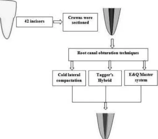
This study was approved by the Research Ethics Committee of the São Jose dos Campos School of Dentistry - São Paulo State University (Protocol number 076/2006). In this study, forty-two uniradicular human teeth (incisors) of patients between 18 and 25 years old, extracted for periodontal or prosthetic reasons, were used. After extraction, the teeth were immersed in distilled water and kept in a freezer at -18°C for a period not exceeding 6 weeks up to the time they were used. The crowns were sectioned with carborundum disks in a high-speed lathe, and the root size was standardized at 14 mm. The roots received
biomechanical preparation, with the working length 1 mm short of the apex, by performing the serial instrumentation technique (classical), with Kerr type endodontic files up to 60 K, alternately irrigating the canals with 1% sodium hypochlorite. To finalize biomechanical preparation, the root canal was flared until 80 K by stepback technique.
After the end of instrumentation, the canals were filled with Ethylenediamine tetraacetic acid (EDTA) solution (Inodon, Porto Alegre, Brazil) for 3 minutes and then irrigated with 1% sodium hypochlorite. The canals were dried with absorbent paper cones (Tanari Industrial Ltd., São Paulo, Brazil).
All roots received AH Plus cement (Dentsply Maillefer, Ballaigues, Switzerland) manipulated according to the manufacturer’s instruction, and introduced into the canal using a size 55-K file operated by hand in a counter-clockwise rotation (Cone Tech, Manaus, Am, Brazil) dipped in the cement.
The roots were randomly divided into three groups (n = 14), according to the root canal obturation technique
(Fig. 1):
Group 1. Cold lateral compaction technique:Briefly, a standard size 60 gutta-percha master cone (Cone Tech, Manaus, Am, Brazil) was inserted to the working length until a “tug back” sensation was felt. The tip of the master cone was then coated with the sealer and seated into position. Lateral compaction was accomplished using a size 25 finger spreader (Dentsply Maillefer, Ballaigues, Switzerland) that could reach within 1 mm short of the working length. Accessory gutta-percha cones R8 (Cone Tech, Manaus, Am, Brazil) were added and similarly compacted. The process was completed when the spreader could not penetrate more than 3 mm into the canal. Finally, excess gutta-percha was removed with a heated plugger at the orifice and vertical condensation was performed.
orifice.
Group 3. E&Q Master system technique:Canals were filled using the E&Q Master system (Meta Dental Corp., Cheongju, Korea), according to the manufacturer’s instructions. Briefly, a 0.04-tapered gutta-percha cone and an appropriate E&Q Pen Tip handpiece were selected to fit the canal 4 mm short of the working length. The sealer was applied and the master cone coated with sealer was inserted into the canal. The heating tip was activated to a setting of 160°C and the excess over the orifice was cut using the E&Q Pen Tip with gutta-percha reaching 4 mm short of the working length. Later, the gun needle was inserted into the root canal and kept in place for 5 seconds. Next, the triggers were pulled slowly and gutta-percha filled in the root canal. After the excess gutta-percha was cut with a heated instrument, vertical condensation was performed. An additional six roots were filled with the three different gutta-percha techniques described previously, for each control group (3 for negative control and 3 for positive control). All roots were subjected to periapical radiographic examination (Kodak, Rochester, New York, USA) in the labial and mesial positions, for comparison with other techniques. All the roots were stored for 7 days in a bacteriological incubator at 37°C with 100% humidity.
Sequentially, all samples were sealed with two layers of nail enamel over the external root surface except in the area corresponding to the apical foramen. The negative control teeth were totally coated with two layers of nail enamel, including the apical foramen. Positive controls were left unfilled.
The specimens were immersed in rhodamine B dye for 16 h at 37°C in a bacteriological incubator. Then, the specimens remained in a vacuum of 430 mmp for 30 min. The specimens remained immersed in dye for 7 days at 37°C. Next, the roots were washed in running water for 1 h, dried, and the impermeable nail enamel was removed manually with sharp instruments.
Next, the roots were sectioned longitudinally in the labio-lingual direction, at low speed with carborundum disks. The specimens were fixed onto glass slides and observed under a Stemi/2000C Stereomicroscope (Carl Zeizz) at ×25 magnification (Fig. 2). The dye penetration
area in the images was assessed by means of Image Tool 3.0 software. The apical leakage was measured as the distance between the root apex to the deepest extent of dye penetration into the coronal portion. The data obtained were subjected to analysis of variance (ANOVA) and Tukey’s tests at a 5% level of significance.
596
Results
The one-way ANOVA showed a P-value of 0.0016, so
the null hypothesis was rejected, indicating significant differences between groups (Table 1).
Table 2 shows the mean values and standard deviations
for the different groups and the results of the Tukey’s test. It can be observed that the groups filled with the CLC technique and E&Q Master system technique did not show significant differences between them, but significantly greater dye leakage was seen in the samples sealed with
Fig. 2 Representative picture of dye leakage.
Table 1 Results of one-way ANOVA
the Hybrid technique.
Discussion
Many techniques have been introduced over the years for efficient and effective filling of the root canal space with gutta-percha. The CLC of gutta-percha is one of the most widely used techniques and often has been used as the standard to which the sealing ability of new filling techniques or materials are compared (14-16). Advantages of the lateral compaction technique are its relative ease of use and controlled placement of the filling material.
The Hybrid technique combines features of CLC with the plasticization and compaction of gutta-percha using compactors designed by McSpadden in 1979 (7). This technique reduces the risk of overfilling, promotes a more cohesive and compact filling, consumes less time and material, and provides a safe and fast method (17).
The E&Q Master was introduced to make root canal filling easier and less time consuming, and is used in a similar manner as the System B technique. This system provides controllable and a precise amount of heat to the gutta-percha in the canal at different times during compaction. However, the new system has some advan-tages: the same pen tip used to apply heat to gutta-percha was utilized to obturate 4-6 mm short of the apex, softening and condensing the apical plug of gutta-percha in the canal, achieving a seal apically. This advantage avoids cooling down of the gutta-percha plug because the heated pen tip almost instantaneously pushes the gutta-percha apically.
The System B presents a tip of the pre-heated System-B plugger and a hand plugger instrument to compact the softened plug of gutta-percha. The gutta-percha plug cools down before the clinician inserts the tip compactor into the root canal. Another advantage of the E&Q master is two independent cordless units (the Master pen and the Gun needle), that facilitates mobility of the system during operation. Currently, the modern trend of simplifying clinical steps and saving time using thermoplasticized operative techniques appears to be efficient and previous evidence has shown it to be superior in adapting to canal walls and filling of lateral canals when compared with CLC (6-8).
In vitrodye leakage tests are very po common pular in
the literature because the laboratory process is easy and cheap. The phenomenon of capillarity is of utmost importance in this passive method used mainly for assessing apical leakage, as the tooth apex is submerged in a dye that penetrates through any space between the canal walls and filling material (18). Although the intensity of the coloration produced after apical infiltration was not an issue in the
present experiment, rhodamine B was used because it does not suffer discoloration by calcium hydroxide-based materials as occurs with methylene blue (19). Vacuum was used based on studies that showed greater infiltration when vacuum was used compared to its nonuse, probably due to formation of air pockets in the filling mass, which hinders penetration of the dye but not bacteria (20). The longitudinal section method enables examination of the exposed filling material and any dye penetration into the material and at the interface of the dentinal wall on one side.
It was observed the results of this study that the negative controls registered no detectable dye penetration. This means that the two coats of nail polish applied to the external root surfaces to avoid dye penetration were effective. The positive controls demonstrated extreme amounts of apical leakage. This indicates that the dye penetration method was correctly executed.
According to the results of this study, the hybrid technique showed a higher degree of apical leakage than the other techniques tested. However, some authors stated that the Tagger’s hybrid technique is considered ideal in terms of practicality of the technique and use of restricted material (5,9). One drawback of the Hybrid technique was that when the handpiece rotated accidentally in an anticlockwise direction, the compactor acted as a corkscrew and went through the tooth and beyond the apex in a fraction of a second. The contraangle handpiece should be set such that it can rotate the compactor only in one direction. Overextension seemed to occur more often with the compactor than with the CLC (8).
The results obtained in this study contradict those obtained by other authors (5,17), which demonstrated that the hybrid technique was superior to CLC, in preventing apical infiltration. In 1983, Tagger et al. (9) evaluated the depth of penetration of the dye leakage of the apical seal after the hybrid technique and the technique of CLC and observed no statistical differences between the two obturation techniques. Hsu and Duh (21) in 1988 and De Moor and Martens (22) in 1999 measured apical dye leakage in roots filled by hybrid root canal filling method and CLC technique and found no significant difference between the two techniques. However, in 1986 Hopkins et al. (8) stated that the apical seal produced by the CLC technique appeared to be significantly better than that produced by Hybrid thermoplastic condensation.
598
rev/min presented less dye leakage and when rotational speed of the engine plugger was increased, air pressure of the device may fluctuate, giving variations of speed when the handpiece is in use. The variations of the speed when the handpiece was used in this study could be responsible for the greater leakage compared to other techniques.
The E&Q Master system showed satisfactory results in this study and demonstrated no significant differences from the CLC technique and was statistically superior to the hybrid technique. This study suggests that the use of thermoplasticized gutta-percha E&Q Master system may have a positive effect on the apical seal, at least initially (7 days after the gutta-percha cools). Although thermo-plasticization occurred about 4 mm short of the apical terminus in the E&Q Master system, plasticization and condensation of the master gutta-percha point in the apical segment of the root canal allowed significant reduction in apical microleakage than the Tagger’s hybrid technique and was similar to the CLC technique, which was considered as the Control Group. The E&Q Master system can be compared to the System B injectable thermoplasticized gutta-percha system, but the practicality of the technique is superior.
In 2007, Inan et al. (11) evaluated different root canal obturation techniques using dye leakage. They reported no statistical differences between the CLC techniques and System B injectable thermoplasticized gutta-percha. Other studies (3,25) confirmed the results of this study; that is, that there were no statistical differences between the CLC techniques and injectable thermoplasticized gutta-percha systems. However, these studies (3,24) comparing the apical sealing ability of root canal obturation techniques employed different leakage test methods. Genço˘glu et al. (25) investigated in vitrothe ability to seal root canals with
System B and CLC techniques and observed that the dye leakage was higher with the use of CLC than using the thermoplasticized technique.
In 2007, Xu et al. (10) evaluated the sealing ability of the E&Q Plus System and CLC using a glucose leakage test. They found that the CLC was as effective as the E&Q Plus Systems only in the short term (up to 1 week). However, greater amounts of leakage were noted for CLC of gutta-percha compared with the other techniques at longer observation times. E&Q Plus system is marketed for use with a technique similar to the continuous wave condensation technique introduced by Buchanan (26) or the System B technique (25). E&Q Plus system presented the same protocol and advantages of the root canal obturation with E&Q Master system.
However, any technique has the possibility of inadequate apical sealing if not executed properly. The main objective
of endodontic obturation is hermetic sealing of the root canal system, minimizing the possibility of failure and microleakage (1,9,12). It is important to note that regardless of technique or modification of the technique adopted, root canal treatment will only be valid if three-dimensional sealing of the canal is achieved, a condition essential to prevent endodontic re-infection. However, longitudinal studies are needed for continual assessment of the sealing ability of the root canal filling over a prolonged period. The selection of the best technique aimed at clinical decision making is an important step in the filling of the root canal. Further in vitroresearch on this subject is still
needed to improve the effectiveness of filling techniques. Based on the results obtained in this study, it can be concluded that:
1. The E&Q Master system presented effective apical sealing and practical handling of the equipment; 2. The CLC technique proved to be effective, but required more clinical time to work;
3. The hybrid technique showed higher degree of apical leakage compared to the E&Q Master system and CLC technique.
References
1. Buckley JP (1929) The pulpless tooth, its pathology and conservation: a new method and technic of filling root canals. J Am Dent Assoc 16, 44-61. 2. Harris GZ, Dickey DJ, Lemon RR, Luebke RG
(1982) Apical seal: McSpadden vs lateral condensation. J Endod 8, 273-276.
3. ElDeeb ME (1985) The sealing ability of injection-molded thermoplasticized gutta-percha. J Endod 11, 84-86.
4. Torabinejad M, Skobe Z, Trombly PL, Krakow AA, Grøn P, Marlin J (1978) Scanning electron microscopic study of root canal obturation using thermoplasticized gutta-percha. J Endod 4, 245-250.
5. Tagger M, Tamse A, Katz A, Korzen BH (1984) Evaluation of the apical seal produced by a hybrid root canal filling method, combining lateral condensation and thermatic compaction. J Endod 10, 299-303.
6. Wu MK, Wesselink PR (1993) Endodontic leakage studies reconsidered. Part I. Methodology, application and relevance. Int Endod J 26, 37-43. 7. Cohen BD (1984) An investigation into the relative
merits of some endodontic filling systems. MSc Thesis, University of Manchester, Manchester. 8. Hopkins JH, Remeikis NA, Van Cura JE (1986)
of apical microleakage. J Endod 12, 198-201. 9. Tagger M, Tamse A, Katz A (1983) Efficacy of
apical seal of Engine Plugger condensed root canal fillings – leakage to dyes. Oral Surg Oral Med Oral Pathol 56, 641-646.
10. Xu Q, Ling J, Cheung GS, Hu Y (2007) A quantitative evaluation of sealing ability of 4 obturation techniques by using a glucose leakage test. Oral Surg Oral Med Oral Pathol Oral Radiol Endod 104, e109-e113.
11. Inan U, Aydemir H, Ta¸sdemir T (2007) Leakage evaluation of three different root canal obturation techniques using electrochemical evaluation and dye penetration evaluation methods. Aust Endod J 33, 18-22.
12. Genço˘glu N (2003) Comparison of 6 different gutta-percha techniques (part II): Thermafil, JS Quick-Fill, Soft Core, Microseal, System B, and lateral condensation. Oral Surg Oral Med Oral Pathol Oral Radiol Endod 96, 91-95.
13. Glickman GN (2001) Injectable thermoplasticized gutta-percha systems. Pract Proced Aesthet Dent 13, 477-482.
14. Wong M, Peters DD, Lorton L (1981) Comparison of gutta-percha filling techniques, compaction (mechanical), vertical (warm), and lateral condensation techniques, Part 1. J Endod J Endod 7, 551-558.
15. Gilbert SD, Witherspoon DE, Berry CW (2001) Coronal leakage following three obturation techniques. Int Endod J 34, 293-299.
16. Dalat DM, Spångberg LS (1994) Comparison of apical leakage in root canals obturated with various gutta percha techniques using a dye vacuum tracing method. J Endod 20, 315-319.
17. Z m e n e r O , G i m e n e s F r í a s J ( 1 9 9 1 ) Thermomechanical compaction of gutta-percha: a
scanning electron microscope study. Endod Dent Traumatol 7, 153-157.
18. Camps J, Pashley D (2003) Reliability of the dye penetration studies. J Endod 29, 592-594.
19. Wu MK, van der Sluis LW, Ardila CN, Wesselink PR (2003) Fluid movement along the coronal two-thirds of root fillings placed by three different gutta-percha techniques. Int Endod J 36, 533- 540. 20. Goldman M, Simmonds S, Rush R (1989) The
usefulness of dye-penetration studies reexamined. Oral Surg Oral Med Oral Pathol 67, 327-332. 21. Hsu TS, Duh BR (1988) Evaluation of the apical seal
produced by a hybrid root canal filling method, c o m b i n i n g l a t e r a l c o n d e n s a t i o n a n d thermomechanical compaction. Zhonghua Ya Yi Xue Hui Za Zhi 7, 97-104. (in Chinese)
22. D e M o o r R J , M a r t e n s L C ( 1 9 9 9 ) A p i c a l microleakage after lateral condensation, hybrid gutta-percha condensation and Soft-Core obturation: an in vitro evaluation. Endod Dent Traumatol 15, 239-243.
23. Saunders EM (1989) The effect of variation in thermomechanical compaction techniques upon the quality of the apical seal. Int Endod J 22, 163-168. 24. Kontakiotis E, Chaniotis A, Georgopoulou M (2007) Fluid filtration evaluation of 3 obturation techniques. Quintessence Int 38, e410-e416.
25. Genço˘glu N, Garip Y, Ba¸s M, Samani S (2002) Comparison of different gutta-percha root filling techniques: Thermafil, Quick-fill, System B, and lateral condensation. Oral Surg Oral Med Oral Pathol Oral Radiol Endod 93, 333-336.