1Senior Neurologist, Head of Cerebrospinal Fluid (CSF) Unit, Head of Multiple Sclerosis (MS) Outpatient Clinical Care, Hospital S. João, Auxiliar Professor of Neuroanatomy, Porto Medical School, Porto, Portugal; 2Technician of CSF Unit, Hospital S. João, Porto, Portugal; 3Senior Neurologist, President of Center for Investigation and Treatment of Multiple Sclerosis of Hospital S. João, Porto, Potugal; 4PhD MD DSc FRCPath FRCP, Department of Neuro i m m u n o l o g y, National Hospital for Neurology & Neuro s u rg e ry, The Institute of Neurology, Queen Square, London, U.K.
Received 24 November 2004. Accepted 21 February 2005.
Maria José Sá, MD - Department of Neurology, Hospital S. João - Alameda Professor Hernâni Monteiro - 4200-319 Porto - Portugal. E-mail: [email protected]
OLIGOCLONAL IgG BANDS IN THE
CEREBROSPINAL FLUID OF PORTUGUESE
PATIENTS WITH MULTIPLE SCLEROSIS
Negative results indicate benign disease
Maria José Sá
1, Lucinda Sequeira
2, Maria Edite Rio
3, Edward J. Thompson
4ABSTRACT - We assessed the frequency of cerebrospinal fluid (CSF) restricted oligoclonal IgG bands (IgG-OCB) in Portuguese multiple sclerosis (MS) patients and its relationship with outcome. Paired CSF/serum samples of 406 patients with neurological disorders were submitted to isoelectric focusing with immunodetection of IgG. Ninety-two patients had definite MS; non-MS cases were assembled in groups inflammatory/infectious diseases (ID, n=141) and other/controls (OD, n=173). We found in the MS group: mean duration, 38.9 months; clinically isolated syndromes, 24%; relapsing/remitting course (RR), 65%; in RR patients the mean EDSS was 2.1 and the mean index of pro g ression was 0.31. Positive patterns significantly p redominated in MS (82.6%; ID, 40.4%; OD, 3.5%). The sensitivity and the specificity of positive IgG-OCB for MS diagnosis was 82.6% and 79.9%, respectively. The sole statistically significant difference in the MS g roup was the lower pro g ression index observed in negative cases. We conclude that the frequency of positive IgG-OCB patterns in our MS patients fits most values reported in the literature, and that negative results indicate benign disease.
KEY WORDS: multiple sclerosis, CSF, oligoclonal bands, IgG, isoelectric focusing.
Bandas oligoclonais da IgG no líquido céfalo-raquidiano de doentes portugueses com esclerose múltipla: resultados negativos indicam doença benigna
RESUMO - Analisamos a frequência de bandas oligoclonais (BOC) restritas ao líquido céfalo-raquidiano (LCR) em doentes portugueses com esclerose múltipla (EM) e sua relação com a clínica. Determinaram-se por focagem isoeléctrica e imunodetecção as BOC da IgG em pares de amostras LCR/soro de 406 doentes com diversas patologias neurológicas: 92 tinham EM definitiva; os casos “não-EM” agruparam-se em doenças infla-matórias/infecciosas (ID; n=141) e outras/controles (OD; n=173). O grupo EM apresentava duração média: 38,9 meses; síndromes clinicamente isolados (CIS), 24%; formas surt o / remissão (RR), 65%, nas quais se encontro u EDSS e índice de progressão médios de 2,1 e 0,31, respectivamente. O perfil positivo predominava signifi-cativamente na EM (82,6%; ID, 40,4%; OD, 3,5%), cuja sensibilidade e especificidade neste diagnóstico foi 82% e 79,9%, respectivamente. A única diferença estatisticamente significativa no grupo EM foi o menor índice de progressão nos casos negativos. Em conclusão, a frequência de BOC positivas nos nossos doentes EM enquadrou-se nos valores da literatura, e a sua negatividade indicou evolução benigna.
PALAVRAS-CHAVE: esclerose múltipla, LCR, bandas oligoclonais, IgG, focagem isoeléctrica.
The intrathecal synthesis of antibodies has been since long recognised as a nospecific marker of i n-flammation in a variety of neurological disorders, p a r-ticularly in multiple sclerosis (MS)1. Namely, the p
re-sence of oligoclonal bands (OCB) of immunoglo-bulin G (IgG) restricted to the cerebrospinal fluid
(CSF) of MS patients is the most sensitive parameter to evidence the inflammatory character of the di-s e a di-s e2 , 3. Yet, the final diagnosis of MS mainly re l i e s
upon the clinical features, due to the absence of s p e-cific biological markers.
-OCB and/or increased IgG Index, was included as a paraclinical exam in the classical study of Poser et a l .4, as well as in the most recent diagnostic criteria
of MS established by McDonald et al.5, essentially
in clinically isolated syndromes (CIS) and primary p ro-gressive forms.
In the literature the frequency of OCB in MS va-ries from 56 to 97%, depending on the methodolo-gy employed and the population studied3 , 6 - 1 1. In o c c
i-dental populations these values are higher than in oriental ones, and this discrepancy has been ascri-bed to genetic diff e re n c e s7 , 1 1. This shows that the p
re-sence of CSF IgG-OCB is not constant in MS, altho-ugh its absence is uncommon, leading some authors to advise an exhaustive diagnostic re v i e w3 , 1 2in “ n
e-gative” cases.
This study was designed to evaluate the re s u l t s obtained in the detection of OCB in the CSF of Por-tuguese patients with definite MS, living in dif-f e rent regions odif-f the country. In addition, we asses-sed the influence of clinical features, such as d u r a t i o n and course of the disease, in the laboratory re s u l t s , in order to identify which factors might eventually be related with the absence of OCB in MS.
METHOD
Patients and study design– All subjects (n = 406) who-se CSF and who-serum samples were conwho-secutively studied in the CSF Laboratory of the Department of Neuro l o g y, Hos-pital S. João, Porto, in 2001 and 2002, to search the pre-sence of IgG-OCB by isoelectric focusing (IEF), entered this study. The CSF samples were always obtained by lum-bar puncture (LP), in patients attended at the Depart-ments of Neurology from 9 Hospitals located in diff e re n t regions of Portugal. In 17 cases more than one CSF sam-ple was studied, but only the first one, which was collec-ted for diagnostic purposes, was considered. The clinical data of all patients were gathered in the requests that a c-companied the samples, in the clinical files of the Depart-ment of Neurology of Hospital S. João, and by question-naires sent to the neurologists, as regards the patients assisted elsewhere. This allowed a confident knowledge of the final diagnosis at discharge and consequent accu-rate distinction between MS and non-MS patients, accor-ding to the criteria of McDonald et al.5, the acquisition
of clinical features in MS patients, and the assembling of the remaining patients in other groups of diagnosis.
Thus, 92 patients (22.7%) were seen to have definite M S5. According to the evolutive course, they were
classi-fied in the subgroups re l a p s i n g / remitting MS (RRMS), se-c o n d a ry pro g ressive (SPMS) and primary pro g re s s i v e (PPMS); all patients with monosymptomatic pre s e n t a t i o n , i.e. clinical evidence of one relapse, plus imaging evidence of dissemination in time and space, were compiled in t h e group of clinically isolated syndromes (CIS).
The following parameters were recorded in the MS group, and in each evolutive subgroup: sex; age at LP; duration of disease as measured in months since the onset symptoms until the time of LP. In the patients with R R M S , the disability and the severity were also determined, as follows: the disability was assessed with the Expanded Disability Status Scale (EDSS) and the severity was calcu-lated by the Poser’s index of progression13, i.e. the EDSS
score divided by the duration of the disease previously expressed in months.
The non-MS patients were assembled in 2 groups ac-cording to the underlying pathology: 141 (34.7%) pre-sented infectious/inflammatory disorders of the nerv o u s system (ID); the remaining 173 (42.6%) displayed other n e u rological diseases/controls (OD). The three most fre-quent diagnosis in groups ID and OD were meningitis, encephalitis and neurosyphilis, and ischaemic cere b ro v a s-cular disease, neurodegenerative disorder and spondilotic myelopathy, respectively. Nine patients conforming to the category of possible MS5w e re included in the ID g ro u p .
In 14 subjects with functional symptoms of the nervous system, the exhaustive study did not disclose any neuro-logical disorder.
L a b o r a t o ry pro c e d u res – The CSF and serum samples w e re collected at the same time in each patient and were immediately sent refrigerated to the CSF Laboratory of the Department of Neurology of Hospital de S. João. Af-ter reception, small aliquots were pre p a red and kept fro-zen at - 20ºC for a few days until processing. To detect t h e IgG-OCB, paired CSF/serum samples of every case were run in parallel by IEF followed by passive protein transfer to a nitrocellulose membrane and immunodetection of IgG with double antibody, according to the method d e s-cribed by Keir et al.1 4. Minor technical adaptations were
done locally, as for instance the refrigeration value, w h i c h was set at +15ºC15. CSF restricted OCB were considered
in case of at least two bands present in the CSF and absent in seru m1 4. The patterns were classified according to the
five types defined by consensus of the Committee of the E u ropean Concerted Action for Multiple Sclerosis (Char-cot Foundation)2, which allows the distinction of local
f rom systemic synthesis of IgG2 , 3 , 1 4 , 1 6: 1. Negative/norm a l
- absence of OCB in the CSF and serum; 2. Positive - CSF restricted OCB; 3. “Greater than” - restricted CSF OCB a n d additional OCB in the CSF and serum; 4. “Mirror” - i d e n t i c a l OCB in the CSF and serum; 5. Paraprotein - monoclone in the CSF and serum. The cases with patterns of types 2 and 3 were joined in the classification “positive”, given that they both traduce intrathecal synthesis of antibodies. The identification of the IEF patterns was done in all cases by two authors (M J S and L S) who were unaware of e a c h other’s results and of the clinical diagnosis.
The internal quality control was perf o rmed accord i n g to the literature2,3,14, recently confirmed by an
Interna-tional Consensus Gro u p1 6: in each run, one CSF sample w i t h
w e re included. For external quality control, seven paire d C S F / s e rum samples of MS cases with negative OCB were sent to a European re f e rence laboratory: Department o f N e u ro i m m u n o l o g y, Institute of Neuro l o g y, National H o s-pital for Neurology and Neuro s u rg e ry, Queen Square, L o n-don, UK; a concordance of the results in both laboratories was seen in 6 cases; the remaining CSF sample was positi-ve for IgG-OCB, instead of negatipositi-ve.
Analysis of CSF results versus clinical data – The num-ber of different OCB patterns was determined, as well as their relative distribution by group of patients. In the MS group, we analysed the distribution of the OCB pat-t e rns by sex, age, pat-type of evolupat-tion or clinical course, du-ration, and in RRMS cases, severity.
The sensitivity of the technique was calculated by the p e rcentage of patients with definite MS and positive CSF p a t t e rns. To assess the specificity of the technique we d e-t e rmined e-the percene-tage of pae-tiene-ts wie-th oe-ther diseases ( c o n t rols) whose CSF was positive for OCB, and then the quotient between the number of cases with negative p a t t e rns and the total number of controls; we did two c a l-culations assuming as control group either the sum of groups ID and OD or the group OD alone.
Statistical analysis – The following tests were perf o r-med: Chi-square (distribution of CSF patterns by gro u p s of patients); Mann-Whitney’s U and Kruskal-Wallis me-thods (relationship between clinical data and CSF OCB p a t t e rn in the MS group); Fisher exact test to determ i n e the influence of duration, severity and pro g ression index in the presence or absence of OCB. Diff e rences were con-sidered statistically significant when P < 0.05.
RESULTS
With respect to the clinical features of the MS g roup, we found a predominance of female (n = 5 7 ) over male (n = 35) sex (ratio: 1.6/1). The mean age of the whole group was 37.1 years ± 12.5 (range: 16 - 73 years); higher values were seen in the fema-les (37.8 years ± 12.5) than in mafema-les (35.9 years ± 12.7), diff e rence that almost reached the significant level (P = 0.0507). As re g a rds the clinical course, the distribution of the cases was as follows: RR, 60 ( 6 5 % ) ; S P, 5 (5%); PP, 4 (4%), CIS, 22 (24%). The re m a i n i n g patient was a woman with Devic’s disease17. No
d i ff e rences were found in the distribution of these forms by sex. We observed that the mean age was higher in patients with PP (63.3 years) and SP (51.5 years) forms than in patients with RR course (48.8 years) or CIS (34 years), but significant differences were detected only between RR and CIS cases (p < 0.03). We found a wide range in the duration of the disease (1 month to 40 years), with mean value of 38.9 months ± 76.2; yet, in 70% cases the
dura-tion was lower than 2 years. Duradura-tion was signifi-cantly higher (P < 0.05) in females (50.8 months) t h a n in males (39.4 months). Moreover, statistically sig-nificant diff e rences were found in the duration v a-lues by MS form (P < 10- 3), the mean value of which
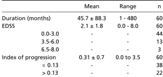
was lowest in patients with CIS (21.9 months) and g reatest in SP cases (69.1 months). The clinical data assessed in the RRMS subgroup, as re g a rds duration (mean: 45.7 months), EDSS scores and the Poser’s index of progression (cut-off value: 0.13) are pre-sented in Table 1.
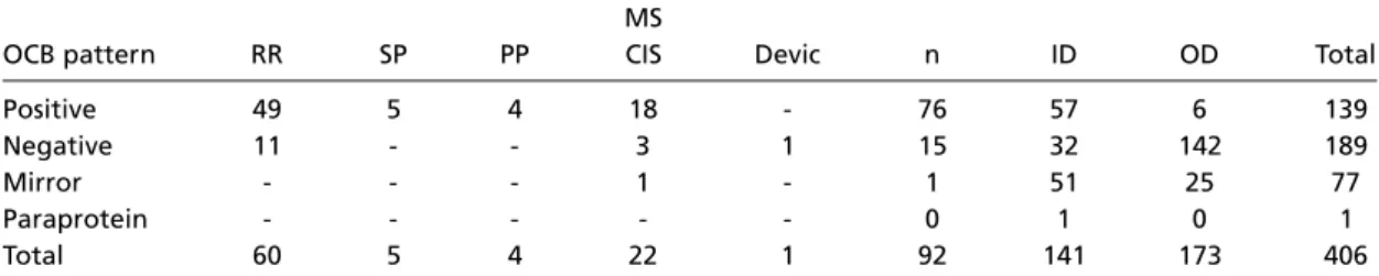
The results obtained in the CSF study are shown in Table 2. We found that the positive type of OCB p a t t e rn predominated both in the MS (82.6%) and ID (40.4%) groups, whereas the negative pattern was mainly noticed in the OD group (82%); these d i ff e rences observed in the distribution of CSF p a t-t e rns according t-to t-the groups of pat-tient-ts were s t-t a-tistically significant (χ2, P < 10-4). The sensitivity of
the positive OCB pattern in MS had a mean value of 82.6%; more o v e r, it was higher in the pro g re s s i v e forms (100%) than in RRMS (81.7%) or in patients with CIS (81.8%). On the contrary, the specificity was lower, since positive OCB were detected in all groups of patients: 3.5% of OD cases and 20% of all non-MS patients. Hence, the specificity values varied according to the control group considered: 79.9% (ID and OD groups) and 96.5% (OD group alone). Conversely, the negative pattern was found in 189 out of 406 cases (46.5%), constituting, as a whole, the most frequent pattern.
As specifically regards the MS group (Table 1), we observed CSF restricted OCB in 76 patients, p re-senting diff e rent types of clinical course as alluded to above, 18 of which had also a few OCB in the s e rum (Type 3 pattern). A negative pattern was seen in 15 patients (16%), including 11 cases with RR cli-nical course. In this RRMS group we found highly significant diff e rences (Fisher exact test, P < 10- 4) in
the correlation between the CSF patterns and the
Table 1. Data of MS patients with relapsing/remitting forms.
Mean Range n
Duration (months) 45.7 ± 88.3 1 - 480 60
EDSS 2.1 ± 1.8 0.0 - 8.0 60
0.0-3.0 - - 44
3.5-6.0 - - 13
6.5-8.0 - - 3
Index of progression 0.31 ± 0.7 0.0 to 3.5 60
≤0.13 - - 38
> 0.13 - - 22
severity of the disease: the patients with negative p a t t e rns presented a more “benign” course, i.e. lo-wer progression indexes, than those with positive ones. In effect, the progression index was smaller than the cut-off level in 8 patients with negative p a t t e rn, whereas it was higher than 0.13 in the re-maining 3 negative cases. In one patient with CIS a “mirror” pattern was detected. Finally, diff e re n c e s w e re also noticed in the correlation of patterns with age, which was higher in negative patients (42.8 years ± 13.4) than in those with CSF restricted IgG-OCB (35.9 years ± 12.1), almost reaching the sig-nificant level (P = 0.053). In contrast, no statistically significant relations were found between the OCB results and any of the other clinical parameters: sex, clinical course and duration.
DISCUSSION
In this study we have for the first time assessed the frequency of locally restricted IgG-OCB in the CSF of Portuguese patients with definite diagnosis of M S . This endeavour was accomplished through the colla-boration that the CSF Laboratory of the Depart m e n t of Neurology of Hospital S. João, Porto, provides to the Departments of Neurology of several Hospitals in the country, in the determination of OCB with IEF and immunodetection of IgG, according to the labo-ratorial pro c e d u res recommended in the literature2 , 3 , 6.
F u rt h e rm o re, it turned feasible to collect a re l e v a n t number of MS cases in 2 years and to obtain pre c i s e and detailed clinical data in all patients.
We have found that 82.6% of MS patients pre-sent positive patterns. This value is higher than tho-se described in oriental populations - 56.1%7 , 1 1- and
in Finland - 76%6, but lower than the values re p o
r-ted in Brazil, 85%9, in Spain, 87.7% 1 0and, part i c
u-l a r u-l y, in nort h e rn European countries2 , 3 , 8 , 1 6. These d i
f-f e rences have been ascribed to immunogenetic f-f a c-tors which influence the intrathecal immune hu-moral functions in human populations7 , 1 1, although
the role of diff e rent laboratory pro c e d u res and te-chniques may also explain, at least in part, the
dis-c repandis-cy of values re p o rted in the literature2 , 3 , 6 - 1 1 , 1 6.
Moreover, we have included patients with CIS in our estimations, whereas Falip et al10set that
spe-cific group of patients apart, so that the reported value of 87.7% positive patterns in their study re-fers exclusively to patients with clinically defined M S1 0. Hence, one might assume that the fre q u e n c y
of positive patterns in our study would be greater if we didn’t include the CIS group, too. Anyway, the frequency of the positive pattern in the CIS g roup that we found (81.8%) is considerably higher than it was found in other series - 54.8%10 and
6 2 . 5 %1 8, which may be somehow explained by
dif-ferences in the criteria of inclusion of these cases. In effect, as previously described, all patients with CIS in our study had imagiological evidence of dis-semination in time and in space.
In 16% MS patients, we did not find local pro-duction of IgG. Intere s t i n g l y, these cases with nega-tive patterns presented lower indexes of progres-sion, indicating a more benign course of the disease, nicely fitting the results of Zeman et al.1 2and s t re n
g-thening the suggestion of other studies1 9 , 2 0that the
absence of OCB at the time of diagnosis indicates a trend towards a better prognosis. Moreover, ali-ke other re p o rts our OCB negative patients were o n the average older than those with positive pattern s ; however, in that study there was also a predomi-nance of male sex, later age of onset and chronic-p ro g ressive forms in OCB negative cases6, diff e re
n-ces which we did not notice. On the contrary, a stu-dy conducted at Japan failed to demonstrate diff e-rences in the clinical course and disability in MS pati-ents with or without OCB in the CSF7. We are
awa-re that the absence of OCB is raawa-re in MS and that “false negative” is a potential situation1 2, although
in the present study it seems unlikely, given the ex-ternal quality control that was provided in a refe-rence European laboratory3,14.
As regards the presence of “mirror” pattern in one patient with CIS, no sound explanation could be advanced to explain this bizarre finding. Indeed,
Table 2. Distribution of IgG oligoclonal patterns in all groups of patients.
MS
OCB pattern RR SP PP CIS Devic n ID OD Total
Positive 49 5 4 18 - 76 57 6 139
Negative 11 - - 3 1 15 32 142 189
Mirror - - - 1 - 1 51 25 77
Paraprotein - - - 0 1 0 1
Total 60 5 4 22 1 92 141 173 406
this type of pattern is very unusual in MS, given t h a t the serum oligoclonal answer which may be found in this disease, as happened in some of our patients, is additional to the typical intrathecal answer21,22.
In this sense, the meaning of the “mirror” pattern is MS is similar to the negative pattern, since they both traduce absence of local synthesis of IgG.
In addition, as concerns the distribution of the CSF OCB patterns in the totality of patients, our re s u l t s a re in accordance with Falip et al.1 0and with a
pre-vious review of our laboratory2 3, stressing that the
p redominance of OCB negative patterns might be explained by the large number of patients without i n f l a m m a t o ry neurological disord e r. On the other hand, the observation of positive patterns in diff e re n t g roups of neurological diseases is a widely re c o g n i z e d f a c t3. However, we found a lower frequency of
po-sitive patterns in all groups of patients than Falip et a l .1 0, mainly in the OD group, which may be due to
d i ff e rences in the composition of control groups. F i n a l l y, the sensitivity value of the positive OCB p a t t e rns for the diagnosis of MS that we found -82.6% - fits most studies8 - 1 0 , 2 4, particularly in those
c a rried out in Spanish - 87.7%1 0and Brazilian
popu-lations - 85% 9. Inversely, the specificity was lower
than the sensitivity, although our values were higher than those re p o rted by others, which varied from 45 to 62%1 0 , 2 4, perhaps reflecting diff e rences in the
c o n t rol groups, too. The well-known lack of specificity of the positive OCB pattern in the diagnosis of MS is due to the considerable number of patients with i n f l a m m a t o ry diseases of the nervous system inclu-ded. Anyway, in agreement with Falip et al.1 0, the
specificity reached higher values when the contro l s w e re confined to the patients of the OD group.
In conclusion, we emphasise that the great ma-jority of Portuguese MS patients have CSF re s t r i c t e d IgG-OCB, the frequency of which is quite similar to the values observed in specific populations. Up to n o w, the role played by the presence of eventual si-milar immunogenetical factors in these results is un-known. More o v e r, this study re i n f o rces the better pro-gnosis of MS patients with negative OCB patterns.
A c k n o w l e d g e m e n t s- We want to thank our collea-gues Dr. Alfredo Sá (Hospital Santo André, Leiria), Dr. António Carn e i ro (Hospital Militar Regional n.º 1, Port o ) , Dr. Armando Morganho (Centro Hospitalar do Funchal, Madeira), Dr. José Figueiredo (Hospital S. Marcos, Braga), Dr. José Pinto Marques (Hospital S. Bernardo, Setúbal), D r.ª Maria Lurdes Rodrigues (Hospital Senhora da Oliveira, Guimarães), Dr.ª Maria Vaz Pato (Centro Hospitalar Cova da Beira, Covilhã) and Dr.ª Susana Pereira (Hospital Pe-dro Hispano, Matosinhos) for making the clinical data
available. Thanks are also due to Professor Joaquim Marques de Sá, Departamento de Engenharia Electro-técnica e de Computadores, Faculty of Engineering, Uni-versity of Porto, as statistical reviewer. Finally, we are grateful to Aventis Pharma Lda., Octapharma, Schering Lusitana, Lda., Schering-Plough-Farma, Lda and Serono P rodutos Farmacêuticos Lda. for their financial support .
REFERENCES
1. Link H. Immunoglobulin G and low molecular weight proteins in hu-man CSF. Chemical and immunological characterization with special reference to MS. Acta Neurol Scand 1967;28(Suppl):S1-S36. 2. Andersson M, A l v a rez-Cermeno J, Bernardi G, et al. Cere b ro s p i n a l
fluid in the diagnosis of multiple sclerosis: a consensus report. J Neuro l Neurosurg Psychiatry 1994;57:897-902.
3. Thompson EJ. Cere b rospinal fluid. J Neurol Neuro s u rg Psychiatry 1995;59:349-357.
4. Poser CM, Paty DW, Scheinberg L, et al. New diagnostic criteria for multiple sclerosis: guidelines for re s e a rch protocols. Ann Neuro l 1983;13:227-231.
5. McDonald WI, Compston A, Edan G, et al. Recommended diagnostic criteria for multiple sclerosis: Guidelines from the international panel on the diagnosis of multiple sclerosis. Ann Neurol 2001;50:121-127. 6. Pirttila T, Nurmikko T. CSF oligoclonal bands, MRI, and the diagnosis
of multiple sclerosis. Acta Neurol Scand 1995;92:468-471.
7. Fukazawa T, Kikuchi S, Sasaki H, et al. The significance of oligoclonal bands in multiple sclerosis in Japan: relevance of immunogenetic back-grounds. J Neurol Sci 1998;158:209-214.
8. S e res E, Bencsik K, Rajda C, Vecsei L. Diagnostic studies of cere b ro s p i n a l fluid in patients with multiple sclerosis. Orv Hetil 1998;139:1905-1908. 9. Puccioni-Sohler M, Passeri F, Oliveira C, Brandão CO, Papaiz-Alvare n g a R. Multiple sclerosis in Brazil: analysis of cere b rospinal fluid by standard methods. Arq Neuropsiquiatr 1999;57:927-931.
10. Falip M, Ti n t o re M, Jardi R, Duran I, Link H, Montalban X. Clinical use-fulness of oligoclonal bands. Rev Neurol 2001;32:1120-1124. 11. Kikuchi S, Fukasawa T, Niino M, et al. HLA-related subpopulations of
MS in Japanese with and without oligoclonal IgG bands. Neurology 2003;60:647-651.
12. Zeman AZJ, Kidd D, McLean BN, et al. A study of oligoclonal band ne-gative multiple sclerosis. J Neurol Neuro s u rg Psychiatry 1996;60:27-30. 13. Poser S, Raun NE, Poser W. Age at onset, initial symptomatology and the course of multiple sclerosis. Acta Neurol Scand 1982;66:355-362. 14. Keir G, Luxton RW, Thompson EJ. Isoelectric focusing of cere b ro s p i n a l
fluid immunoglobulin G: an annotated update. Ann Clin Biochem 1990;27:436-443.
15. Sá MJ, Sequeira L, Rio ME, et al. Prevalência de bandas oligoclonais da IgG no líquido céfalo-raquidiano de doentes com esclerose múltipla. Sinapse 2003;2:10-16.
16. Reiber H, Thompson EJ, Grimsley G, et al. Quality assurance for cere-b rospinal fluid protein analysis: international consensus cere-by an internet-based group discussion. Clin Chem Lab Med 2003;41:331-337. 17. Wingerchuk DM, Hogancamp WF, O'Brien PC, Weinshenker BG. The
clinical course of neuromyelitis optica (Devic's syndrome). Neurology 1999;53:1107-1114.
18. Ti n t o re M, Rovira A, Brieva L, et al. Isolated demyelinating syndro m e s : comparison of CSF oligoclonal bands and diff e rent MR imaging criteria to predict conversion to CDMS. Mult Scler 2001;7:359-363.
19. Avasarala JR, Cross AH, Trotter JL. Oligoclonal band number as a mar-ker for prognosis in multiple sclerosis. Arch Neurol 2001;58:2044-2045. 20. M e s a ros S, Drulovic J, Levic Z. Clinical characteristics and neuro-physiologic findings in patients with multiple sclerosis without oligo-clonal IgG in cerebrospinal fluid. Srp Arh Celok Lek 2003;131:122-126. 21. Zeman A, McLean B, Keir G, Luxton R, Sharief M, Thompson E. The significance of serum oligoclonal bands in neurological diseases. J Neurol Neurosurg Psychiatry 1993;56:32-35.
22. Zeman AZ, Keir G, Luxton R, Thompson EJ. Serum oligoclonal IgG is a common and persistent finding in multiple sclerosis and has a systemic source. QJM 1996;89:187-193.
23. Carvalho M, Nadais G, Sequeira L, Campos MM, Sá MJ. Detecção de bandas oligoclonais da IgG no LCR por focagem isoeléctrica num labo-ratório de LCR: casuística de um ano. Sinapse 2001;1:55.