1Biologist, Master Science Student, Neurology Service / Gaffrèe Guinle University Hospital / Federal University of Rio de Janeiro
State (HUGG/UNIRIO), Neurolife Laboratory, Rio de Janeiro RJ, Brazil;2Biologist, Neurolife Laboratory;3B i o c h e m i s t ry, Neuro l i f e
L a b o r a t o ry, CSF Laboratory / Clinical Pathology Service / Clementino Fraga Filho University Hospital / Federal University of Rio de J a n e i ro (SPC/HUCFF/UFRJ);4Biologist, CSF Laboratory / SPC / HUCFF / UFRJ;5Ti t u l a r- P ro f e s s o r, PhD, Institute of Micro b i o l o g y / U F R J ;
A d j u n c t - P ro f e s s o r, PhD, CSF Laboratory / SPC / HUCFF / UFRJ;6N e u rology Service / HUGG / UNIRIO; Neurolife Laboratory, Rio de
Janeiro/RJ. Support: FAPERJ (Fundação de Amparo á Pesquisa do Rio de Janeiro). Received 25 April 2005, received in final form 22 August 2005. Accepted 5 October 2005.
Dra. Marzia Puccioni-Sohler - Rua Dezenove de Fevere i ro 185 / 705 - 22280-030 Rio de Janeiro RJ - Brasil. E-mail: mpuccioni@hucff . u f r j . b r
INFLUENCE OF THE CEREBROSPINAL
FLUID LABORATORY PARAMETERS IN
THE ELISA TEST FOR NEUROCYSTICERCOSIS
USING A TOTAL CYSTICERCI ANTIGEN
Cristiane S. Casanova
1, Maria José S.P. Ribeiro
2, Reizer R. Gonçalves
3,
Luiz Cláudio Faria
4, José Mauro Peralta
5, Marzia Puccioni-Sohler
6ABSTRACT - To evaluate if the cere b rospinal fluid (CSF) parameters may influence the cysticercosis immunore-activity response in the CSF. CSF samples of 109 patients were analyzed and classified in three gro u p s , a c c o rding to the neurological manifestations and the reactivity in antibody-enzyme linked immunosor-bent assay (Ab-ELISA) testing in CSF for neuro c y s t i c e rcosis (NC): group A, 18 patients with neurological o rders compatible with NC and reactive Ab-ELISA in CSF for NC; group B, 50 patients with neurological orders non-compatible with NC and reactive Ab-ELISA for NC; group C, 41 patients with neurological dis-orders non-compatible with NC and non-reactive Ab-ELISA in CSF for NC. The CSF analysis in group A was compatible with NC. The group B in comparison to the groups A and C presents higher frequency and intensity of hypercytosis, presence of red blood cells in CSF, protein concentration and immunological re a c-tive test for other etiological agents (p0.05). Based on the present data, we suggest that the inflamma-t o ry process and high proinflamma-tein conceninflamma-trainflamma-tion may deinflamma-termine false posiinflamma-tive reacinflamma-tions in inflamma-the Ab-ELISA inflamma-tesinflamma-t for NC in the CSF.
KEY WORDS: cerebrospinal fluid, enzyme linked immunosorbent assay, neurocysticercosis.
Influência dos parâmetros laboratoriais do líquido cefalorraquidiano no teste ELISA para neu-rocisticercose utilizando antígeno total de cisticerco
RESUMO - Avaliar se os parâmetros do líquido cefalorraquidiano (LCR) podem influenciar na re a t i v i d a d e da resposta imune específica do LCR na neuro c i s t i c e rcose (NC). Amostras de LCR de 109 pacientes foram analisadas e classificadas em três grupos, de acordo com as manifestações neurológicas e reatividade do teste de Ab-ELISA para NC no LCR. Grupo A, 18 pacientes com enfermidades neurológicas compatíveis com NC e reatividade do teste Ab-ELISA para NC no LCR; grupo B, 50 pacientes com enfermidades neuro l ó g i-cas não compatíveis com NC e reatividade do teste Ab-ELISA para NC no LCR; grupo C, 41 pacientes com e n f e rmidades neurológicas não compatíveis com NC e na ausência de reatividade do teste de Ab-ELISA para NC no LCR. A análise do LCR do grupo A foi compatível com NC. O grupo B apresentou maior fre-qüência e intensidade da pleocitose, da presença de hemácias no LCR, hiperpro t e i n o rraquia, re a t i v i d a d e imune para outros agentes etiológicos em comparação aos grupos A e C (p0.05). Os dados indicam que o processo inflamatório e os elevados níveis de concentração da proteína no LCR podem influenciar na ocorrência de reações falso positivas de Ab-ELISA para NC. Destacamos a importância da correlação clíni-co-laboratorial para o diagnóstico de neurocisticercose e o uso de testes laboratoriais confirmatórios.
PALAVRAS-CHAVE: líquido cefalorraquidiano, ensaio imunoenzimático, neurocisticercose.
N e u ro c y s t i c e rcosis (NC) is a common parasitic dis-ease of the human central nervous system (CNS) cau-sed byC y s t i c e rcus cellulosaethe larval stage ofTa e n i a solium.The distribution of the disease is universal
manifesta-tions of the disease. Epilepsy and increased intracra-nial pre s s u reare the most common neurological ma-nifestations associated with NC1. The cere b ro s p i n a l
fluid (CSF) syndrome of NC was described by Lange (1940) (apud Machado et al.2). It included the pre
s-ence of pleocytosis, eosinophils, and antibodies anti-c y s t i anti-c e ranti-cus for NC in CSF. In the last deanti-cades new diag-nostic parameters such as the combined use of epi-demiological, clinical and neuroimaging studies have been added to complement the CSF syndrome of NC. Thus, the diagnostic criteria for NC is based on the clinical and epidemiological data, neuroimaging and laboratories studies3,4.
Several immunological tests for detecting antibo-dies toT. soliumantigens in serum and CSF have been also proposed for the laboratory diagnosis of NC5 - 8.
H o w e v e r, most of them are limited in value because of poor sensitivity and/or specificity and the perf o-mance of the test which depends on the number, lo-cation and stage of development of the parasite8.
A c c o rding to published data, Ab-ELISA assay pre s-ents a lower level of sensitivity and specificity in de-tecting serum antibodies anti-T. soliumc o m p a red to e l e c t roimmunotransfer blot (EITB) assay, but the val-ues increase up to 97.6% and 98.9% re s p e c t i v e l y when perf o rmed on CSF5 , 7 , 9. On the base of its
sensi-tivity (98%) and specificity (100%) values, EITB assay is today considered as the best immunological test available for serum. For this reason, Del Brutto et al.4
p roposed that the presence of serum antibody
anti-T. soliumshould be considered as a major criteria but only if detected by EITB assay, while Ab-ELISA re a c-tivity detected in the CSF should be a minor criteria ( s e rum Ab-ELISA positivity is not taken into account). N e v e rtheless due to the lower cost and time needed to perf o rm the test, Ab-ELISA assay is still largely per-f o rmed both in serum and CSF to support the diag-nosis of NC. On the other hand, the sensitivity and specificity of the test for NC may be lower accord i n g the presence of a single lesion or calcifications4.
Re-cently studies have demonstrated false positive re a c-tions for NC in CSF samples. This situation usually occurs in the presence of a chronic inflammatory pro-cess such as neuro t u b e rculosis or because of cross re-activity to helminth infections, such as echinococco-sis10,11.
In this study we investigated if the CSF findings may interf e rer with the reactivity of Ab-ELISA test-ing evaluated for NC. The analysis of the albumin quotient and the IgG intrathecal fraction (IgGI F) were also included to study the blood-CSF barrier function and the intrathecal synthesis of IgG, respectively12.
METHOD
Population –We selected 109 cases among patients
under laboratory investigation in a re f e rence CSF Laboratory in Rio de Janeiro city (Neurolife Laboratory) and in CSF L a b o r a t o ry of the Clinical Pathology Service of the Clemen-tino Fraga Filho University Hospital of the Federal University of Rio de Janeiro (HUCFF/UFRJ), during the period between J a n u a ry 1999 and May 2002. The study received appro v a l of the Hospital Ethical Comitee Board. After the diagnos-tic hypothesis was established, according Sotelo et al.3, the
patients were classified in three groups:
G roup A – 18 patients with diagnosis of established NC based on the presence of neurological manifestations
com-patible with NCand reactive Ab-ELISA for NC in CSF3,13;
G roup B – 50 patients with neurological diseases non-compatible with NC and reactive Ab-ELISA for NC in CSF: Guillain Barrè syndrome (four patients), chronic inflammato-ry demyelinating polyradicul oneuropathy (t en patients ), viral meningitis (five patients), bacterial meningitis (22 ents), stroke (four patients), cerebral tumor (five pati-ents)3,11,13;
G roup C – 41 patients with neurological diseases non-compatible with NC and non-reactive Ab-ELISA for NC in CSF: multiple sclerosis (four patients), chronic inflammato-ry demyelinating polyradiculoneuropathy (29 patients) and cerebral tumor (eight patients)13,14.
CSF analysis –CSF specimens were evaluated for white
and red blood cell counts, protein and glucose concentra-tion and reactivity for HIV, HTLV-I/II, cytomegalovirus (CMV),
Herpes simplexv i rus (HSV) and Toxoplasma gondiiu s i n g
Ab-ELISA commercial kits for antibodies detection. Pre s e n c e of bacteria and fungi was evaluated by conventional meth-ods for culture isolation and identification. Antibodies to
Taenia soliumantigens were detected using BioELISA
neu-rocisti kit (BioMérieux Brasil S.A., RJ, Brazil) following the manufacturer instructions. Ab-ELISA relative index for NC was determined by dividing the optical density (OD) re a d-ing obtained for the sample testd-ing by the cut-off OD val-ue. CSF and corresponding serum samples were analyzed for total IgG and albumin by nephelometry (Dade Behring, Inc., Marburg GmbH, Germany). The evaluation of CSF
local-ly synthesized immunoglobulin was based on the IgGI F
a c c o rding to Reiber and Felgenhauer1 2. Blood-CSF barr i e r
dysfunction was determined by using the albumin quo-tient12.
Data analys is – The Mann-Whitney stati stical method
was used to compare the quantitative data and the test of Fisher or chi-square for the pro p o rtions; p values <0.05 were considered statistically significant.
RESULTS
The median and the standard deviation of the ag e in the groups A, B and C were 40.83±18.26, 44.72± 21.84 and 32.68±16.26 yrs., re s p e c t i v e l y. The most fre-quent clinical manifestations of the patients fro m group A (NC) are shown in Table 1.
Significant diff e rences in the CSF parameters were found in the laboratory analysis among the three stu-died groups (Table 2). In group A, most of CSF sam-ples presented laboratory findings compatible with NC, such as mild pleocytosis with a predominance of lymphocytes and monocytes associated with eosino-phils (11%) and hyperpro t e i n o rrachia. A significant number of patients (60%) had intrathecal synthesis of total IgG. All had antibodies anti-cysticercus of T. soliumin CSF by ELISA. The presence of pleocytosis, eosinophils and antibodies anti-cysticercus re p re s e n t s the classical syndrome of NC (Table 2). Group B pre-sented a high number of patients with intense pleocy-tosis, presence of red blood cells, higher concentra-tion of protein and CSF-blood barrier dysfuncconcentra-tion in comparison with samples from groups A and C. CSF samples composing group C were normal, except for a discrete pleocytosis and the presence of ery t h ro-cytes in two patients. All CSF samples in this gro u p had negative results when tested for antibodies to NC by ELISA (Table 2).
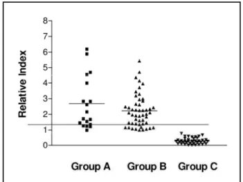
No diff e rence was found in the distribution of the ELISA relative index between samples from gro u p s A and B (Fig 1).
Table 1. Clinical manifestations, relation of active and inactive forms of NC in group A, (n=18).
Clinical manifestations Active forms Inactive forms Headache Seizures Cysts Hydrocephalus Calcifications
(8) 28% (10) 72% (6) 33% (2) 11% (10) 56%
Table 2. Results of laboratory analysis of CSF samples from patients with neuro l o g i c a l manifestations and positive (groups A and B) or negative (group C) results in ELISA test -ing for neurocysticercosis in CSF.
Groups of CSF samples Statistical analysis
Laboratory A B C AxB AxC BxC
Findings n=18 n=50 n=41 p value p value p value
Pleocytosis >4/mm3 33% 66% 5% <0.05 <0.05 –
Erythrocytes >0/mm3 44% 68% 58% <0.05 >0.05 >0.05
Protein = >40 mg/dL 55% 88% 0% <0.05 <0.05 <0.05
(IgGIF) >10% 60% 27% 0% >0,05 <0.05 <0.05
Q. Albumin >7x10⁻³ 33% 73% 0% <0.05 – –
R.E.T. Cysticercosis 100% 100% 0% – – –
R . E . T., (reactive ELISA test) for cysticercosis in CSF; IgGI F(intrathecal fraction of igG) >10% indicates intrathecal synthesis of IgG; Q. Albumin >7x10⁻³ indicates blood-CSF barrier dysfunction.
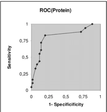
The ROC curve (Fig 2) shows the best cutpoint for the protein concentration in the group A (patients with the diagnosis of NC). The best cutpoint for the p rotein concentration was60 mg/dL, with a sensi-tivity of 83% and a specificity of 80%. The best cut-point for pleocytosis was 7 cells/mm3, with a
sensi-tivity of 77.8% and specificity of 64%. A cutpoint was not found for the other CSF parameters in the gro u p A and B.
DISCUSSION
Assuming that patients enrolled in group A are the true positive (patients affected by NC) and gro u p C the true negative, the problem with false re s u l t s arises from the patients enrolled in group B (patients with neurological disorders not compatible with NC and Ab-ELISA reactivity for NC in CSF).
Significant diff e rences were found among the CSF parameters in the NC group (group A) and the t rol groups (groups B and C). It included: protein con-centration, pleocytosis, albumin quotient and the pre-sence of ery t h rocytes. The high concentration of pro-tein associated with the blood-CSF barrier dysfunc-tion among CSF samples included in group B indicat-ed that part of this protein was not synthesizindicat-ed in the CNS1 5. False-positive results for Ab-ELISA testing
in serum have been detected probably due to the se-rum proteins that bind to cysticercusT. soliuma n t
i-g e n s5 , 1 6. Glycoproteins ofT. soliumhave been
conside-red as very sensitive and specific antigens for NC diag-nosis when western blot (WB) assay is used with se-rum samples but they did not show the same eff i c i e n-cy on Ab-ELISA testing5. The occurrence of
false-pos-itive reactions in Ab-ELISA CSF test may be related to blood-CSF barrier dysfunction allowing the passage of serum antibodies and other proteins to the CNS.
In the group B, elevated pleocytosis associated with other infectious disease agents (viral and bacte-rial) indicated meningeal inflammatory process not related with NC. In this group all CSF samples were reactive in the Ab-ELISA for NC but without clinical and imaging findings for NC. Another explanation of false positive Ab-ELISA test for NC could be associa-ted to cross reactivity to other infectious agents1 1.
This hypothesis was not acceptable because the pa-tients in this group presented diff e rent kinds of disea-ses: infectious (viral and bacterial meningitis) but also non-infectious (stroke, tumor and polineuropathy).
We used the ROC curve to compare the sensitivi-ty and specificisensitivi-ty of the CSF parameters for the gro u p s A (NC) and B (false-positive for NC), in sense of to choose a best cut-point. Based on our findings, it was demonstrated a greater probability of CSF false-pos-itive reactions in Ab-ELISA test for NC (usingT. soli -umantigens) in the cases of pleocytosis higher than 7 cells/mm3or protein concentration in CSF higher
than 60 mg/dL.
Several studies have been conducted in order to i m p rove the diagnosis of NC2 , 6. Analysis of the
integri-ty of the blood-CSF barrier function has contributed to determine the intrathecal synthesis of antibody. In addition, purified larvalTaeniaantigens from ho-mologous( T. solium)or heterologous( T. crassiceps)
parasites have been obtained and characterized in order to obtain reagents with high degree of speci-f i c i t y6 , 1 7 - 1 9. Antigen preparations with contamination
of pig proteins, a common problem whenT. solium
cysts are used, are avoided using antigens obtained from larvae of T. crassiceps2,19,20.
As a re t rospective study, we had some limitations, such as the absence of paired serum samples to com-p a re the scom-pecific serum and CSF antibodies. However, the Ab-ELISA assay (usingT. soliumantigens) in seru m is not a good test for the diagnosis of NC, due the p resence of high frequency of false-positive re a c-tions. We used the Sotelo´s criteria for the diagnosis of NC, considering it more appropriate for the kind of study. It is based predominantly on clinical and imaging features1.
H e re, we demonstrated that the CSF findings may influence the immunoreactivity of the CSF ELISA test for NC. CSF reactive patients without NC had higher f requency of blood-CSF dysfunction, elevated level of protein concentration, pleocytosis and red blood cells count in comparison to the CSF reactive patients with NC (true positive).The most frequently findings w e re the high level of protein and blood-CSF barr i-er dysfunction. These may contribute to the si-eru m p roteins that bind to cysticercusT. soliuma n t i g e n s5
passage to the CSF, conducting to false positive re a c-tion. In addition, we also proposed a cut-point for the protein concentration and pleocytosis. The chance of false-positive Ab-ELISA for NC increases with the concentration of protein higher than 60 mg/dL (sen-sitivity of 83% and a specificity of 80%) and pleocy-tosis higher than 7 cells/mm3(sensitivity of 77.8% and
specificity of 64%). Thus, the results of the Ab-ELISA tests should be carefully considered in together with other clinical, laboratorial and epidemiological data for the diagnosis of NC. Confirm a t o ry tests for CSF should be included in the routine diagnosis criteria for NC.
Acknowledgments– The authors thank Mrs. Rosânge-la Noé from “Comissão de Investigação Científica do HUCFF/ UFRJ”for the statistical analysis of this study.
REFERENCES
1. Sotelo J, Del Butto OH. Review of neuro c y s t i c e rcosis. Neuro s u rg Focus 2002;12:1-7.
2. Machado LR, Livramento JA, Vaz AJ, et al. IgG intrathecal synthesis and specific antibodies index in patients with neurocysticercosis. Arq Neuropsiquiatr 2002;60:395-399.
3. Sotelo J, Guerrero V, Rubio F. Neurocysticercosis: a new classification base on active and inactive forms. Arch Intern Med 1985;145:442-445. 4. Del Brutto OH , Rajsherkhar V, White A C J r, et al. Proposed diagnostic
criteria for neurocysticercosis. Neurology 2001;57:177-183. 5. Tsang VCW, Brand JA, Boyer AE. An enzyme-linked immoelectro t r a n
s-fer blot assay glycoprotein antigens for diagnosing human cysticerc o-sis. J Infec Dis 1989;159:50-59.
6. Ito A, Plancarte A, Ma L, et al. Novel antigens for neuro c y s t i c e rc o s i s : simple method for preparation and evaluation for serodiagnosis. Am J Trop Med Hyg 1998;59:291-294.
7. P roaño-Narvez JV, Meza-Lucas A, Mata-Ruiz O, Garc i a - J e ronimo RC, C o r rea D. Laboratory diagnosis of human neuro c y s t i c e rcosis: double-blind comparison of enzyme-linked immunosorbent assay and elec-troimmunotransfer blot assay. J Clin Microbiol 2002;40:2115-2118. 8. Dorny P, Brandt J, Zoli A, Geerts S. Serodiagnostic tools for human and
porcine cysticercosis. Acta Trop 2003;87:79-86.
9. Vaz AJ, Livramento JA. Neurocisticercose. In Ferreira AW, Avila SLM (eds). Diagnóstico laboratorial das principais doenças infecciosas, 2.Ed. Rio de Janeiro: Guanabara Koogan, 2001:316-322.
10. Katti MK. Assessement of antibody responses to antigens of M y c o b a c -terium tuberculosisandC y s t i c e rcus cellulosaein cere b rospinal fluid of c h ronic meningitis patients for definitive diagnosis as TBM/NCC by passive hemmaglutination and immunoblot assays. FEMS Immnol Med Microbiol 2002;33:57-61.
11. Gekeler F, Einchenlaub S, Mendonza EG, Sotelo J, Hoelscher M, Hoschen T. Sensitivity and specificity of ELISA and immunoblot for diagnosing neuro c y s t i c e rcosis. Eur J Clin Microbiol Infec Dis 2002; 21:227-229.
12. Reiber H, Felgenhauer K. Protein transfer at the blood cere b ro s p i n a l fluid barrier and the quantification of the humoral immune re s p o n s e within the central nervous system. Clin Chem 1987;163:319-328. 13. Adams RD, Victor M, Ropper AH. Principles of neuro l o g y. International
Edition New York: MC Graw-Hill, 2001.
14. Mc Donald WI, Compston A, Edan G, et al. Recommended diagnostic criteria for multiple sclerosis: guidelines from the international panel on the diagnosis of multiple sclerosis. Ann Neurol 2001;50:121-127. 15. Reiber H, Peter JB. Cere b rospinal fluid analysis: disease-related
pat-terns and evaluation programs. J Neurol Sci 2001;184:101-122. 16. Vaz AJ, Ferreira AW, Silva MV, et al. Teste imunoenzimático para
pes-quisa de anticorpos anti-C y s t i c e rcus cellulosaeem líquidos cefalorra-quianos de pacientes com meningites de etiologia indeterminada. Rev Inst Med Trop Med São Paulo 1990;32:196-203.
17. Peralta RH, Vaz AJ, Pardini A, et al. Evaluation of an antigen fromTa e n i a crassicepsc y s t i c e rcus for the serodiagnosis of neuro c y s t i c e rcosis. A c t Trop 2002;83:159-168.
18. P a rdini AX, Peralta RH, Vaz AJ et al. Use ofTaenia crassicepsc y s t i c e r-cus antigen preparations for detection of antibodies in cere b ro s p i n a l fluid simples from patients with neuro c y s t i c e rcosis. Clin Diag Immunol 2002;9:190-193.
19. Larralde C, Sotelo J, Montoya RM, et al. Immunodiagnosis of human cysticercosis in cerebrospinal fluid: antigens from from murine Taenia crasssicepsc y s t i c e rci effectively substitute those from porcineTaenia soli -um.Arch Pathol Lab Med 1990;114:926-928.