DOI: 10.1590/0004-282X20160140
VIEW AND REVIEW
Understanding dystonia: diagnostic issues
and how to overcome them
Compreendendo a distonia: questões diagnósticas e como superá-las
Sarah Camargos, Francisco Cardoso
INTRODUCTION AND HISTORICAL ASPECTS
Etymologically, the word dystonia comes from the Greek language and means altered muscle tone. According to the irst Oppenheim deinition in 1911, the “muscle tone was hypotonic at one occasion and in tonic muscle spasm at another, usually, but not exclusively, elicited upon voluntary movements”1.
he irst description of dystonia dates back to the end of the 19th century and is by Barraquer-Roviralta although
not recognized as such1. Later, in 1911, Herman Oppenheim
coined the term “dystonia musculorum deformans” for the
description of this movement disorder in a Jewish patient1.
Contrary to the prevailing view of that time, he proposed an organic origin of the disease. As time progressed, several pieces of evidence supported the organic nature of dystonia: the hereditary mechanism, limited eicacy of psychotherapy,
good response to thalamotomy or pallidotomy and, inally,
the onset of dystonia following brain lesions in monkeys2.
In June of 1975, Stanley Fahn and Roswell Eldridge chaired an International Symposium on Dystonia in New York City. At the conference, David Marsden described sporadic forms of dystonia and deined its focal forms ( for instance, cervi-cal dystonia, blepharospasm, oromandibular dystonia and writer’s cramp) as ‘formes frustres’ of generalized dystonia. He, and other authors who attended this meeting, empha-sized the organic foundation of dystonia. At that conference, Masaya Segawa reported patients with dopa-responsive dys-tonia (DRD), for the irst time outside Japan2. In 1984, the Ad Hoc Committee of the Dystonia Medical Research Foundation deined dystonia as “a syndrome of sustained muscle contrac-tions frequently causing twisting and repetitive movements
or abnormal postures”3. Other important hallmarks in the
Universidade Federal de Minas Gerais, Clínica de Distúrbios de Movimento, Departamento de Clínica Médica, Belo Horizonte MG, Brasil.
Correspondence: Francisco Cardoso; Av Pasteur 89/1107; 30150-290 Belo Horizonte MG, Brasil; E-mail: [email protected]
Conflict of interest: There is no conlict of interest to declare.
Received 16 May 2016; Received in inal form 13 June 2016; Accepted 07 July 2016.
ABSTRACT
The diagnosis and treatment of dystonia are challenging. This is likely due to gaps in the complete understanding of its pathophysiology, lack of animal models for translational studies, absence of a consistent pathological substrate and highly variable phenotypes and genotypes. The aim of this review article is to provide an overview of the clinical, neurophysiological and genetic features of dystonia that can help in the identiication of this movement disorder, as well as in the differential diagnosis of the main forms of genetic dystonia. The variation of penetrance, age of onset, and topographic distribution of the disease in carriers of the same genetic mutation indicates that other factors – either genetic or environmental – might be involved in the development of symptoms. The growing knowledge of cell dysfunction in mutants may give insights into more effective therapeutic targets.
Keywords: dystonia; apoptosis.
RESUMO
O diagnóstico e o tratamento da distonia podem ser desaiadores. Isso se dá provavelmente a pouca compreensão da isiopatologia, a falta de modelos animais para estudos translacionais, ausência de um substrato patológico consistente e genótipo e fenótipo altamente variáveis. O objetivo deste artigo de revisão é fornecer uma visão geral dos aspectos clínicos, neuroisiológicos e genéticos de distonia que podem ajudar na identiicação deste distúrbio do movimento, bem como no diagnóstico diferencial das principais formas de distonia hereditária. Há uma ênfase particular na nova deinição e classiicação da Internacional das Distonias, bem como as recentes descobertas dos mecanismos moleculares subjacentes em algumas formas de distonia primária. A variação de penetrância, idade de início, e distribuição topográica da doença em portadores da mesma mutação genética indica que outros fatores - genéticos ou ambientais podem estar envolvidos no desenvolvimento dos sintomas. O conhecimento crescente sobre a disfunção celular em mutantes pode gerar insights sobre alvos terapêuticos mais eicazes.
history of dystonia were the discovery and description of the DYT1 locus and gene in, respectively, 1989 and 19974.
During the last two decades, there has been a remarkable growth of research in the ield of dystonia with a multitude of studies encompassing genetics, molecular biology, clinical aspects, as well as pharmacological and surgical treatment. For instance, the word dystonia has 15,774 PubMed citations, 1,366 being in the last two years. Recently, Broussolle et al.
shed light on the history of the geste antagoniste phenom
-enon in dystonia5. Figure 1 shows a brief timeline in dystonia.
MOVING TOWARDS A NEW DEFINITION AND NEW CLASSIFICATION
Fahn classiied dystonia according to age at onset, topo-graphic distribution and etiology. he latter included primary, secondary and psychogenic dystonia3. Later, Bressman6 de -ined primary dystonia as those cases where dystonia is the sole phenomenon and there is no structural brain lesion or in -born error of metabolism. In her classiication, secondary dys-tonia is characterized by the combination of dysdys-tonia and oth -er abnormalities in the neurological examination. It includes dystonia plus (genetic forms where dystonia is combined with
another movement disorder such as parkinsonism and my
-oclonus), heredodegenerative dystonia (genetic degenerative diseases where dystonia is part of the picture), acquired dys-tonia (the movement disorder results from a deined acquired cause such as stroke, use of neuroleptics and many others) and psychogenic dystonia6. Recently, a consensus committee spon-sored by the Dystonia Medical Research Foundation, Dystonia Coalition and European Dystonia Cooperation in Science and Technology has proposed a new deinition and classiication of
dystonia7. he new deinition, based on some pivotal features
of the abnormal movement, attempts to overcome shortcom
-ings from the past:
“Dystonia is a movement disorder characterized by
sus-tained or intermittent muscle contractions causing abnor
-mal, often repetitive, movements, postures, or both. Dystonic movements are typically patterned, twisting, and may be
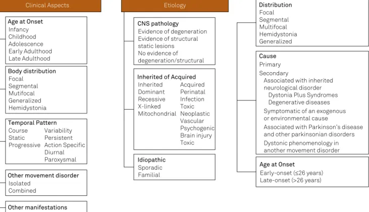
tremulous. Dystonia is often initiated or worsened by volun-tary action and associated with overlow muscle activation7.” In the new classiication system, there are two axes: clini-cal features and etiology, as shown in Figure 2. For cliniclini-cal fea-tures, one should note age at onset, topographic distribution, temporal pattern and other associated movement disorders or manifestations. With reference to etiology, important aspects to be deined are: whether dystonia is inherited, acquired or idiopathic, as well as whether there is evidence of central ner -vous system pathology (degeneration or static lesion) 7.
From a clinical point of view, the new classiication has not introduced substantial changes. he most notable difer-ence is the acknowledgement that tremor may be the main manifestation of dystonia. However, there are aspects that were not received with unanimous approval, such as the use of the word “idiopathic”, a previously abandoned term7.
HOW DOES IT MOVE? PHENOMENOLOGY
he irst and most important question related to the
clinical aspect of dystonia is how to identify the phenome
-non. Sometimes this is a diicult task, especially when there are other movement disorders involved. he new
classiica-tion, and occasionally resorting to neurophysiological stud
-ies, provides guidance on how to diagnose dystonia, even in challenging cases.
Clinical Features
Postures and movements
One of the most important features of dystonia is the pre -dictability and patterned nature of the muscle contractions. his makes it unique when compared to other hyperkinetic disorders. Contractions can be sustained, ixed or intermit-tent and sometimes mixed with tremor. his tremor can be rhythmic or irregular with occasional jerks8. Dystonic pos-tures are when a body part is lexed or twisted along its lon-gitudinal axis (except for blepharospasm and laryngeal dys-tonia). Dystonic movements, are usually twisting in nature
Figure 1. Timeline of dystonia. Oppenheim
described “dystonia musculorum
deformans”
Onset of dystonia following brain lesion
International Symposium on Dystonia
Dopa responsive dystonia outside Japan
Definition of focal dystonia
DBS for Cervical Dystonia
Definition of Dystonia
Ad Hoc Committee of the Dystonia Medical Research
Foundation
FDA approaval for use of Botulinum
Toxin in dystonias
DYT5a/b genes
DYT1 gene DBS for Generalized
Dystonia
1911
DYT11 gene DYT12, DYT8 genes
DYT16, DTY18 genes
DYT6 gene DYT10
gene DYT23, DYT24 genes
DYT4, DYT25 genes
New Classification
Consensus Committee on
Dystonia
DYT2, DYT26 DYT27 genes
or a pull in a preferred direction. Movements are sustained
at their peak and lessen when a given posture is reached8.
According to the new deinition, dystonia is often initiated or worsened by voluntary action.
Sensory trick or geste antagoniste
Sensory tricks are often, but not exclusively, tactile stim-uli, usually in the body part afected by the movement
dis-order, that produce a meaningful alleviation of dystonia5.
hey are usually simple natural movements, never forceful, involving the body region afected by dystonia. Physiological studies with electromyography (EMG) have shown a modi-ication in recruitment during the efect of sensory tricks. However, the underlying mechanism remains to be
deter-mined. Loyola et al. hypothesized that sensory tricks may in
-duce a rebalancing of central processing by reducing the acti -vation of the supplementary motor area and primary sensory motor cortex9.
Overflow
Overlow is deined as the extension of muscle contrac-tion into an adjacent area anatomically distinct from the primary movement when dystonic posture reaches a peak.
A voluntary compensatory posturing may not easily be dis
-tinguishable from overlow7,8,10,11,12. Usually, voluntary move -ments are slower and more variable. According to Hallett, overlow is the clinical representation of impairment of
nor-mal surrounding inhibition present in dystonia12. According
to this hypothesis, there is an imbalance of abnormal senso
-rimotor integration circuitry and cortical excitability. As a result, aferent inputs are inadequately processed at several levels of the central nervous system. his remodeled system creates an eferent output of abnormal co-contraction with the absence of surrounding inhibition11.
Mirroring
Mirror dystonia occurs on the afected body side when a speciic task (e.g., writing, inger sequence, piano-like move-ments) is performed by the homologous opposite normal
body part. It could be considered as a subset of motor over
-low. he existence of simultaneous activation of the cortical
spinal pathway, mediated by the impaired inhibition of trans
-callosal commissure, has been hypothesized7,8,10,11,12,13.
DIFFERENTIAL DIAGNOSIS
he recognition of dystonia is sometimes a diicult task,
at least in part due to a combination of more than one hy
-perkinetic movement disorder and even to the overlapping of hyperkinesia and bradykinesia. Diagnosis is essentially
a clinical one and should be preceded by meticulous infor
-mation and observation of movement, including the onset,
spread, duration, rhythmicity, topography and predictabili
-ty8. Dystonia may mimic several movement disorders: phasic
movements can resemble tremor, myoclonus and even tics.
Figure 2. New Classiication of dystonia based on two axes (Adapted from Albanese et al in Mov Disord. 2013;28:863-873) and the previous Classiication of Dystonia (Geyer & Bressman, Lancet Neurol. 2006; 5: 780–90)
Clinical Aspects Etiology
Other manifestations Age at Onset
Infancy Childhood Adolescence Early Adulthood Late Adulthood
Body distribution Focal
Segmental Mutifocal Generalized Hemidystonia
Temporal Pattern Course
Static Progressive
Variability Persistent Action Specific Diurnal Paroxysmal
Other movement disorder Isolated
Combined
Distribution Focal Segmental Multifocal Hemidystonia Generalized
Cause Primary Secondary
Associated with inherited neurological disorder
Dystonia Plus Syndromes Degenerative diseases Symptomatic of an exogenous or environmental cause
Associated with Parkinson’s disease and other parkinsonian disorders Dystonic phenomenology in another movement disorder
Age at Onset
Early-onset (≤26 years) Late-onset (>26 years) CNS pathology
Evidence of degeneration Evidence of structural static lesions No evidence of degeneration/structural
Inherited of Acquired Inherited
Dominant Recessive X-linked Mitochondrial
Acquired Perinatal Infection Toxic Neoplastic Vascular Psychogenic Brain injury Toxic
Tremor is an involuntary, patterned and rhythmic oscillation
of a body region around a joint axis, generating a symmet
-ric velocity in both directions at midpoint of the movement8. Diferently, dystonic tremor can be arrhythmic, has irregu-lar amplitude and superimposed jerks. Myoclonus is an in-termittent condition, a sudden, brief, shock-like movement caused by muscular contractions or inhibitions. Dystonia has a sustained component not seen in myoclonus. Tics are
par-oxysmal, stereotyped muscle contractions, temporally sup
-pressible and usually have a premonitory sensation and re
-lief after performance. Partial suppression and a premonitory sensation are hallmarks that diferentiate tics from dystonia.
Abnormal postures can be present in several disorders with
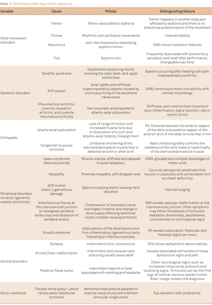
-out representing an involuntary dystonic muscle contraction. hese contractions, called pseudodystonias, have a myriad causes, including orthopedic, ocular, vestibular, inlammato-ry, rheumatologic, peripheral (soft tissues, muscle, ligaments) or central problems14,15. Table 1 lists the speciic causes of pseudodystonia. Striatal hands, camptocormia and Pisa
syn-drome are examples of abnormal postures usually seen in pa
-tients with Parkinson’s disease (PD) with an unknown etiol-ogy. Diferent from the majority of dystonias, striatal hands are ixed, don’t worsen with activity nor do they disappear during sleep. Camptocormia and Pisa syndrome are postural
deformities in which, respectively, there is an abnormal an
-teroposterior lexion of the trunk or its marked lateral lex-ion. hese conditions have been observed in several diseas-es such as PD, multiple system atrophy, diferent dystonias, Tourette syndrome, myotonic dystrophy, osteo and musculo-skeletal disorders, myopathies, amyotrophic lateral sclerosis, and drug-induced conditions. An interesting feature is that these postures are reversible when the patient stands against a wall, uses a high frame walker or lies lat15. Some may
in-terpret such facts as sensory tricks. Other evidence suggests
they have a dystonic etiology with a few studies demonstrat
-ing EMG changes associated with dystonia and its occasional
improvement with botulinum toxin14. However, it has been
proposed that the dystonic phenomenon, if it exists, has a short presentation as an early phenomenon. Ultimately, in the later stages, dystonia disappears with soft tissue, mus -cle and spine problems dominating the picture14,15.
Fixed dystonia is characterized by a sustained
abnor-mal limb posture regardless of other factors16. It is most
commonly associated with severe cases of primary or sec
-ondary dystonia, corticobasal syndrome or in associa
-tion with complex regional pain syndrome (types I and II without/with preceding nerve injury). This condition was previously labeled as “reflex sympathetic dystrophy” or “causalgia-dystonia”16. Interestingly, in 1892, Charcot
de-scribed two patients with a combination of pain, edema,
discoloration of skin associated with paralysis, contrac
-tures and an hysterical condition16. Whether fixed dysto
-nia is a functional/psychogenic movement disorder or not is still a matter of debate. Several studies have shown that
a number of patients fulfill the criteria for psychogenic or
somatoform disorder16. Typically, fixed dystonia develops
subacutely in the distal limb of young women, sometimes spreading to other limbs in a characteristic flexed position
without overflow or geste antagoniste. However, a small
percentage of patients with fixed dystonia in the context
of complex regional pain syndrome remain without a psy
-chiatric diagnosis16.
A challenging diferential diagnosis is psychogenic
dys-tonia. Usually, movement is both inconsistent and incongru
-ous in an organic movement disorder. Electrophysiological testing is a valuable clinical tool (see below). Movement
may disappear with distraction and be enhanced by sugges
-tion. Commonly, psychogenic dystonias are ixed at onset, have excessive pain or fatigue and potential for secondary gain. Patients frequently have a history of multiple somati-zations and psychiatric disturbance. Deinite diagnosis re-quires repeated and extensive evaluations. he prognosis is usually poor2,16.
Tremor and Dystonia
Dystonia may erroneously be interpreted as tremor. Dystonic tremor is frequently misdiagnosed as essential tremor or Parkinson disease.
Dystonic tremor is deined as dystonia that manifests itself mostly with tremor17. Isolated head tremor, presenta
-tion of head tremor before arm tremor and more severe head tremor than arm tremors are virtually all manifestations of dystonic tremors. An interesting clue is that in essential tremor, head tremor often disappears when the patient lies
down but persists in cervical dystonia18. Cases of isolated
voice tremor predating the onset of hand tremor, and being more severe than hand tremor, is considered to be “tremu-lous dystonia”17. Usually dystonic tremors have irregular am
-plitudes and superimposed jerks.
When tremor occurs in the dystonia site, it is called dystonic tremor. his is a phenomenon that has received more attention lately. Tremor can be present in approxi-mately 30% of subjects with dystonia. According to Erro et
al., the prevalence of resting tremor among tremulous dys
-tonic patients is 12% of all dys-tonic patients17. his study
demonstrated that rest tremor in dystonic patients is most
-ly unilateral or asymmetric and, remarkab-ly, does not have re-emergent tremor. he latter is a postural rest tremor in PD that reappears after a variable delay while maintaining posture. Not surprisingly, a diagnosis of PD is quite com-mon in this group of patients. Diferent from PD, SPECT
imaging of dopamine transporter shows an intact nigros
Table 1. Conditions that may mimic dystonia.
Variable Cause Pitfalls Distinguishing feature
Other movement disorders
Tremor Tremor associated to dystonia
Tremor happens in another body part affected by dystonia and there is no directional predominance of the movement
Chorea Rhythmic and oscillatory movements Unpredictability
Myoclonus Jerk-like movements resembling
dystonic tremor EMG shows myoclonic features
Tics Dystonic tics
Frequently associated with premonitory sensation and relief after performance.
Changeable over time.
Systemic disorders
Sandifer syndrome
Opisthotonic posturing mainly involving the neck, back, and upper
extremities
Spasms occurring after feeding with pain. Videotelemetry conirms.
Stiff person
Axial rigidity and stiffness superimposed by spasms caused by
continuous iring of the peripheral nerve axons
EMG: continuous motor unit activity with normal morphology.
Rheumathoid arthritis, juvenile idiopathic arthritis, and juvenile
rheumatoid arthritis
Non traumatic anteroposterior atlanto-axial subluxation
Stiffness, pain and limited movement plus inflammatory signs (swollen, red or
warm) joints
Orthopedic
Atlanto axial subluxation
Loss of range of motion and increased muscle tone due to dislocation of a joint and atlanto-axial rotatory misalignment
RX: Distance between the anterior aspect of the dens and posterior aspect of the anterior arch of the atlas is more than 3 mm
Congenital muscular torticollis
Unilateral shortening of the sternocleidomastoid muscle that is
detected at birth or after birth
Neck ultrasonography conirms the existence of the neck mass or hypertrophy
of the sternocleidomastoid muscle
Peripheral disorders (muscle, ligaments, vessels and bones)
Isaacs syndrome (Neuromyotonia)
Muscle cramps, stiffness and delayed muscle relaxation.
EMG: grouped and complex discharges of motor units
Myopathy Proximal myopathy with dropped neck
Cervical paraspinal weakness that results in passively and correctable chin
on chest deformity
Soft nuchal mass/ Ligamentous
damage
Space occupying lesion causing neck
deviation Cervical bulging
Arteriovenous istula at the craniocervical junction
(or elongated vertebral artery loop and dilatation of
vertebral artery)
Compression of accessory nerve, meningeal irritation and change in
bone supply affecting vestibular nuclei complex causing torticollis
MRI reveals vascular malformation at the craniocervical junction. Other symptoms
may follow the attacks of torticollis: headache, drowsiness, papilledema, corticobulbar or corticospinal signs
Grisel’s syndrome
Subluxations of the atlantoaxial joint from inlammatory ligamentous laxity
following an infectious process
RX reveals subluxation. Radicular and medullar signs can occur
Central disorders
Epilepsy Intermittent tonic contractions EEG shows epileptiform abnormalities
Arnold Chiari malformation Intermittent and unusual neck posturing usually associated
Usually associated with posterior fossa dysfunction signs and pain
Posterior fossa tumor Intermittent head tilt or twist associated with vomiting and headache
Other neurological signs such as increased intracranial pressure and localizing signs. Torticollis can be the first
sign of central nervous system tumor. Brain image reveals the diagnosis
Oculo-vestibular
Troclear nerve palsy/ Lateral rectus palsy/ Vestibular
torticollis
Abnormal head posture adopted to improve visual acuity and maintain
binocular single vision
tremor with some peculiarities revealed by the underlying
dystonic nature of the movement disorder19:
1) position/task speciicity 2) Jerkiness
3) presence of tremor-lurries, thumb hyperextension 4) pronation-supination type rather than vertical 5) absence of remarkable response to levodopa 6) Static rather than progressive disease.
Although SWEDD patients may have a slow gait, small stride length and reduced arm swing, they have a normal trunk and elbow posture, normal stride length variability and normal bilateral step phase coordination. hese features dif-ferentiate them from patients with PD20. A long-term follow-up study of 16 patients with dystonic tremor resembling PD and SWEDD, at least ive years after initial scan,
demonstrat-ed rdemonstrat-educdemonstrat-ed striatonigral uptake in two patients and thus con
-version to PD21. In summary, the majority of SWEDDs turned
out to have dystonic tremor, but a small proportion of them may still develop PD after some time.
SPECIAL ISSUES
Neurophysiology as a clinical tool
According to some authors, EMG mapping is the “only clinical tool with a proven value for the diagnosis of dystonia
to complement clinical examination”8. Although the previous
statement is debatable, neurophysiologic studies can some
-times help deine the nature of the movement. he usual fea-tures of dystonia on neurophysiological studies are:
1) Prolonged EMG bursts (200-500ms)
2) Simultaneous contractions (co-contraction) of agonist and antagonist muscles
3) Abnormal voluntary control of muscles with somatotop
-ic contiguity (overlow): contraction of surrounding? muscles through impaired inhibition of spinal and medulla relexes12.
If dystonic postures and movements have all the phenome
-nological features described above; or if at least two of the acti -vation/deactivation features (tricks, mirroring, overlow) diag-nostic criteria are met; or if one of the above plus EMG mapping demonstrating features of dystonia (such as abnormal activa-tion, co-contraction and overlow), this can help clinical diag-nosis8. hese proposed criteria need to be validated before they
can be incorporated into regular clinical practice8.
Non-motor features
It is widely known that several genetic presentations of dystonia are associated with psychiatric abnormalities. For
instance, alcohol addiction, anxiety, depression and, partic
-ularly, obsessive-compulsive disorder have been associated with DYT11 (myoclonus-dystonia). Interestingly, patients
present with more compulsions than obsessive symptoms22.
Recently, mild executive dysfunction was also described as part of the clinical spectrum of DYT1122. Mood disorders and
psychoses have a higher prevalence in patients with rapid onset dystonia-parkinsonism (DYT12) than in non-mutant carriers23,24. Psychiatric non-motor features are also found in
sporadic forms of primary dystonia. In one study comparing primary dystonia patients (blepharospasm and cervical
dys-tonia) with matched controls, it was demonstrated that anxi
-ety, panic disorder, agoraphobia, obsessive-compulsive disor-der, alcohol abuse and drug dependence were more common in the patients with dystonia25. Moreover, these psychiatric disorders frequently preceded the movement disorder. here
is still debate whether these symptoms result from dysfunc
-tion of the brain or are a psychological reac-tion to a disabling condition. he onset of the psychiatric abnormalities be-fore the development of motor indings of dystonia suggest the former hypothesis is true. Studies of cognition in dysto-nia have yielded conlicting results. A recent investigation of non-depressed adult-onset primary cranial-cervical dys-tonia patients showed diferences in working memory, pro-cessing speed, visual motor ability and short-term memory. he impairment did not correlate with the disease severity or duration of dystonia26. hese indings could be partially explained by the involvement of the fronto-striatal circuits previously demonstrated by voxel-based morphometry stud-ies26. Further studies are necessary to conirm these data and
also to assess whether these abnormalities are persistent or even progressive. From a clinical perspective, the impression is that the majority of patients with primary dystonia do not have meaningful cognitive abnormalities25,26.
ETIOLOGY
he etiology of primary dystonia is assumed to be a
com-plex combination of intrinsic metabolic properties, environ
-mental and genetic factors27,28.
Risk factors
In a recent study from Queensland, Australia, several pu
-tative associations have been made with isolated idiopathic dystonias. Anxiety disorders, tremor, cigarette smoking and
head injuries with a loss of consciousness were statistical
-ly associated with increased risk of developing dystonia27.
Scoliosis and soft tissue trauma appear to increase the risk of cervical dystonia in genetically predetermined individ-uals28,29. Sunlight exposure has been linked to a higher risk
factor for the development of blepharospasm. It has been
hypothesized that high insolation induces excessive blink
-ing and this overuse results in sustained orbicularis oculii
spasms30. Repetitive highly-skilled manual performance may
important clue to the etiopathology of dystonia31. Although controversial, peripheral neuro-musculoskeletal injury ap-pears also to be a risk factor for task speciic dystonias. A pos-sible explanation could be overcompensation to the deicit generating a lack of inhibition leading to dystonia32.
Pathophysiology
The pathophysiology of dystonia lies in the basis of lack of inhibition. Basal ganglia filter and modulate inputs to improve the precision of fine movements. The failure of surrounding suppression is probably related to deficient inhibition by basal ganglia gabaergic interneurons and
output31. One emerging theory is that sensorimotor sys
-tems have Hebian-like plasticity33. In a dystonic endophe
-notype, the summation of abnormal sensorimotor plastic
-ity and the inabil-ity to control homeostatic mechanisms results in a chaotic reorganization of sensory-motor maps. For instance, an abnormal plastic change occurs without a downregulation to inhibit it34.
It is speculated that the cerebellum is also involved in the deicit of sensorimotor integration presented in dystonia. he cerebellum might process aferent proprioceptive
infor-mation and modify the threshold of the somatosensory cor
-tex through the cerebello-thalamo-cortical loop. It could also inluence the cortex plasticity35.
Genetic factors
here is a relatively large body of knowledge of genetic fac-tors in dystonia. Since the irst description, in 1997, of a gene linked to familial dystonia, DYT1, several other genes have been reported either by linkage analyses or high throughput assays. his has led to new insights of the cellular pathways associated with this neurological dysfunction6.
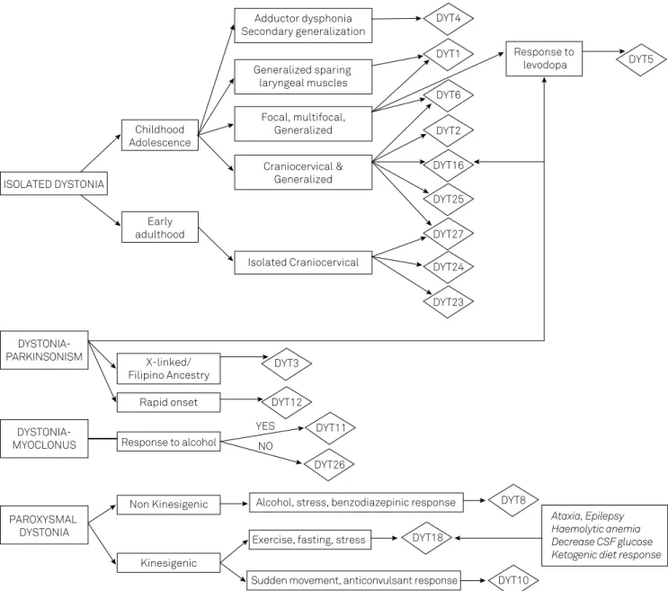
he practitioner faces the daunting task of how to cor-relate the large number of dystonia-cor-related genes with clini-cal features. To overcome this challenge, a wide-ranging algo-rithm, constructed on the clinical basis for the diagnosis of hereditary dystonias in which genetic testing is possible, was created (Figure 3). In a didactic and pragmatic approach, dys-tonia was categorized into pure dysdys-tonia, dysdys-tonia associated
with other movement disorders or paroxysmal dystonias36.
To make a speciic diagnosis where dystonia is the sole neurologic manifestation, one should take into consideration the age at onset, spread pattern and involvement of cranial and laryngeal muscles. DYT1, the most common hereditary
dystonia, is also the most common cause of pure genetic dys
-tonias4. Patients usually have childhood or adolescence
on-set, with initial focal involvement of one limb (usually lower) spreading to other limbs and muscles, becoming generalized
but sparing the larynx and cranial muscles. Although phe
-notypically similar to DYT1, DYT6 onset is later (average 19 years of age), typically in the cranial and cervical muscles, and those who have limb onset (often arms) later develop crani-al or cerviccrani-al dystonia. In a patient with foccrani-al, segmentcrani-al or
generalized dystonia with cranial muscle involvement, DYT6 is the most likely the cause37. However, if negative, the next gene that should be tested is DYT1. It is very important to emphasize that DYT1 and DYT6 have an incomplete pene-trance (30% and 60%, respectively), which can easily lead to
misinterpretation as sporadic, or autosomal recessive inheri
-tance. he same mutation in DYT6, even within a family, can present with diferent phenotypes38.
Isolated craniocervical dystonia can be related to two recently-described forms of dystonia: DYT23 and DYT24. he former was described in a family of German origin and is still to be conirmed39. DYT24 was described in a British family and three unrelated patients. he patients usual-ly present with adult-onset dystonia involving the neck and/or face. DYT27 was recently described as a cause of segmental dystonia in three German families. he patients present with cranial, laryngeal and segmental dystonia with cervical, upper limbs and trunk involvement. he mode of inheritance is recessive. DYT25 is another dystonia that can overlap clinically with DYT1 and DYT6. It may present as fo-cal, segmental or generalized dystonia, with onset mostly in adulthood (mean age 31 years)40. DYT2 is another cause of
recessive segmental dystonia, often with slow progression to generalized dystonia, therefore also overlapping with DYT1 and DYT6. Its causative gene was only recently de-scribed (COL6A3) and it is related to Bethlem myopathy41. “Whispering dysphonia” (adductor spasmodic dysphonia, DYT4) was described in a large family in North Queensland, Australia in 1985. Dysphonic symptoms may improve with alcohol. Patients typically have a thin body and face,
hol-lowed cheeks and a bradykinetic tongue as well as psychi
-atric symptoms (anxiety, aggressive behavior and alcohol abuse). Dystonia progresses to the generalized form and
pa-tients often exhibit a peculiar hobby horse gait with atax
-ia. Recently the putative gene of this condition has been described at 19p13.3 (TUBB4)42. Other primary dystonias, whose genes have not been identiied yet, are DYT7, DYT13, and DYT17. With the exception of DYT7, whose clinical fea-tures resemble DYT23 and DYT24, these dystonias have a phenotype overlapping with DYT1 and 637,38.
In the case of dystonia associated with other movement
disorders, the phenomenology guides us through the diag
-nosis. If there is myoclonus, DYT11 or DYT26 are probably the cause. In DYT11, myoclonus, highly responsive to al-cohol, is usually a signiicant symptom and the dystonic manifestation is often represented by spasmodic torticollis
or writer’s cramp43. Occasionally, dystonia may be the only
manifestation of the disease and may precede myoclonus. A high incidence of psychiatric disturbances is described in DYT11. Otherwise, in DYT27 patients do not present with
psychiatric disturbances or myoclonus responsive to alco
only in DYT11 and DYT27, the encoding proteins of which are, respectively, epsilon sarcoglycan and potassium channel tetramerization domain-containing protein 1744,45.
In the case of parkinsonism associated with dystonia,
levodopa response, mode of inheritance and mode of on
-set provide clues to the correct etiologic diagnosis. If dys
-tonia-parkinsonism responds to levodopa, DRD is probably the cause. Two genes are related to DRD: GCH1 and TH46,47. Mutations of GCH1 (GTP cyclohydrolase 1) are transmit-ted in an autosomal dominant manner (DYT5a). Patients may develop worsening of the symptoms at the end of the
day and there is complete remission of symptoms after ad
-ministration of low doses of levodopa48. Importantly, unlike individuals with PD, patients with DYT5a usually do not develop complications from the chronic use of levodopa. he phenotype spectrum of CGH1 mutations has expand-ed and includes spasticity and spastic paraplegia. DYT5b is
the other gene encoding mutation of the tyrosine hydroxy
-lase enzyme. It causes the recessive Segawa syndrome, also characterized by marked improvement with administration of levodopa, and diurnal luctuation. However, unlike the dominant form, patients usually present with motor and
speech delay, hypotonia, encephalopathy, ataxia, and auto
-nomic failure, as well as changes of the sleep-awake cycle47. In a dystonia-parkinsonism patient not responsive to le-vodopa with a recessive mode of inheritance, DYT16 should be considered. Some DYT16 patients may not exhibit
par-kinsonism but, instead, present with pure generalized dys
-tonia with predominantly axial features involving speech and a sardonic smile49. If a pedigree shows only afected males, X-linked inheritance is likely. he X-linked dystonia occurs predominantly in Filipino families from the Panay Island. In addition to parkinsonism, myoclonus, chorea and tremor were also described48.
Figure 3. Algorithm for the diagnosis of hereditary dystonias.
ISOLATED DYSTONIA
Childhood Adolescence
Early adulthood
Adductor dysphonia Secondary generalization
Generalized sparing laryngeal muscles
Focal, multifocal, Generalized
Craniocervical & Generalized
Isolated Craniocervical
Response to levodopa DYT4
DYT1
DYT6
DYT2
DYT16
DYT25
DYT27
DYT24
DYT23
DYT5
DYSTONIA-PARKINSONISM
DYSTONIA-MYOCLONUS
PAROXYSMAL DYSTONIA
X-linked/ Filipino Ancestry
Rapid onset
Response to alcohol
Non Kinesigenic
Kinesigenic
DYT3
DYT12
DYT11
DYT26 YES
NO
Alcohol, stress, benzodiazepinic response
Exercise, fasting, stress
Sudden movement, anticonvulsant response
DYT8
DYT18
DYT10
he onset of dystonia can be quite informative about the underlying genetic cause as well. DYT12 typically has an acute onset (minutes to 30 days) after a stressful event, with
a rapid craniocaudal evolution and prominent bulbar par
-kinsonism, characterized by bradykinesia and postural insta
-bility with no tremor. hese individuals fail to respond to le-vodopa. here are also reports of patients with a less abrupt mode of onset51`.
If dystonia is paroxysmal, it is important to distinguish between kinesigenic and non-kinesigenic forms. Either
pro-longed exercise or sudden movements can precipitate kinesi
-genic dyskinesia. In the former, paroxysmal exercise-induced dyskinesia (DYT18) is likely the cause. his condition has a strong association with epilepsy, mostly absence seizures,
as well as ataxia, mild cognitive impairment, hemolytic ane
-mia, reticulocytosis and hypoglycorrhachia52,53. Attacks usu -ally last 10-40 minutes. It is also named GLUT1 deiciency syndrome type 2, which represents the less severe phenotype.
A ketogenic diet can help some patients53. GLUT1 deiciency
syndrome results from the impairment of glucose transport to the brain. he classical form includes epilepsy, develop-mental delay, acquired microcephaly, hypotonia, spasticity
and movement disorders53. Paroxysmal movement disorders,
alternating hemiplegia, ataxia and migraine have broadened the phenotype. Another form of exercise-induced dystonia is DYT9. In addition to paroxysmal movements, patients usu-ally have progressive spastic paraparesis, cognitive decline, epilepsy, migraine and episodic ataxia. Alcohol, cafeine and
stress can also trigger symptoms. Improvement with acet
-azolamide is reported in some patients54. here is genetic
heterogeneity in kinesigenic dystonia, with many patients testing negative for currently known genes.
If sudden movements induce dystonia, DYT10 is the most probable cause. he attacks last a few seconds (usually less
than a minute), without loss of consciousness, with good re
-sponse to antiepileptic drugs. Seizures (benign childhood ep-ilepsy) are common55.
In non-kinesigenic paroxysmal dyskinesia, DYT8, the at-tacks can be precipitated by alcohol, fatigue, hunger, stress and cafeine. hey last from minutes to hours, occurring sev-eral times a week with good response to benzodiazepines and sleep beneit56. All these paroxysmal dystonias have au
-tosomal dominant inheritance.
Protein Functions and Interactions
An interesting growing ield of knowledge is the in-teraction between proteins linked to hereditary dystonia (Figure 4). he irst described protein related to dystonia is torsinA, a putative member of AAA+ protein superfamily (ATPases associated with diverse cellular activities). AAA+ ATPases work as “molecular machines” with chaperone
func-tion, ultimately guaranteeing multimerizafunc-tion, protein fold
-ing eiciency and prevent-ing abnormal aggregation57,58,59. TorsinA localizes in the endoplasmic reticulum (ER) lumen
through its C-terminal domain. he hydrophobic N-terminal domain is thought to be linked to the ER membrane and nuclear envelope (NE) through another membrane protein, possibly LULL159. he critical substrate for the protein relat-ed to DYT1 function has not been clearly establishrelat-ed. Several studies demonstrate that it interacts with other proteins (LAP1, LULL1, KLC1, vimectin, snapin, nesprin, actin, cine-sin59-62, organizing both the NE and ER. his function
sug-gests that it plays a role in secretory pathways and synaptic recycling. TorsinA can modulate stress response either acting as a classical chaperone impairing the cell’s ability to clear missfolded proteins or modulating the response of cells to ER
stress63. TorsinA mutation (an inframe GAG deletion) causes
a loss of a glutamic acid near the carboxyl terminus, resulting in structural change of protein and redistribution of torsinA from the ER to the NE, which leads to a loss of function and modiication of torsinA-mediated pathways. Mutated tor-sinA has other possible mechanisms: its altered form could modify its own oligomerization and degradation, since the site of the mutation is located within the C-terminal AAA+ subdomain, which supports oligomerization of other AAA+ ATPases. In addition, the mutated form appears to interact with tyrosine hydroxylase (DYT5b) while the wild type does not. Such interaction results in an enhancement of tyrosine
hydroxylase activity and possibly a disruption of the regula
-tory mechanisms of tyrosine hydroxylase64. Dystonia might
be explained by a functional imbalance of neuronal activity. DYT1 mutation could afect the protein process in
dopami-nergic neurons of the substantia nigra, which have the high
-est levels of torsinA message expression in the human brain65. DYT6 was described in two nonrelated Mennonite families in 1997 and the locus was narrowed down in 2007 with a third Amish-Mennonite family; and, two years later, hanatos-associated protein (THAP1) gene mutations were described as responsible for DYT6 dystonia37,66. THAP1 has a conserved protein motif, the THAP domain, that is a zinc-dependent DNA binding domain located at N-terminus of the protein. Human THAP1 protein may function as se-quence-speciic DNA binding factors with roles in prolifera-tion, apoptosis, cell cycle and transcriptional regulation. It is
a nuclear proapoptotic factor that enhances apoptosis via tu
-mor necrosis factor (TNF-alpha) and interacts with another proapoptotic factor, prostate-apoptosis-response-4 (Par4)67. Par4 is a well-known proapoptotic gene and functions as a transcriptional repressor. Cells are usually resistant to apop -tosis by Par4; however, they are greatly sensitized by Par4 in neurodegenerative diseases. Par4 can also act as a regulator of D2R (dopamine receptor subtype 2), interfering with the inhibitor input to adenylate cyclase. It is not yet clear wheth -er and how the int-eraction of THAP1 with Par-4 can afect the ability of Par-4 to bind D2R. Some studies have demon-strated that THAP proteins may play a role in the control of cell proliferation and cell-cycle progression68. THAP1
such as RRM1, a gene required in the S phase of DNA
synthe-sis. It remains to be determined, however, how these cell ab
-normalities lead to dystonic movements.
DYT3 dystonia is caused by a speciic retrotransposon insertion in intron 32 of the TAF1 gene. his is associated with deregulation of the cell cycle. TAF1 (TATA-binding pro-tein-associated factor-1 gene or DYT3) is part of the tran-scriptional complex (TFIID), which is a DNA binding com-plex required for RNA polymerase II, mediates transcription of many protein-encoding genes and also induces the G1/S phase progression. DYT3 patient postmortem brain studies have shown decreased expression of TAF1 and dopamine re-ceptor D2 gene69. Once more, it is unclear how these changes
result in dystonia.
A surprising link was found between THAP1 and TAF-1 (DYT3): both share protein partners (HCF-1, a cell cycle fac-tor and potent transcriptional coactivafac-tor, and OGT, an en-zyme that mediates O-GlcNAcylation of nucleocytoplasmic
proteins)70. his study raised the possibility that OGT and
glucose metabolism may play a role in the pathophysiology of both DYT6 and DYT3 dystonias. here is another possible link between DYT1 and DYT6: THAP1 (DYT6) may regulate transcription of torsinA (DYT1) as a transcriptional repres-sor. DYT6 mutants may lead to an enhancement of torsinA (DYT1) expression. TorsinA, in high levels, has been proven to be deleterious to neuron cells, thus leading to loss of func -tion and then to dysfunc-tion71. THAP1 may regulate other potential gene targets, which could also inluence the difer-ent phenotype between DYT1 and DYT6.
A mutation in the PRKRA gene (protein kinase,
inter-feron-inducible double stranded RNA dependent activator, DYT16) was irst described in three unrelated Brazilian
fam-ilies50. he same mutation (p.P222L) was found to segregate
with the disease in a Polish family with dystonia72. DYT16 en-codes PKR, an interferon-induced serine/threonine kinase ex-pressed ubiquitously. PKR has been consistently implicated in Figure 4. The dystonic cell.
The Dystonic Cell
Glucose
Na+ Glucose-binding site
Glucose Na-binding
site
Threonine Lipids Acetone
O2 Tetrahydrobiopterine
H2O Dihydrobiopterine
SNAP25 MPRT2
Synaptic Vesicle
Channel L-dihydroxyphenylalanine
CO2
Dopamine L-Tyrosine
Tyrosine Hydroxylase
HAGH-MR1? D-Lactate+ Gluthathione
GP-αolf
Dopamine
GTP-tubulin
GDP GTP
GTP-tubulin
Vesicle recycle
26s Proteasome degradation
Ubiquitin
mutation
Pol ll TAF3 TAF1
Transcription initiation
TNFα
THAP1 apoptosis
G (phase) S (phase)
PKR - PACT apoptosis Glut 1
Na+ DYT 18
ATP
Cl
-Ca++
Anoctamin (SL 9)
DYT 24
DYT 5a
DYT 10
Ca++
Methylglyoxal + H2O
DYT 8
Microtubule
DR1
DYT 25
GNAL
Polymerisation (TUB4A) DYT 4
Depolymerisation
Catastrophe
Rescue
TOR1A
Mutated
DYT 1
ε-sarcoglycan
ε-sarcoglycan
DYT 11
ε-sarcoglycan
K+
Na+ Na+
K+
ATPase α3
DYT 12 DYT 1
Translocation mutation DYT 1 to nuclear envelope TFIID complex
DYT 3
DYT 6 P21Cip 1
DYT 23
DYT 16 neurotransmission release receptor
turnover Ca++ effectors
Ca++
VGCC
Ca++
ATPase α3
Agrin
DYT 5b
several diverse cellular functions such as growth regulation, cellular stress response, apoptosis, diferentiation and signal-ing pathways73,74.
Stress induces the phosphorylation of PACT. PACT binds to PKR at a double strand RNA (dsRNA) binding motif lead-ing to PKR activation. Activated PKR phosphorylates transla-tion initiatransla-tion factor (eIF2α, which is responsible for the inhi -bition of protein synthesis and apoptosis. Recent studies with lymphoblast cell lines comparing activities of wild type and P222L mutants demonstrated that the homozygous mutant activates PKR with slower kinetics albeit more robustly and for longer duration. Also, in mutants the ainity interactions PACT-PACT dimer and PACT-PKR are enhanced, intensifying PKR activation resulting in cellular death by apoptosis75. he
mechanisms of the increment in apoptosis and dystonia are uncertain although it is reasonable to assume that the stress response is a common pathway in DYT1, DYT6 and DYT1663.
TREATMENT
While the pathogenesis of dystonia still needs to be
unraveled, pharmacological treatment options are lim
-ited to symptomatic relief of the abnormal movement. Anticholinergics, GABA agonists, dopamine precursor, dopa-mine agonists, dopadopa-mine antagonists and also MAO
deple-tors have been used for treatment of several types of dysto
-nia76. We performed a literature review based on MEDLINE
and the Cochrane Library to identify publications on phar
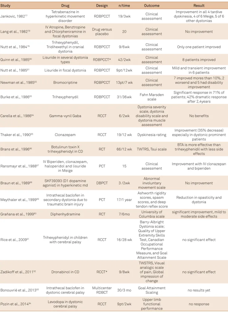
-macological treatment (not including botulinum toxin in-jections) published between 1973 and 2015. Of note, there is only one evidence-based review addressing the issue77. As shown in Table 2, there are 13 randomized double blind placebo controlled trials (RCT). Trihexyphenidyl is the only proven efective anticholinergic in a randomized double-blinded placebo controlled crossover trial (RCCT) for gener-alized and segmental dystonia. he beneits for cranial or fo-cal dystonias were uncertain (Table 2). In a study of patients with cerebral palsy, trihexyphenidyl did not demonstrate beneits in any outcome measure. Regarding dopamine pre-cursors, a recent trial with levodopa failed to demonstrate a better upper limb functional performance in patients with dystonic cerebral palsy. Other than this study, levodopa has never been tested in a RCT for generalized or focal dystonias. Nevertheless, clinical experience shows that levodopa has a dramatic response in DRD. For this reason, a trial of levodopa in generalized dystonia is mandatory to exclude DRD in all
patients with early onset dystonia. Lisuride, a dopamine ago
-nist, was tested in two trials with inconsistent efects in both generalized and focal dystonias. Tetrabenazine was tested only once in a RCCT and was shown to be efective in all four tardive dyskinesias, four of six Meige syndromes and ive of six other dystonias. Intrathecal baclofen injection has been tested in a placebo-controlled study in secondary dystonia
due to traumatic brain injury with reduction in spasticity and dystonia; there are no results for other RCCT studies. he only RCT evaluating benzodiazepines in dystonias stud-ied clonazepam in tardive dyskinesia. In this small group of patients, 35% of the 19 patients with a predominantly dyston-ic clindyston-ical pdyston-icture experienced decrease of dystonia severity. Apomorphine, bromocriptine in high doses (18–150mg/d), oral baclofen, clonazepam and dopamine antagonists have been used in uncontrolled studies, case reports, case series or open trials with unproven efects77-94.
Some recent experimental studies have focused on the ef-fect of antimuscarinic therapy in dystonia. In a DYT1 mice model, high-frequency stimulation in striatal spiny neurons failed to induce long-term depression, whereas low-frequen-cy stimulation did not depotentiate corticostriatal synapses in very speciic brain areas95. Also, cholinergic interneurons responded abnormally to D2R activation with potentiation rather than inhibition95. Anticholinergic drugs with selective muscarinic receptor antagonism are able to ofset the synap-tic plassynap-ticity deicit96. In summary, there is a rationale for the
use of anticholinergic agents in the management of dystonia. Botulinum toxin therapy revolutionized the treatment of focal dystonias. It acts by inducing chemodenervation of the afected muscles. Currently, several botulinum toxin formu-lations are available and widely used in the treatment of dys -tonias, and are the irst line treatment for focal and segmen-tal dystonias97. A recent evidence-based review of botulinum
toxin in movement disorders supports the use of botulinum toxin in several types of dystonias. For blepharospasm, the recommendation is level A for onabotulinumtoxinA (Botox®) (onaA) and incobotulinumtoxinA (Xeomin®) (incoA); level B for abobotulinumtoxinA (Dysport®)(aboA) and level U for rimabotulinumtoxinB (rimaB). In cervical dystonias, the
ev-idence supports level A for all formulations. In limb dysto
-nias, the recommendation is level B for aboA, onaboA and level U for incoA and rimaB. For oromandibular dystonia the recommendation is level C for aboA and onaA. For adductor dysphonia evidence supports level C for onaA and level U for other formulations98.
Table 2. Controlled studies in dystonia since 1973.
Study Drug Design n/time Outcome Result
Jankovic, 198277
Tetrabenazine in hyperkinetic movement
disorder
RDBPCCT 19/3wk Clinical assessment
Improvement in all 4 tardive dyskinesia, 4 of 6 Meige, 5 of 6
other dystonias
Lang et al., 198278
IV Atropine, Benztropine and Chlorpheniramine in
focal dystonias
Drug versus placebo 20
Clinical
assessment No improvement
Nutt et al., 198479
Trihexyphenydil, Tridihexethyl in cranial
dystonia
RDBPCCT 9/6wk Clinical
assessment Only one patient improved
Quinn et al., 198580 Lisuride in several dystonia
types RDBPCCT* 42/2wk
Clinical
assessment 8 patients improved
Nutt et al., 198581 Lisuride in focal dystonia RDBPCCT 9pt/12wk Clinical
assessment
Mild and transient improvement in 6 patients
Newman et al., 198582 Bromocriptine RDBPCCT 13pt/7 wk Clinical
assessment
7 improved mores than 10%, 2 worsend and 5 had disability
improvement
Burke et al., 198683 Trihexyphenydil RDBPCCT 31/36wk Fahn Marsden
scale
Signiicant response in 71% of patients; 42% dramatic response
after 2,4years
Carella et al., 198684 Gamma-vynil Gaba RCCT 6/2wk
Dystonia severity scale, dystonia disability scale and
dystonia muscle assessment
No beneits
Thaker el al., 199085 Clonazepam RCCT 19/12 wk Dyskinesia rating
Improvement (35% decrease) especially in dystonic prominent
patients
Brans et al., 199686 Botulinun toxin X
trihexyphenidyl in CD RCT 66/12 wk TWTRS, Tsui scale
BTA is more effective than trihexyphenidil with less side
effects
Ransmayr et al., 198887
IV Biperiden, clonazepam, haloperidol and lisuride
in Meige
PCT 15 Clinical
assessment
Improvement with IV clonazepan and biperiden
Braun et al., 198988 SKF39393 (D1 dopamine
agonist) in hyperkinetic md DBPCT 3 /2wk
Abnormal involuntary movement scale
No improvement
Meythaler et al., 199989
Intrathecal baclofen in secondary dystonia due to
traumatic brain injury
PCT 17/1 year
Ashworth rigidity scores, spasm scores, and deep tendon relex score
Reduction in spasticity and dystonia
Grañana et al., 199990 Diphenhydramine RCT 7/6mo University of
Columbia scale
signiicant improvement, mild to moderate side effects
Rice et al., 200991 Trihexyphenidyl in children
with cerebral palsy RCCT 16/28 wk
Barry-Albright Dystonia scale; Quality of Upper
Extremity Skills Test, Canadian
Occupational Performance Measure, and Goal
Attainment Scale
no signiicant effect
Zadikoff et al., 201192 Dronabinol in CD RCCT* 9/8wk
TWSTRS, Visual analogic scale of pain, Global impression of
change
no signiicant effect
Bonouvrié et al., 201393 Intrathecal baclofen in
dystonic cerebral palsy
Multicenter
RDBCT 30/3 mo
Goal Attainment
Scaling no results yet
Pozin et al., 201494 Levodopa in dystonic
cerebral palsy RCCT 9pt/2wk
Upper limb functional performance
no response
for up to 96 months in four patients, others have found pro-gression of disability in eight patients with the need for new lead implantation. his led to signiicant improvement in four patients102,103. Regarding DYT6, Vidailhet et al. showed that the
site of dystonia itself is a better predictor than genetic status
but patients with spasmodic dysphonia and cranial involve
-ment tend to have a limited response to DBS101. Individual cor -tical plasticity, ixed skeletal deformities, presence of myelopa-thy and placement of the electrodes have major inluences on the outcome of surgery in both cervical and generalized dysto -nia. Finally, DBS is considered efective in myoclonus-dystonia (DYT11) and tardive dyskinesia101. he role of surgery in sec-ondary dystonias (cerebral palsy, inherited metabolic disor-ders) is still debatable. One study of a small group of patients
demonstrated that deep anterior cerebellar stimulation reduc
-es spasticity and symptoms of secondary dystonia in cerebral palsy patients104. Other treatments have been studied for dys -tonia. Subthalamic nucleus stimulation and simultaneous GPi
and subthalamic nucleus stimulation are emerging as promis
-ing in the surgical treatment of dystonia105. Apparently subtha
-lamic nucleus stimulation does not impair working memory and attention, as has been reported in PD106. Patients should be
aware of the risk of inherent complications of the surgery, such as stimulation-related dysarthria, parkinsonism, gait disorders and depression (including suicide attempts).
COMMENTS
he new deinition of dystonia has created a conceptual framework that helps clinicians diagnose and classify diferent forms of dystonia. Nevertheless, clinical features remain the cor-nerstone of identiication and diferential diagnosis. With a few exceptions, ancillary tests, as well as neurophysiological stud -ies, play a limited role in diagnosing dystonia. Dystonia is a syn-drome, with clinical and etiological heterogeneity, probably with a inal common pathway. It seems to be the resulting force of a
complex network of genetic, epigenetic and environmental vec
-tors. Penetrance is the result of adding individual vectors6,27,28,29,30. Sensory abnormalities may drive dystonia, as well as so-matosensory receptive ields being abnormally enlarged and disorganized in patients, with sensitive stimuli modulating dystonic movement. Motor and sensory systems mutually integrate and are abnormal in dystonia patients. he patho-physiology of dystonia lies in the lack of inhibition. Basal gan-glia act as iltering and modulating inputs to improve the precision of ine movements. Abnormal dynamic
homeostat-ic plasthomeostat-icity could be derived from an imbalance of dopami
-nergic and choli-nergic reciprocal signaling in the striatum. he cerebellum may have a role in processing proprioceptive information, and regulating or integration and cortex plastic -ity through cerebello-thalamo-cortical loops33,34,35 .
A great number of new monogenic dystonias have been described since the irst gene was recognized in 1997. To date,
28 loci and 18 genes are known. Whole genome association studies, high-throughput sequencing and the loss of hetero-geneity map have certainly accelerated the pace of gathering new information. In the last ive years, eight genes related to familial dystonias have been described. All of them used new technology, mainly associated with linkage studies. Putative genes related to Mendelian forms of dystonia impair sever-al pathways: chaperone-like (DYT1); apoptosis regulation (DYT6, DYT16); dopamine formation (DYT5a and b); syn-aptic transport (DYT10); synsyn-aptic recycling (DYT1); neuro-nal structure and transport (DYT4); transcription syndrome (DYT3, DYT6), redox status maintenance (DYT8); reduced D2 receptor availability (DYT1, DYT3, DYT6 and DYT11), trans-membrane traicking (DYT11), gradient ion maintenance (DYT12 and DYT24), glucose transport (DYT18), transduc-tion signal pathway (DYT25), and cell cycle control (DYT23). None of these genes have shown an obvious common end-point. Functional cell studies are emerging, especially shared partners among the described genes related to dystonia63.
A large bulk of knowledge about dystonia has been pro
-duced in recent years. Noticeably half of all PubMed publi-cations are concentrated over the last 15 years. Non-motor
features have increasingly been recognized as part of dysto
-nia pathophysiology. In the next years, more studies in the ield will clarify the spectrum of the dystonic syndromes. Deep brain stimulation has become a landmark in the treat-ment of generalized dystonias. New potential targets may arise in the near future. Modern next-generation sequencing technologies have increased awareness of new phenotypes in dystonia-related genes. For instance, diferent mutations in ATP1A3 genes can cause cerebellar ataxia, arelexia, pes ca-vus, optic atrophy and sensorineural hearing loss (CAPOS) syndrome; rapid onset dystonia-parkinsonism and alternat-ing hemiplegia syndrome; and the same mutation can cause either rapid onset dystonia parkinsonism or alternating hemiplegia syndrome. Several theories such as environment, other modifying genes, non-coding variations and epigenetic factors explain the phenomenon but are still lacking conir-mation. Possibly, a greater understanding of genetic support-ers will overtake “the single gene disease concept” and this might lead to more individualized and rationale therapeutic targets providing patients with better treatment.
An improved understanding of pathogenetic pathways
common to several dystonias could possibly lead to the dis
-covery of new therapeutic targets to provide better symptom
-atic and, hopefully, etiological treatment. Further studies with more homogenous and bigger samples of patients with the same etiological category, and standardized examination, are needed to increase our knowledge of this cryptic disease.
Acknowledgements
References
1. Lanska DJ. Chapter 33: the history of movement disorders. Handb Clin Neurol. 2010;95:501-46. doi:10.1016/S0072-9752(08)02133-7
2. Munts AG, Koehler PJ. How psychogenic is dystonia? Views from past to present. Brain. 2010;133(5):1552-64. doi:10.1093/brain/awq050
3. Fahn S. Concept and classiication of dystonia. Adv Neurol. 1988;50:1-8. doi:10.1212/WNL.50.5_Suppl_5.S1
4. Ozelius LJ, Hewett JW, Page CE, Bressman SB, Kramer PL, Shalish C et al. The early-onset torsion dystonia gene (DYT1) encodes an ATP-binding protein. Nat Genet. 1997;17(1):40-8. doi:10.1038/ng0997-40
5. Broussolle E, Laurencin C, Bernard E, Thobois S, Danaila T, Krack P. Early ilustrations of geste antagoniste in cervical and generalized dystonia. Tremor Other Hyperkinet Mov (N Y) 2015;5:332-44. doi:10.7916/D8KD1X74
6. Bressman SB. Dystonia genotypes, phenotypes, and classiication. Adv Neurol. 2004:94:101-7.
7. Albanese A, Bhatia K, Bressman SB, Delong MR, Fahn S, Fung VS et al. Phenomenology and classiication of dystonia: a consensus update. Mov Disord. 2013;28(7):863-73. doi:10.1002/mds.25475
8. Albanese A, Lalli S. Is this dystonia? Mov Disord. 2009;24(12):1725-31. doi:10.1002/mds.22597
9. Loyola DP, Camargos S, Maia D, Cardoso F. Sensory tricks in focal dystonia and hemifacial spasm. Eur J Neurol. 2013;20(4):704-7. doi:10.1111/ene.12054
10. Sitburana O, Wu LJ, Shefield JK, Davidson A, Jankovic J. Motor overlow and mirror dystonia. Parkinsonism Relat Disord. 2009;15(10):758-61. doi:10.1016/j.parkreldis.2009.05.003
11. Hallett M. Pathophysiology of writer’s cramp. Hum Mov Sci. 2006;25(4-5):454-63. doi:10.1016/j.humov.2006.05.004
12. Sohn YH, Hallett M. Disturbed surround inhibition in focal hand dystonia. Ann Neurol. 2004;56(4):595-9. doi:10.1002/ana.20270
13. Cox BC, Cincotta M, Espay AJ. Mirror movements in movement disorders: a review. Tremor Other Hyperkinet Mov (NY). 2012;2. pii: tre-02-59-398-1. doi:10.7916/D8VQ31DZ
14. Castrioto A, Piscicelli C, Pérennou D, Krack P, Debû B. The pathogenesis of Pisa syndrome in Parkinson’s disease. Mov Disord. 2014;29(9):1100-7. doi:10.1002/mds.25925
15. Doherty KM, van de Warrenburg BP, Peralta MC, Silveira-Moriyama L, Azulay JP, Gershanik OS et al. Postural deformities in Parkinson’s disease. Lancet Neurol. 2011;10(6):538-49. doi:10.1016/S1474-4422(11)70067-9
16. Schrag A, Trimble M, Quinn N, Bhatia K. The syndrome of ixed dystonia: an evaluation of 103 patients. Brain. 2004;127(10):2360–72. doi:10.1093/brain/awh262
17. Erro R, Rubio-Agusti I, Saifee TA, Cordivari C, Ganos C, Batla A et al. Rest and other types of tremor in adult-onset primary dystonia. J Neurol Neurosurg Psychiatry. 2014;85(9):965-8. doi:10.1136/jnnp-2013-305876
18. Agnew A, Frucht SJ, Louis ED. Supine head tremor: a clinical comparison of essential tremor and spasmodic torticollis patients. J Neurol Neurosurg Psychiatry. 2012;83(2):179-81. doi:10.1136/jnnp-2011-300823
19. Quinn NP, Schneider SA, Schwingenschuh P, Bhatia KP. Tremor: some controversial aspects. Mov Disord. 2011;26(1):18-23. doi: 0.1002/mds.23289
20. Mian OS, Schneider SA, Schwingenschuh P, Bhatia KP, Day BL. Gait in SWEDDs patients: comparison with Parkinson’s disease patients and healthy controls. Mov Disord. 2011;26(7):1266-73. doi:10.1002/mds.23684
21. Batla A, Erro R, Stamelou M, Schneider SA, Schwingenschuh P, Ganos C et al. Patients with scans without evidence of dopaminergic deicit: a long-term follow-up study. Mov Disord. 2014;29(14):1820-5. doi:10.1002/mds.26018
22. Peall KJ1, Smith DJ, Kurian MA, Wardle M, Waite AJ, Hedderly T et al. SGCE mutations cause psychiatric disorders: clinical and genetic characterization. Brain. 2013;136(1):294-303. doi:10.1093/brain/aws308
23. Tricht MJ1, Dreissen YE, Cath D, Dijk JM, Contarino MF, Salm SM et al. Cognition and psychopathology in myoclonus-dystonia. J Neurol Neurosurg Psychiatry. 2012;83(8):814-20. doi:10.1136/jnnp-2011-301386
24. Brashear A, Cook JF, Hill DF, Amponsah A, Snively BM, Light L et al. Psychiatric disorders in rapid-onset dystonia-parkinsonism. Neurology. 2012;79(11):1168-73. doi:10.1212/WNL.0b013e3182698d6c
25. Fabbrini G, Berardelli I, Moretti G, Pasquini M, Bloise M, Colosimo C et al. Psychiatric disorders in adult-onset focal dystonia: a case-control study. Mov Disord. 2010;25(4):459-65. doi:10.1002/mds.22983
26. Romano R, Bertolino A, Gigante A, Martino D, Livrea P, Defazio G. Impaired cognitive functions in adult-onset primary cranial cervical dystonia. Parkinsonism Relat Disord. 2014;20(2):162-5. doi:10.1016/j.parkreldis.2013.10.008
27. Newman JR, Boyle RS, O’Sullivan JD, Silburn PA, Mellick GD. Risk factors for idiopathic dystonia in Queensland, Australia. J Clin Neurosci. 2014;21(12):2145-9. doi:10.1016/j.jocn.2014.03.032
28. Molloy A, Kimmich O, Williams L, Butler JS, Byrne N, Molloy F et al. An evaluation of the role of environmental factors in the disease penetrance of cervical dystonia. J Neurol Neurosurg Psychiatry. 2015;86(3):331-5. doi:10.1136/jnnp-2014-307699
29. Defazio G, Abbruzzese G, Girlanda P, Buccafusca M, Currà A, Marchese R et al. Primary cervical dystonia and scoliosis: a multicenter case control study. Neurology. 2003;60(6):1012-15. doi:10.1212/01.WNL.0000049932.22065.60
30. Hutchinson M. Sun exposure is an environmental factor for the development of blepharospasm. J Neurol Neurosurg Psychiatry. 2016;87(4):420-4. doi:10.1136/jnnp-2014-310266
31. Lin PT, Hallett M. The pathophysiology of focal hand dystonia. J Hand Ther. 2009;22(2):109-14. doi:10.1016/j.jht.2008.10.008
32. Leijnse JN, Hallett M. Etiological musculo-skeletal factor in focal dystonia in a musician’s hand: a case study of the right hand of a guitarist. Mov Disord. 2007;22:1803-8. doi:10.1002/mds.21636
33. Weise D, Schramm A, Stefan K, Wolters A, Reiners K, Naumann M et al. The two sides of associative plasticity in writer’s cramp. Brain. 2006;129(10):2709-21. doi:10.1093/brain/awl221
34. Quartarone A, Morgante F, Sant’angelo A, Rizzo V, Bagnato S, Terranova C et al. Abnormal plasticity of sensorimotor circuits extends beyond the affected body part in focal dystonia. J Neurol Neurosurg Psychiatry. 2008;79(9):985-90. doi:10.1136/jnnp.2007.121632
35. Lehéricy S, Tijssen MA, Vidailhet M, Kaji R, Meunier S. The anatomical basis of dystonia: current view using neuroimaging. Mov Dis. 2013;28(7):944-57. doi:10.1002/mds.25527
36. Camargos S, Cardoso F. New algorithm for the diagnosis of hereditary dystonia. Arq Neuropsiquiatr. 2012;70(9):715-7. doi:10.1590/S0004-282X2012000900013
37. Saunders-Pullman R, Raymond D, Senthil G, Kramer P, Ohmann E, Deligtisch A et al. Narrowing the DYT6 dystonia region and evidence for locus heterogeneity in the
Amish-Mennonites. Am J Med Genet A. 2007;143A(18):2098-105. doi:10.1002/ajmg.a.31887