VIEWS AND REVIEWS
Movement disorders emergencies: a review
Emergências em distúrbios do movimento: uma revisão
Renato P. Munhoz1,2, Mariana Moscovich1, Patrícia DareAraujo1, Hélio A. G. Teive2
Movement disorders (MD) encompass disorders charac-terized by involuntary movements and/or loss of control or eiciency in voluntary movement1.Patients with MD
usu-ally have disabling symptoms; however, most cases are fol-lowed and treated in ambulatory care settings. A MD emer-gency is deined as a neurological disorder evolving acutely or subacutely, in which the clinical presentation is dominated by MD and in which failure to accurately diagnose and man-age the patient may result in signiicant morbidity or even mortality1.In this review, we used these criteria to divide MD
emergencies into six main topics.
I. Emergencies in Parkinson’s disease (PD)
a. Severe motor luctuations and the super OFF phenomenon
b. Parkinsonism-hyperpyrexia and dyskinesia-hyperpy-rexia syndromes
c. Acute parkinsonism
d. Acute psychosis in Parkinson’s disease
II. Acute drug reactions a. Acute dystonia
b. Neuroleptic malignant syndrome c. Serotonergic syndrome
d. Malignant hyperthermia III. Acute exacerbation of chronic MDs
a. Status dystonicus
b. Neurological and orthopedic complications of dysto-nia and pseudodystodysto-nia
c. Laryngeal dystonia in multiple system atrophy and other conditions
d. Tic status and neurological complications of tics e. Wilson’s disease emergencies
f. Others conditions
IV. Acute chorea and hemiballism-hemichorea V. Stif-person syndrome
VI. Lethal catatonia
1Department of Neurology, Pontifical Catholic University of Paraná (PUCPR), Curitiba PR, Brazil;
2Movement Disorders Unit, Neurology Service, Internal Medicine Department, Federal University of Paraná (UFPR), Curitiba PR, Brazil.
Correspondence: Renato P. Munhoz; Avenida Sete de Setembro 4698/1507; 80240-000 Curitiba PR - Brasil; E-mail: renatopuppi@gmail.com
Conflict of interest: There is no conflict of interest to declare.
Received 14 January 2012; Received in final form 27 January 2012; Accepted 06 February 2012
ABSTRACT
Movement disorders (MD) encompass acute and chronic diseases characterized by involuntary movements and/or loss of control or ef-ficiency in voluntary movements. In this review, we covered situations in which the main manifestations are MDs that pose significant risks for acute morbidity and mortality. The authors examine literature data on the most relevant MD emergencies, including those related to Parkinson`s disease, acute drug reactions (acute dystonia, neuroleptic malignant syndrome, serotonergic syndrome and malignant hyper-thermia), acute exacerbation of chronic MD (status dystonicus), hemiballism and stiff-person syndrome, highlighting clinical presentation, demographics, diagnosis and management.
Key words: movements disorders, dystonia, neuroleptic malignant syndrome, serotonergic syndrome, malignant hyperthermia, status dystonicus, dyskinesias, stiff person syndrome.
RESUMO
Os distúrbios do movimento (DM) englobam doenças agudas e crônicas caracterizadas por movimentos involuntários e/ou perda do controle ou eficiência em movimentos voluntários. Nesta revisão, incluímos situações nas quais as principais manifestações são DM que represen-tam risco devido à alta morbidade e mortalidade. Os autores revisaram aspectos relacionados às principais emergências em DM, incluindo aquelas relacionadas a doença de Parkinson; reações causadas por drogas (distonia aguda, síndrome neuroléptica maligna, síndrome se-rotoninérgica, hipertermia maligna); exacerbação aguda de DM crônicos (status distonicus); hemibalismo e síndrome da pessoa rígida. São destacados a apresentação clínica, os dados demográficos, o diagnóstico e o tratamento.
I. EMERGENCIES IN PARKINSON’S DISEASE (PD)
a. Severe motor fluctuations and the super OFF phenomenon
Motor luctuations (MF) develop in 70% of Parkinson’s disease patients treated with levodopa for nine years or lon-ger and in nearly all patients with early onset PD after less than ten years of treatment2. he deinition of MF is closely
related to the deinition of ON and OFF periods: an ON pe-riod is the time of levodopa responsiveness, with adequate control of motor symptoms, while an OFF period is the time when parkinsonian symptoms are signiicantly present. Both usually correlate directly or indirectly with levodopa plasma levels, however the mechanisms that lead to MF are com-plex and involve the reduction of striatal synthesis and stor-age of dopamine from exogenous levodopa and subsensitiza-tion of postsynaptic dopaminergic receptors2. he “wearing
of ” phenomenon describes the presence of end dose OFF periods, mainly due to the loss of stratial dopamine stor-age capacity and short levodopa half-life2. he ON-OFF
phe-nomenon and unpredictable OFFs describe the occurrence of sudden and disabling OFF periods with no clear relation-ship with timing of levodopa intake. he “delayed ON” (in-creased time latencies from dose intake to the start-up of an ON period) and “no-ON” phenomenon (complete failure of a levodopa dose to exert an ON response) are typically related to impaired absorption of oral levodopa2.
In rare instances, MF can represent a MD emergency, es-pecially in the case of frequent and distressful ON-OFF pe-riods. Typically, management involves various combined approaches, such as administration of small multiple daily doses of levodopa, controlled release, dispersible and solu-ble levodopa formulations, subcutaneous dopamine ago-nists, MAO-B and COMT inhibitors, and surgical approach-es. Administration of crushed levodopa tablets may also be helpful in certain circumstances2.
he “super OFF” phenomenon is a rare complication found in patients with PD deined as an increase in motor disability scores in comparison to basal (OFF period) mo-tor scores described in those experiencing MF3. As the
de-nomination implies, it means that patients afected by this phenomenon have the sensation of being more symptom-atic from most parkinsonian motor standpoints than their usual OFF levodopa dose status. he super OFF state can be divided into a beginning-of-dose and an end-of-dose inhibi-tory efect, depending on whether it occurs before or after levodopa has reached a therapeutic plasma concentration4.
he phenomenon is often very distressing and disabling, lasting from minutes to hours. he pathophysiology of su-per OFF episodes is not entirely clear, but is probably linked either to presynaptic inhibition of iring and release of dop-aminergic neurons, or an efect mediated by the preferential stimulation of a subpopulation of postsynaptic DA receptors that have an inhibitory action on motor activity caused by
low levels of levodopa in the rising or declining phase of the plasma concentration curve3. Gershanik et al.4 reported a
re-duction in the severity of super OFF phenomenon in patients treated with clozapine, while Dziewczapolski et al.3 found
the same beneicial efects of clozapine in an animal mod-el of parkinsonism. hese authors speculate on the possibil-ity that clozapine could inhibit the inhibitory action of low plasma levodopa levels, leading to an apparent antiparkin-sonian efect. Another study5 explored the pathophysiology
of super OFF episodes by examining the efects of subthresh-old, threshold and suprathreshold infusions of apomorphine in a randomized, double-blind, placebo controlled, crossover study. hey concluded that these episodes may be present only in certain subgroups of PD patients. hey also suggested that the main mechanism involved is not likely to be a pre-synaptic or autoreceptor efect of dopaminergic agents, thus leaving the possibility of a post synaptic cause5.
b. Parkinsonism-hyperpyrexia and dyskinesia-hyperpyrexia syndromes
Parkinsonism-hyperpyrexia syndrome (PHS), also known as neuroleptic malignant-like syndrome, is a rare complica-tion of PD6, characterized by hyperthermia, autonomic
dys-function, altered consciousness, severe rigidity and elevat-ed serum creatine kinase (CK) levels. PHS may be triggerelevat-ed by infections, reduction in dopaminergic drug dosage, hot weather or dehydration. Potentially life-threatening compli-cations include deep venous thrombosis and pulmonary em-bolism, aspiration pneumonia and renal failure. Treatment is based on intravenous luids infusion, antithermics and use of levodopa and dopamine agonists. Occasionally, dantrolene may be necessary.
Another MD emergency related to PD, recently desig-nated dyskinesia-hyperpyrexia syndrome7, consists of
se-vere dyskinesias (dyskinetic status), leading to muscle ex-haustion, rhabdomyolysis, hyperthermia and confusion. his complication shares some of the clinical characteris-tics of PHS, but difers in those dyskinesias – instead of ri-gidity, dominates the clinical picture. Additionally, the dys-kinesia-hyperpyrexia syndrome, contrarily to PHS, should be treated by reducing the dosage of dopaminergic drugs, particularly dopamine agonists.
c. Acute parkinsonism
Acute severe de novo parkinsonism is an uncommon form
Acute worsening of motor symptoms in patients with PD may also occur independently of disease progression. his situation typically occurs due to additional medical con-ditions, as urinary or respiratory infections and metabolic disturbances (hypothyroidism and so on), or even due to a concomitant neurological disease, for example, subdural he-matomas, compressive spinal cord lesion or brain tumor8,10.
Treatment of both de novo and worsening of PD should be focused on resolution of the etiologic process and occasion-ally symptomatic treatment of parkinsonism using dopamin-ergic agents11.
d. Acute psychosis in Parkinson’s disease
Psychosis is relatively common among advanced PD pa-tients, often associated with some degree of cognitive dys-function. he acute occurrence of psychosis is frequently as-sociated and triggered by the same agents used to treat the motor symptoms of PD (levodopa, dopamine agonists, an-ticholinergics, amantadine, COMT and MAO-B inhibitors), however other comorbidities, such as infections (urinary and respiratory tract), metabolic or neurological disturbances, may play a role10,11.he manifestations typically include
visu-al hvisu-allucinations, persecutory delusions, confusion and psy-chomotor agitation. After treatment of potential comorbidi-ties, management recommendations begin with the stepwise withdrawal of potentially associated drugs, starting with anticholinergics, followed by MAO-B inhibitors, dopamine agonists, amantadine and COMT inhibitors. Frequently, the introduction of a second generation antipsychotic,as clozap-ine or quetiapclozap-ine, is necessary. Other options include risperi-done or olanzapine, with a signiicant risk of motor symp-toms worsening8,11.
II. ACUTE DRUG REACTIONS
a. Acute dystonia
By deinition, dystonia is characterized by involuntary sustained muscle contractions, frequently causing twisting and repetitive movements or abnormal postures12.While
dystonias can be classiied as focal, segmental, multifocal, hemidystonia or generalized, their etiologies are typically de-ined as primary (including genetic forms and idiopathic) or secondary.
Drug induced acute dystonia (DID) is one of the com-monest forms of secondary dystonia, along with tardive dy-stonia. his complication occurs in wide frequency range, depending on the speciic drugs prescribed, indications and studied populations13,14.he causative drugs are commonly
antidopaminergic, not restricted to the “classic” neurolep-tics, including haloperidol, risperidone, olanzapine and an-tiemetics15. Less frequently, other drug classes, such as
anti-convulsants and antidepressants, are involved in this type of complication. he contractions can occur immediately after
ingestion of a single dose, over the course of several days of therapeutic doses administration, after dose increase or as a manifestation of overdosage15. Most cases occur in the irst
72 hours of medication use with often dramatic symptoms, especially in children15. Patients at higher risk for DID include
those with a younger age, of male gender, a previous history of similar reactions, recent use of cocaine, comorbidity with mood disorders and metabolic abnormalities (hypokalemia, dehydration, hypoparathyroidism, etc)13,14.
Clinical presentation commonly includes craniocervical distribution with blepharospasm, bucolinguomandibular, face and neck dystonia, and oculogyric crisis with contrac-ture of the extraocular muscles leading to conjugate eyes de-viation, usually with a predominance of the superior rectus muscle and consequent upward eye deviation. A peculiar form of drug-induced acute dystonia is laryngeal spasm due to dystonia of vocal cords related muscles (laryngospasm), leading to acute upper airway obstruction. Appendicular symptoms tend to be more common among elderly patients. Typically, symptoms and signs are action induced, following treatment initiation or dose increase14,16.
Finally, tardive dyskinesias may became a MD emergency, particularly in the cases of severe respiratory/diaphragmat-ic dyskinesias and the case of so-called “withdrawal-emer-gent” dyskinesias (severe dyskinesias after withdrawal from neuroleptics)8.
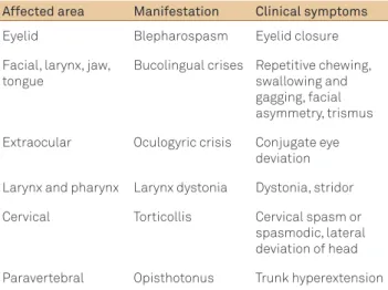
Table 1 shows the clinical manifestations of acute dysto-nia according to the afected muscle group.
he diferential diagnoses of acute dystonia include other neurological and systemic disorders, such as partial seizures, meningitis, tetanus, electrolyte disturbances and strychnine poisoning. he treatment options includeintramuscular or intravenous anticholinergics (e.g., biperiden 5 mg IV, followed by PO maintenance for up to seven days). Alternatively, or in milder cases, promethazine 50 mg IM may be efec-tive. Oculogyric crisis usually improves signiicantly with clonazepam 2 mg PO or diphenhydramine 25 or 50 mg IM. “Prophylaxis” with anticholinergics or amantadine is contro-versial and should not be routinely used1,8,12,15-18.
b. Neuroleptic malignant syndrome
Neuroleptic malignant syndrome (NMS) is an idiosyncrat-ic abrupt reaction to antidopaminergidiosyncrat-ic drug treatment with characteristic clinical and paraclinical indings19,20.Classically,
the causative agents include the “classic” neuroleptics, how-ever after the widespread use of newer agents, the “atypi-cal” agents have also been implicated21.Additionally, NMS
has been described during treatment with other dopamine-blocking drugs or after abrupt withdrawal of dopaminergic agents1,8,9,11,22,23.
male gender, young age, concomitant use of lithium and se-lective serotonine reuptake inhibitors (SSRIs), high ambient temperatures, hyponatremia, dehydration, physical exhaus-tion, mental retardaexhaus-tion, extrapyramidal syndromes, psycho-motor agitation and a previous episode of NMS24.NMS
oc-curs in all age groups, from 9 months to 78 years, with mean ages around the fourth and ifth decades across studies20-24.
Mortality remains relatively high, varying from 20 to 30% of cases20-22. Most cases that survive acute NMS recover without
sequelae; however, some patients remain with cognitive dei-cits and motor abnormalities, including rigidity, tremor and dystonic contractures25.
Diagnostic criteria include the tetrad of hypertherm-ia, rigidity, altered mental status and dysautonomia. hese main indings can be associated with other signs such as ac-tion and/or rest tremor, dystonia, chorea, myoclonus, sei-zures, ataxia, hyporelexia and extensor plantar relexes19,20.
Additionally, NMS may be associated with laboratory chang-es that include raised serum CK (>1000 IU/L), impaired liver, renal and coagulation status tests, leukocytosis, electrolyte disturbances, proteinuria and rhabdomyolysis with myo-globinuria20-22.he most important diferential diagnoses in
most cases are serotonergic syndrome, malignant hyper-thermia and malignant catatonia20-25. Also, sudden
withdraw-al of intrathecwithdraw-al baclofen, used for the treatment of spasticity, can lead to an acute syndrome similar to NMS1.
Clinical approaches to treatment involve early recogni-tion, exclusion of the diferential diagnoses, medication with-drawal and pharmacological and nonpharmacological inter-ventions22-24. he choice of these later is mainly determined
by the severity of NMS and the presence of additional com-plications. Supportive care includes correction of volume and electrolyte imbalance, prevention of infection, thrombosis and pressure ulcers, and body cooling. Patients with severe symptoms often require management in an intensive care facility24.
Pharmacological interventions should be focused on re-ducing muscle rigidity with, for example, benzodiazepines or
dantrolene; reversing the hypothesized excessive dopamin-ergic blockade using bromocriptine, levodopa or amanta-dine; and controlling agitation22,26. Electroconvulsive
thera-py (ECT) has been used in selected cases27.Finally, patients
should be withdrawn of neuroleptics for, at least, two weeks after the termination of management of the acute phase of NMS before treatment of the background psychiatric condi-tion can be restarted19. his should be done with low doses,
slow titration, avoiding concomitant use of lithium, and close observation to prevent dehydration and the initial symptoms of NMS.
c. Serotonergic syndrome
Serotonin syndrome (SS) is a potentially fatal disorder that results most often from the combined use of two or more agents that enhance serotonin activity or concentra-tion in the central nervous system1,9,11,12,28. his under
recog-nized syndrome is usually manifested by acute development of agitation, mental status changes, myoclonus, incoordina-tion, postural instability, hyperrelexia, rigidity and dysau-tonomic features (diaphoresis, fever, diarrhea, tachycardia and hemodynamic instability) after a change in serotonergic drug regimen29.he most commonly used diagnostic
crite-ria require at least three of these clinical features after oth-er possible causes have been excluded, as discussed in de-tail below. Although onset is typically abrupt, some patients report insidious or recurrent symptoms of subtle cognitive decline, behavioral abnormalities and tremor with mild pos-tural disorders days to weeks before the full-blown syndrome develops29-31.
SS is not an idiosyncratic reaction, but a predictable one. In theory, the ofending drugs or, more often, the combina-tion of drugs are those that cause excessive brainstem and spinal cord serotonin or 5-hydroxytryptamine type 1A re-ceptor (5-HT1A) stimulation30,31. Evidence of
pathophysio-logic roles for 5-HT2 and possibly 5-HT3 subtypes have also been speculated30,32.he implicated agents listed in previous
reviews include inhibitors of serotonin reuptake (SSRIs, tri-cyclic antidepressants, dextromethorphan, amphetamine and cocaine), inhibitors of serotonin metabolism (mono-amine oxidase inhibitors [MAOIs]), serotonin precursors (L-tryptophan), enhancers of serotonin release (amphet-amines, cocaine, fenluramine), serotonin agonists (risperi-done, sumatriptan, ergotamines) and nonspeciic enhancers of serotonin activity, as lithium and electroconvulsive ther-apy29-33.Any of these agents alone can potentially cause SS,
however a combination is usually needed, with greater risk for patients using MAOIs or SSRIs concomitantly with any other serotonergic agent30. Although doses taken are usually
within therapeutic limits, almost 20% of the cases reviewed presented after medication overdose30-32,34. Other
circum-stances in which these agents may cause SS include rapid dose titration, addition or switch to a new agent without a proper washout period, liver or renal disease, endogenously
Table 1. Clinical manifestations of acute dystonia according to the affected muscle group.
Affected area Manifestation Clinical symptoms
Eyelid Blepharospasm Eyelid closure
Facial, larynx, jaw, tongue
Bucolingual crises Repetitive chewing, swallowing and gagging, facial asymmetry, trismus
Extraocular Oculogyric crisis Conjugate eye deviation
Larynx and pharynx Larynx dystonia Dystonia, stridor
Cervical Torticollis Cervical spasm or spasmodic, lateral deviation of head
reduced monoamine oxidase A activity (e.g., primary enzyme deicit, chronic schizophrenia, certain personality disorders), cytochrome P450 2D6 enzyme inhibition and old age32.
he prevalence and incidence of SS is largely unknown, however it is probably increasing mirroring the wide use of serotonergic agents in medical practice for various purpos-es29. As serum or cerebrospinal luid (CSF) serotonin levels
are useless for the diagnosis of SS, diagnosis is established on clinical grounds30,31.he starting point is obviously a
his-tory of exposure to agents that direct or indirectly interfere with the serotonergic system. Additionally, a number of clini-cal manifestations are required: altered mental status, auto-nomic instability and motor abnormalities, including tremor, myoclonus, gait and appendicular ataxia, hyperrelexia and muscle rigidity. Seizures, renal failure and disseminated in-travascular coagulopathy have also been described in more severe cases28-30,33. Finally, laboratory abnormalities include
metabolic acidosis, rhabdomyolysis, elevated serum ami-notransferase, creatinine levels and white blood cell count. he most widely used and validated diagnostic criterion is the Hunter Serotonin Toxicity Criteria34 that includes the use
of a serotonergic agent plus any of the following: (i) myoclo-nus, agitation or diaphoresis; (ii) tremor and hyperrelexia; (iii) hypertonia; (iv) temperature above 38 degrees Celsius. Also, other possible causes have to be ruled out, including infections, other forms of intoxication, metabolic and hor-monal abnormalities, and drug withdrawal. Some symptoms of SS can mimic those due to an overdose of cocaine or lith-ium30-34. Other diferential diagnoses include anticholinergic
poisoning, malignant hyperthermia and NMS, each of these readily distinguished from SS based on medication exposure history29,30,34.
he key principles for successful treatment are preven-tion, early detection and acute intervention. Withdrawal of the ofending agents is the mainstay of therapy, enough to control signs and symptoms in almost half of all cases30,34.For
persistent or severe symptoms, speciic pharmacologic treat-ment may be necessary. Although the role of theoretically speciic therapy is still uncertain and possibly does not have a direct impact on survival, it may reduce symptoms dura-tion33. Nonspeciic 5-HT receptor blockers, such as
cyprohep-tadine and methysergide, have their use documented by sev-eral case reports and have been credited with shortening the syndrome’s duration and with preventing the onset of experi-mentally induced SS in animals32,33,35. Other potential
thera-peutic agents include benzodiazepines, chlorpromazine and propranolol28,29,33. Supportive measures include treatment of
seizures, cardiac arrhythmias, disseminated intravascular coagulation, muscle rigidity and hyperthermia33.Prognosis
is generally good, provided early recognition, and adequate management occurs. In previous reports and reviews, mor-bidity has not been formally assessed, and mortality rates ranged from 2.4 to 12%, with the caveat that these data rep-resent series of known patients with SS29,30. After recovery,
cautious reintroduction of a serotonergic agent may be nec-essary, with close observation of symptoms and signs. It is also essential for clinicians to be reminded that, in general, these agents must be started at the lowest eicacious dose with slow titration, especially in the elderly, in whom polyp-harmacy is unavoidable28,31.
d. Malignant hyperthermia
Malignant hyperthermia (MH) is a rare, potentially fa-tal complex genetic disorder of skelefa-tal muscle that occurs when a patient with an inherited MH-susceptible mutation is exposed to triggering anesthetic agents1,36-38. MH usually
manifests as a hypermetabolic crisis characterized by rapid onset of hyperthermia, luctuations in blood pressure, hyper-carbia, hyperkalemia, metabolic acidosis, muscle rigidity and rhabdomyolysis. he episodes are usually associated with the administration of inhalation anesthetics or depolarizing muscle relaxants36,37.
he clinical syndrome results from uncontrolled calcium lux across skeletal muscle membrane, and in more than half of afected families an autosomal dominant linkage to a gene encoding a skeletal muscle intracellular Ca2+ release channel
receptor (type 1 ryanodine receptor) is detected38.Treatment
with dantrolene is efective, combined with discontinuation of the triggering agents and correction of metabolic and elec-trolyte abnormalities37.In the 1970s, mortality was greater
than 80%, however with the current management and diag-nostic awareness, mortality is less than 5%37,38.
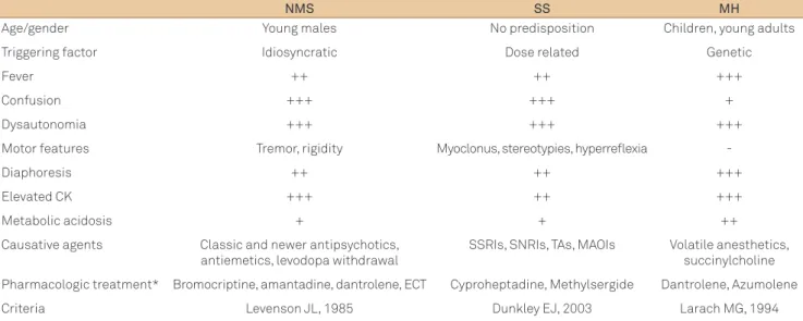
A comparison of features and management of NMS, SS and MH is presented in Table 2.
III. ACUTE EXACERBATION OF CHRONIC MDS
a. Status dystonicus
Status dystonicus (SD), or dystonic storm, is a life threat-ening condition associated with both primary and secondary dystonia. SD was irst recognized by Jankovic and Penn39 in
1982 and is deined as “(…) increasingly frequent and severe episodes of generalized dystonia, which necessitate urgent hospital admission”. hese episodes are usually refractory to traditional pharmacological therapy and may be triggered by trauma, surgery, infection, fever and abrupt introduction, withdrawal or change in medical treatment, including lithi-um, tetrabenazine and clonazepam40-42. he muscle
contrac-tures are painful, interfering with respiratory function and causing metabolic complications as hyperpyrexia, dehydra-tion, respiratory insuiciency, rhabdomyolysis and acute renal failure. SD is rare and no deinitive consensus exists regarding the ideal treatment strategy that most often include a com-bination of several drugs and even surgical interventions40-42.
admission in intensive care units to avoid metabolic, renal and ventilatory complications.
As the speciic pharmacological substrate of dystonia is ill deined, to date, treatment of SD is mainly empirical and variable. Agents used in previous case series and reports in-clude tetrabenazine, baclofen, chlormethiazole, anticho-linergics (benzhexol and biperiden), dopamine receptor blockers (haloperidol and pimozide), anticonvulsants (car-bamazepine, primidone and valproic acid), levodopa, ac-etazolamide, benzodiazepines (midazolan, clonazepam and diazepam), deep sedation with propofol, barbiturate anes-thesia and muscle blockers39-42. hese drugs were used in
an-ecdotal reports as combinations with variable eicacy, often requiring additional interventions, for example, continu-ous infusion of intrathecal baclofen and stereotactic surgery (thalamotomy, pallidotomy or DBS electrodes placement). As already outlined, SD is a potentially life threatening com-plication and its course and outcome is highly variable, in-cluding cases with complete remission, deterioration of clini-cal condition and death40-42.
b. Neurological and orthopedic complications of dystonia and pseudodystonia
Severe dystonic contractures, particularly in generalized dystonia, may lead neurological and orthopedic complica-tions, including spinal cord compression (particularly cervi-cal myelopathy) and even lesions to nervous plexi or periph-eral nerves. From an orthopedic standpoint, patients with severe generalized dystonia may also develop severe and dis-abling scoliosis8,43,44.
he term pseudodystonia encompasses conditions that mimic dystonia, including some that may present acute or subacutely, including infectious and neoplastic torticol-lis, atlantoaxial rotatory subluxation and localized tetanus.
Infectious torticollis as known as Grisel’s syndrome may be secondary to pharyngitis, tonsillitis or retropharyngeal abscess1,8,9,17.
c. Laryngeal dystonia in multiple system atrophy and other conditions
Multiple system atrophy (MSA) is a form of atypical kinsonism characterized by the presence of a MD (either par-kinsonism or cerebellar ataxia) and dysautonomia. Some of these patients may present developed disturbing and poten-tially dangerous laryngeal stridor, predominantly during the night, with an increased risk of sudden death due to upper airway obstruction1,8. his complication is caused by
poste-rior cricoarytenoid muscles dysfunction (which abduct the vocal cords), either by dystonia or paresis.
Acute laryngeal dystonia may also be a very rare form of task-speciic dystonia (Gerhardt’s syndrome), in which the af-fected individual presents spasm of the adductor muscles of the vocal cord during inspiration but while performing other activities, such as speaking. Clinically patients developed se-vere stridor with risk of upper airway obstruction. Immediate treatment with intravenous diphenhydramine (25 mg to 50 mg) is efective in most cases. Occasionally, tracheostomy or even botulinum toxin injection is necessary1,8,10,16,17.
d. Tic status and neurological complications of tics Patients with chronic motor, vocal tics or Tourette’s syn-drome may present with continuous disabling tics that can-not be suppressed, interfering dramatically with daily life activities and leading to cardiorespiratory diiculties, au-tonomic signs and even rhabdomyolysis45.his condition
is known as tic status and represents a rare MD emergen-cy. Triggering factors described in the literature include withdrawal of drugs used to treat tics (e.g., haloperidol or
Table 2. Comparison of features and management of neuroleptic malignant syndrome, serotonergic syndrome and malignant hyperthermia.
NMS SS MH
Age/gender Young males No predisposition Children, young adults
Triggering factor Idiosyncratic Dose related Genetic
Fever ++ ++ +++
Confusion +++ +++ +
Dysautonomia +++ +++ +++
Motor features Tremor, rigidity Myoclonus, stereotypies, hyperreflexia
-Diaphoresis ++ ++ +++
Elevated CK +++ ++ +++
Metabolic acidosis + + ++
Causative agents Classic and newer antipsychotics, antiemetics, levodopa withdrawal
SSRIs, SNRIs, TAs, MAOIs Volatile anesthetics, succinylcholine
Pharmacologic treatment* Bromocriptine, amantadine, dantrolene, ECT Cyproheptadine, Methylsergide Dantrolene, Azumolene
Criteria Levenson JL, 1985 Dunkley EJ, 2003 Larach MG, 1994
clonidine), mycoplasma infection and extreme reactive stress45. Treatment includes readministration of previously
withdrawn drug, dosage increase of previous therapy, addi-tion of benzodiazepines or switching to second generaaddi-tion antipsychotics. Also, discontinuation of agents know to in-duce tics, such as methylphenidate, is essential. Botulinum toxin injections may be particularly helpful in patients with malignant vocal tics or coprolalia8,16,45.
Finally, occasional patients may have motor tics that, sim-ilar to dystonia and other hyperkinetic MD, may cause neuro-logical complications like cervical myelopathy in the case of violent neck motor tics8,17,45.
e. Wilson’s disease emergencies
Wilson’s disease (WD) is an autosomal recessive disease of copper metabolism caused by mutations in the ATP7B gene. he most common neurologic symptoms are tremor, dysar-thria, dystonia, ataxia and parkinsonism. Emergencies in WD are related to hepatic involvement ( fatal acute or fulminant liver failure) or sudden worsening of neurological symptoms, particularly after use of D-penicillamine8.
f. Others conditions
Other several clinical conditions that can present as MD emergencies include hypocalcemia, tetanus, rabies and strychnine toxicity1.Tetanic muscle spasm can
devel-op secondary to hypocalcemia. Tetanus, caused by the exo-toxin tetanopasmin, produced by the anaerobic bacterium Clostridium tetani, presents traditionally with muscle spasms (triggered by touch, visual, auditory stimuli), stifness, auto-nomic instability and respiratory insuiciency. Management in intensive care unit involves the use of tetanus immune globulin, benzodiazepines and ventilatory support. Rabies and strychnine toxicity represent a rare form of infection and toxic causes of MD emergencies1.
IV. ACUTE CHOREA AND HEMIBALLISM-HEMICHOREA
Hemiballism is a relatively rare hyperkinetic disorder, characterized by irregular, wide amplitude, vigorous involun-tary movements of the limbs, primarily due to involuninvolun-tary activity of the proximal limb and associated axial muscles46.
Ballism is considered a very severe form of chorea, in which the movements have a violent, linging quality. he term de-rives from the Greek word ballismos, which means “jump-ing about or danc“jump-ing.” his MD most often involves one side of the body, but, occasionally, bilateral movements occur, a condition then deined as biballism46-48. he most
consis-tent neuropathological inding in hemiballism is a lesion of the contralateral subthalamic nucleus (STN), usually of vas-cular origin49. However, lesions in diferent locations are not
uncommon, including the ipsilateral STN, caudate nucleus,
putamen, thalamus, substantia nigra, and the premotor and motor cortex50. Also, other causes have been described, as
metabolic disorders (nonketotic hyperglycemia, hypoglyce-mia, hyperthyroidism), Sydenham’s chorea (chorea insatiens), systemic lupus erythematosus/antiphospholipid syndrome, post-operative period of cardiac surgery with extracorpore-al circulation (post-pump chorea), infectious diseases of the central nervous system (e.g., human immunodeiciency vi-rus infection, toxoplasmosis, syphilis, abscess), tumors and multiple sclerosis47-49.Frequently, hemiballism is associated
to hemichorea and, then, the term hemiballism-hemichorea (HBC) is used to describe this clinical spectrum48,49.
A particular form of HBC associated to nonketotic hy-perglycemia was described by Lin and Chang51 in three
di-abetic patients. In two of them, the hyperkinesia was the initial presenting symptom of diabetes. Soon after this de-scription, Lee et al.52 reported on eight female diabetic
pa-tients with HB-HC, further characterizing the clinical syn-drome with magnetic resonance imaging (MRI) correlation (contralateral high signal intensity putaminal lesion with-out edema or swelling on T1-weighted MRI).Finally, Mestre et al.53 presented the pathological correlate of the imaging
indings, deining that putaminal petechial haemorrhages in a fatal case of nonketotic hyperglycemia chorea.
Prognosis is benign in most cases. Mild chorea often does not require treatment; however, when the hyperkinet-ic movements are of suhyperkinet-icient severity to cause functional disability and exhaustion, several diferent approaches have been advocated47,50-54. A combination of a benzodiazepine
with either haloperidol, olanzapine or tetrabenazine, titrat-ed over days to maintain a balance between control of the movement disorder and side efects, such as sedation, can be efective47,50,51.Other options include gabapentin, valproic
ac-id55, and, as a last resort, stereotactic surgery51.
V. Stiff-person syndrome
Stif person syndrome (SPS) is a rare neurological dis-order characterized by the presence of luctuating muscle rigidity and spasms of the trunk and more proximal body parts56-58.Patients with SPS may present as an emergency
with severe pain and spasms of the lumbar paraspinal mus-cles and lower limbs. he spasms, often associated with intense pain, typically begin with an abrupt jerk followed by tonic activity that slowly subsides over seconds to, less commonly, minutes56,57.Rarely, these spasms last days
(sta-tus spasticus)58. he afected muscles are typically extremely
hard to palpation with a board-like appearance, leading to hyperlordosis56. Muscle spasms can occur spontaneously or
be a potential complication of the excessive muscle con-tractions in SPS. Sudden death can also occur due to an acute autonomic failure56. In a signiicant number of cases,
SPS is believed to be mediated by autoantibodiesto glutam-ic acid decarboxylase (anti-GAD)56,57 that limit the gamma
amino-butyric acid (GABA) neuronal activity and lower the threshold for muscle spasms, other neurologic and psychi-atric features of the disorder. SPS with elevated serum anti-GAD levels may occur with other autoimmune disorders, includingdiabetes mellitus, Graves’ disease, Hashimoto’s thyroiditis and pernicious anemia. Ten percent of cases with normal levels of this antibody may be related to au-toantibodies againstamphiphysin which commonly rep-resent a paraneoplastic syndrome related to breastcancer, mediastinal tumors, small cell lung cancer,Hodgkin’s dis-ease and colon cancer56.
he management of SPS is based on the use of drugs that promote GABAergic inhibition, for example, benzodi-azepines (diazepam and clonazepam) and baclofen56,57.
High-dose benzodiazepines can abolish the excessive motor unit activity. Oral baclofen provides relatively modest relief, while intrathecal administration seems to be much more efective. Finally, variable degrees of beneit have been reported with the use of antiepileptic drugs, such as valproic acid, levetirac-etam and gabapentin56-58.
VI. Lethal catatonia
Lethal or malignant catatonia (MC) is an uncommon and probably under-recognized clinical syndrome that usually occurs in the setting of schizophrenia, but may also follow infections as meningitis and encephalitis, head trauma, drug intoxication and metabolic disturbances, for example, ure-mia, porphyria and Wernicke’s encephalopathy1,59. he
diag-nosis is clinical, and no paraclinical tests conirm the speciic diagnosis. Also, little is known about its pathophysiology59.
From a clinical standpoint, MC is characterized by hyper-thermia, autonomic instability and severe rigidity, which, if prolonged, can be followed by exhaustion, coma, renal fail-ure, cardiovascular collapse and death. Violent behavior, including unprovoked assaults and suicide attempts, may occur occasionally1. Although MC difers from NMS by the
severity of the behavioral abnormalities in the early stages, both syndromes may become indistinguishable in their more advanced stages59.
Other diferential diagnoses include non-psychiatric stupor, encephalopathy, locked-in syndrome, malignant hy-perthermia, nonconvulsive status epilepticus or autism59.
Treatment includes high dose intravenous benzodiazepines, barbiturates and ECT1. Other interventions, such as
dant-rolene, bromocriptine, amantadine and anticonvulsants (e.g., carbamazepine), may have some utility59.
1. Kipps CM, Fung VSC, Grattan-Smith P, de Moore GM, Morris JGL. Movement disorder emergencies. Mov Disord 2005;20:322-334.
2. Melamed E, Ziv I, Djaldetti R. Management of motor complications in advanced Parkinson’s disease. Mov Disord 2007;22(Suppl):S379-384.
3. Dziewczapolski G, Menalled LB, Savino MT, Mora M, Stefano FJE, Gershanik O. Mechanism of action of clozapine-induced modification of motor behavior in an animal model of the ‘super-off’ phenomenon. Mov Disord1997;12:159-166.
4. Gershanik O, Lera G, Gomez Arevalo G. Clinical profile of parkinsonian patients with super off phenomenon. Neurology 1994;44:248.
5. Gunzler SA, Koudelka C, Carlson NE, Pavel M, Nutt JG. Effect of low concentrations of apomorphine on parkinsonism in a randomized, placebo-controlled, crossover study. Arch Neurol 2008;65:193-198.
6. Mizuno Y, Takubo H, Mizuta E, Kuno S. Malignant syndrome in Parkinson’s disease: concept and review of the literature. Parkinsonism Relat Disord 2003;9:S3-S9.
7. Gil-Navarro S, Grandas F. Dyskinesia-hyperpyrexia syndrome: another Parkinson’s disease emergency. Mov Disord 2010;25:2691-2692.
8. Poston KL, Frucht SJ. Movement disorders emergencies. J Neurol 2008;255:S2-S13.
9. Sá DS, Teive HA, Troiano AR, Werneck LC. Parkinsonism associated with neurocysticercosis. Parkinsonism Relat Disord 2005;11:69-72.
10. Robottom BJ, Weiner WJ, Factor SA. Movement disorders emergencies part 1. Hypokinetic disorders. Arch Neurol 2011;68:567-572.
11. Tousi B. Movement disorder emergencies in the elderly: recognizing and treating an often-iatrogenic problem. Cleve Clin J Med 2008;75:449-457.
12. Fahn S. Medical treatment of dystonia. In: Tsui JKC, Calne DB (eds). Handbook of dystonia. New York: Marcel Dekker; 1995:317-328.
13. Fabiani G, Teive HA, Germiniani F, Sá D, Werneck LC. Clinical and therapeutical features in 135 patients with dystonia: experience of movement disorders unity of the Hospital de Clínicas of the Universidade Federal do Paraná. Arq Neuropsiquiatr 1999;57:610-614.
14. Casey DE.Neuroleptic-induced acute dystonia. In: Lang AE (Ed). Drug-Induced movement disorders. Mount Kisco: Futura Publishing Co; 1992:21-41.
15. Barreira R, Magaldi RB. Acute dystonia after use of bromopride in pediatric patients. Rev Paul Pediatr 2009;27:110-114.
16. Robottom BJ, Factor SA, Weiner WJ. Movement disorders emergencies part 2. Hyperkinetic disorders. Arch Neurol 2011;68:719-724.
17. Frucht SJ. Hyperkinetic movement disorders emergencies. American Academy of Neurology. Minneapolis: Sillabus; 2002.
18. Miyasaki JM, Lang AE. Treatment of drug-induced movement disorders. In: Kurlan R (Ed). Treatment of movement disorders. Philadelphia: Lippincott; 1995:429-474.
19. Levenson JL. Neuroleptic malignant syndrome. AM J Psychiatry 1985;142:113.
20. Henderson VW, Wooten GF. Neuroleptic malignant syndrome: a pathogenic role for dopamine receptor blockade? Neurology 1981;31:132-137.
21. Moscovich M, Nóvak FT, Fernandes AF, et al. Neuroleptic malignant syndrome. Arq Neuropsiquiatr 2011;69:751-755.
22. Trollor JN, Chen X, Sachdev SP. Neuroleptic malignant syndrome associated with atypical antipsychotic drugs. CNS Drugs 2009;23: 477-492.
23. Rosebush PI, Garside S, Mazurek MF. Recognizing neuroleptic malignant syndrome. CMAJ 2004;170:1645.
24. Berardi D, Dell’Atti M, Amore M, De Ronchi D, Ferrari G. Clinical risk factors for neuroleptic malignant syndrome. Hum Psychopharmacol 2002;17:99-102.
25. Levinson DF, Simpson G. Sequelae of neuroleptic malignant syndrome. Biol Psychiatry 1987;22:273-278.
26. Rosebush PI, Stewart T, Mazurek MF. The treatment of neuroleptic malignant syndrome. Are dantrole and bromocriptine useful adjuncts to supportive care? Br J Psychiatry 1991;159:709-712.
27. Trollor JN, Sachdev PS. Electroconvulsive treatment of neuroleptic malignant syndrome: a review and report of cases. Aus N Z J Psychiatry 1999;33:650-659.
28. Sternbach H. The serotonin syndrome. Am J Psychiatry 1991;148: 705-713.
29. Boyer EW, Shannon M. The serotonin syndrome. N Engl J Med 2005; 352:1112-1120.
30. Lane R, Baldwin D. Selective serotonin reuptake inhibitor-induced serotonin syndrome: review. J Clin Psychopharmacol 1997;17:208– 221.
31. Munhoz RP. Serotonin syndrome Induced by a combination of bupropion and SSRIs. Clin Neuropharmacol 2004;27:219–222.
32. Turkel SB, Nadala JG, Wincor MZ. Possible serotonin syndrome in association with 5-HT(3) antagonist agents. Psychosomatics 2001;42:258–260.
33. Ables AZ, Nagubilli R. Prevention, recognition, and management of serotonin syndrome. Am Fam Physician 2010;81:1139-1142.
34. Dunkley EJ, Isbister GK, Sibbritt D, Dawson AH, Whyte IM. The Hunter Serotonin Toxicity Criteria: simple and accurate diagnostic decision rules for serotonin toxicity. QJM 2003;96:635-642.
35. Graudins A, Stearman A, Chan B. Treatment of the serotonin syndrome with cyproheptadine. J Emerg Med 1998;16:615–619.
36. Saidman W, Havard ES, Eger EI. Hyperthermia during anesthesia. JAMA 1964;190:1029-1032.
37. Ali SZ, Taguchi A, Rosenberg H. Malignant hyperthermia. Best Pract Res Clin Anaesthesiol 2003;17:519-533.
38. Mathews KD, Moore SA. Multiminicore myopathy, central core disease, malignant hyperthermia susceptibility, and RYR1 mutations: one disease with many faces? Arch Neurol 2004;61:27-29
39. Jankovic J, Penn AS. Severe dystonia and myoglobinuria. Neurology 1982;32:1195-1197.
40. Mariotti P, Fasano A, Contrarino MF, et al. Management of status dystonicus: our experience and review of the literature. Mov Disord 2007;22:963-968.
41. Manji H, Howard RS, Miller DH, et al. Status dystonicus: the syndrome and its management. Brain 1998;121:243-252
42. Teive HA, Munhoz RP, Souza MM, et al. Status dystonicus: study of five cases. Arq Neuropsiquiatr 2005;63:26-29.
43. Teive HA, Sá DS, Grande CV, Antoniuk A, Werneck LC. Bilateral pallidotomy for generalized dystonia. Arq Neuropsiquiatr 2001; 59:353-357.
44. Spitz M, Gonçalves L, Silveira L, Barbosa ER. Myelopathy as a complication of cervical dystonia. Mov Disord 2006;21:726-735.
45. Krauss JK, Jankovic J. Severe motor tics causing cervical myelopathy in Tourette´s syndrome. Mov Disord 1996;11:563-566.
46. Shannon KM. Ballism. In: Jankovic J, Tolosa E (Eds). Parkinson’s disease and movement disorders. 3 ed., Baltimore: Williams and Wilkins; 1998:365-375.
47. Coral P, Teive HAG, Werneck LC. Hemibalismo: relato de oito casos. Arq Neuropsiquiatr 2000;58:698-703.
48. Dewey RB, Jankovic J. Hemiballism-hemichorea. Clinical and pharmacologic findings in 21 patients. Arch Neurol 1989;46:862-867.
49. Whittier JR. Ballism and the subthalamic nucleus (nucleus hypothalamicus; corpus Luysi) review of the literature and study of 30 cases. Arch Neurol Psychiatry 1947;58:672-692.
50. Lang AE. Persistent hemiballismus with lesions outside the subthalamic nucleus. Can J Neurol Sci 1985;12:125-128.
51. Lin JJ, Chang MK. Hemiballism-hemichorea and non-ketotic hyperglycemia. J Neurol Neurosurg Psychiatry 1994;57:748-750.
52. Lee BC, Hwang SH, Chang GY. Hemiballismus-hemochorea in older women: A clinical syndrome with MRI correlation. Neurology 1999;52:646-648.
53. Mestre TA, Ferreira JJ, Pimentel J. Putaminal petechial haemorrahage as the cause of non-ketotic hyperglycaemic chorea: a neuropathological case correlated with MRI findings. J Neurol Neurosurg Psychiatry 2007;78:549-550.
54. Zétola VF, Verschoor B, Lima FM, et al. Hemibalismo-hemicoréia em estado hiperglicêmico não cetótico: distúrbio do movimento associado ao diabete melito. Arq Bras Endocrinol Metab 2010;54:335-338.
55. Sethi KD, Patel BP. Inconsistent response to divalproex sodium in hemichorea/hemiballism. Neurology 1990;40:1630-1631.
56. Espay AJ, Chen R. Rigidity and spasms from autoimmune encephalomyelopathies: stiff-person syndrome. Muscle Nerve 2006;34:677-690.
57. Munhoz RP, Fameli H, Teive HA. Stiff person syndrome as the initial manifestation of systemic lupus erythematosus. Mov Disord 2010;25:516-517.
58. Brown P, Marsden CD. The stiff man and stiff man plus syndromes. J Neurol 1999;246:648-652.