ARTICLE
Language impairment in Huntington’s disease
Alterações de linguagem na doença de Huntington
Mariana Jardim Azambuja1, Marcia Radanovic2, Mônica Santoro Haddad3, Carla Cristina Adda4, Egberto
Reis Barbosa5, Letícia Lessa Mansur6
Huntington’s disease (HD) is an autosomal dominant degenerative disorder, determined by a mutation on the short arm of the fourth chromosome (4p16.3), leading to an unstable expanded CAG trinucleotide repeat in the huntingtin gene (between 39 and 86). The huntingtin gene encodes the huntingtin protein, a large protein thought to be important for gene transcription, energy homeostasis and vesicle function. The larger the number of repetitions, the earlier and the faster is the progression of the disease. HD is characterized by progressive mental and motor
alterations. The prevalence of HD is about 5-10/100,000 in populations of European origin, but somewhat less com-mon in non-European populations. Chorea is the more prominent among the motor alterations, and may be pres-ent in up to 90% of affected patipres-ents. Other frequpres-ent altera-tions in HD are dysarthria, postural imbalance, ocular dys-function, hypotonia and bradikinesia, associated or not to rigidity. Regarding mental alterations, dementia, personal-ity changes, humor disturbances and, more rarely, psycho-sis occur in these patients1,2.
Movement Disorders Unit, Neurology Division, Hospital das Clínicas, University of São Paulo School of Medicine, São Paulo SP, Brazil.
1MSc, Department of Neurology, University of São Paulo School of Medicine, São Paulo SP, Brazil; 2MD, MSc, PhD, Department of Neurology, University of São Paulo School of Medicine, São Paulo SP, Brazil; 3MD, MSc, Department of Neurology, University of São Paulo School of Medicine, São Paulo SP, Brazil;
4MSc, Psychology Division, Hospital das Clínicas, University of São Paulo School of Medicine, São Paulo SP, Brazil; 5MD, PhD, Associate Professor, Department of Neurology, University of São Paulo School of Medicine, São Paulo SP, Brazil;
6MSc, PhD, Associate Professor, Department of Physiotherapy, Speech-Hearing-Language Pathology and Occupational Therapy, University of São Paulo School
of Medicine, São Paulo SP, Brazil.
Correspondence: Marcia Radanovic; Rua Dr. Eneas de Carvalho Aguiar 255 / 5º andar; 05403-000 São Paulo SP - Brasil; E-mail: radanovic@hcnet.usp.br
Support: This work was supported by FAPESP – Fundação de Amparo à Pesquisa do Estado de São Paulo (Process number: 03/04048-6).
Conflict of interest: There is no conflict of interest to declare. Received 03 January 2012; Accepted 12 January 2012
ABSTRACT
Language alterations in Huntington’s disease (HD) are reported, but their nature and correlation with other cognitive impairments are still under investigation. This study aimed to characterize the language disturbances in HD and to correlate them to motor and cognitive aspects of the disease. We studied 23 HD patients and 23 controls, matched for age and schooling, using the Boston Diagnostic Aphasia Examina-tion, Boston Naming Test, the Token Test, Animal fluency, Action fluency, FAS-COWA, the Symbol Digit Modalities Test, the Stroop Test and the Hooper Visual Organization Test (HVOT). HD patients performed poorer in verbal fluency (p<0.0001), oral comprehension (p<0.0001), repeti-tion (p<0.0001), oral agility (p<0.0001), reading comprehension (p=0.034) and narrative writing (p<0.0001). There was a moderate correlarepeti-tion between the Expressive Component and Language Competency Indexes and the HVOT (r=0.519, p=0.011 and r=0.450, p=0.031, respectively). Language alterations in HD seem to reflect a derangement in both frontostriatal and frontotemporal regions.
Key words: Huntington disease, language, movement disorders, frontal lobe, caudate nucleus.
RESUMO
Alterações de linguagem são descritas na doença de Huntington (DH), mas sua natureza exata e a correlação com outras funções cognitivas ainda estão em investigação. Os objetivos deste estudo foram caracterizar o prejuízo de linguagem na DH e correlacioná-lo aos aspectos motores e cognitivos da doença. Foram estudados 23 pacientes com DH e 23 controles, equiparados quanto à idade e escolaridade. Usamos os testes de Boston para Diagnóstico da Afasia, de Nomeação de Boston, Token, Modalidades de Símbolos e Dígitos, Stroop, Organização Visual de Hooper (TOVH), fluência de animais, fonêmica e verbos. Pacientes com DH apresentaram pior desempenho na fluência verbal (p<0,0001), compreensão oral (p<0,0001), repetição (p<0,0001), agilidade oral (p<0,0001), compreensão de leitura (p=0,034) e narrativa escrita (p<0,0001). Houve correlação moderada entre os índices Componente de Expressão e Competência de Linguagem e o TOVH (r=0,519, p=0,011 e r=0,450, p=0,031, respectiva-mente). Alterações de linguagem na DH parecem refletir prejuízos nas regiões frontostriatais e frontotemporais.
Except for verbal fluency, which has been well stud-ied3, there are few descriptions of other language
altera-tions in HD and there is no consensus about its determi-nants. Impairment in motor speech production has been reported more frequently4, and the linguistic aspects are
less well understood. This may be due, in part, to the fact that most patients have dysarthria and develop dementia or psychotic symptoms, thus hampering cognitive evalua-tion. This may also be due to language alterations occur-ring late in the course of the disease, when an appropriate evaluation is far more difficult because of the aforemen-tioned reasons.
To the best of our knowledge, there are no studies spe-ciically addressing language disturbances in HD taking into account all concurrent motor, psychiatric and cognitive (ex-tralinguistic) alterations. he aims of this study were: a) to characterize the language alterations in a sample of HD pa-tients; b) to correlate such language alterations with cogni-tive (extralinguistic), psychiatric and motor indings.
METHODS
We studied 23 HD patients diagnosed by a neurologist taking into consideration the clinical indings and a positive family history for HD, according to the criteria established by Folstein et al.5. In 15 cases the diagnosis was
genetical-ly conirmed. Patients who presented other neurological or psychiatric disorders, hearing deiciency or disturbances in the acquisition of language were not included in this study. To determine the duration of the disease, we considered the reports by patients and family members regarding the ear-liest symptoms whether motor, cognitive or psychiatric. In one case, the clinical manifestations appeared prior to the age of 20 (juvenile HD).
At the time of evaluation, ive patients (21.7%) were not on medication. Sixteen patients were taking antipsychotic drugs: 13 (56.5%) were taking haloperidol (doses ranging from 2 to 25 mg) and three (13%) were taking olanzapine (2.5 mg). Fifteen patients (62.5%) were taking SSRI antide-pressants: 13 (56.5%) were taking sertraline (doses ranging from 50 to 200 mg) and one patient was taking luoxetine (40 mg). One patient was taking a tryciclic anitdepressant (nortriptyline, 10 mg). hree patients were taking clonaze-pam in doses ranging from 0.5 to 2 mg.
he control group was composed by 23 normal subjects matched for gender, age and schooling. he subjects en-rolled as controls were required to achieve normal scores in
the Mini Mental State Examination (MMSE)6 and fulill the
“Mayo Older American Normative Studies” (MOANS)7
cri-teria, adapted for non-elderly adults. he control group was recruited from caregivers who were not genetically related to the patients and from the community. All subjects were
Brazilian Portuguese native speakers; one patient and two controls were left-handed.
he motor and cognitive evaluation was performed us-ing the Brazilian version of the Uniied Huntus-ington’s Disease Rating Scale (UHDRS)8,9, which addresses four domains of
clinical and functional performance: motor function, be-havioral abnormalities, cognitive function and functional capacity. he cognitive evaluation included the MMSE, the
FAS-COWA, the Digit Symbol Modalities Test (DSMT)10,
the Stroop Test11 and the Hooper Visual Organization Test
(HVOT)12. he HVOT was adapted so as to make it possible
to diferentiate between visual (discriminative) errors and naming errors. hus, if the subject replied incorrectly in the visual organization task, the pictures were then organized and re-presented, only to be named. In this “visual confron-tation naming task”, semantic cues and phonemic cues were also given. Depressive symptoms were assessed using the
Montgomery-Asberg Depression Rating Scale (MADRS)13.
Language was examined addressing oral comprehen-sion, oral production, written comprehension and writ-ten production through the following batteries: the Token Test, Reduced Version14, the Boston Diagnostic Aphasia
Examination (BDAE)15, Short Version, complemented by
the subtests Word Discrimination, Following Commands, Complex Ideational Material, Naming of Special Categories, and Syntactic Processing from the Extended Version, the
Boston Naming Test (BNT)16, Animal luency, and Action
lu-ency. A qualitative analysis of naming errors was performed according to the criteria proposed by Hodges et al.17. he
ex-istence of dysarthria and its severity was evaluated following the criteria proposed by Yorkston18.
hree language indexes proposed by Goodglass et al.15
were employed in order to perform the correlation between language performance and motor, cognitive and psychiatric symptoms, as well as with the duration of disease: Expression Component (mean scores on the BNT and Grammatical Form), Comprehension Component (mean scores on the subtests Word Discrimination, Following Commands and Complex Ideational Material), and the Language Competency Index (composed by the mean scores of the lan-guage Expression and Comprehension indexes).
All evaluations were performed by the same investigator (MJA) in a silent room. Language tests were performed with-out time restriction, in order to prevent that motor diicul-ties or dysarthria could hamper the patients’ performance in those tasks when such abilities were not being speciically addressed. he UHDRS (motor and functional subscales) and the MADRS scales were administered by a neurologist, and a neuropsychologist was responsible for the cognitive (extra-linguistic) evaluation.
Statistical analysis
he two groups (controls and HD) were compared re-garding demographical and clinical characteristics using Student’s t-test and the χ2 test, as appropriate. he
per-formance of both groups on neuropsychological and
lan-guage tests was compared by means of the Student’s t-test.
Pearson correlations were used to analyze associations between the indexes of language performance and motor, functional, cognitive, and psychiatric alterations, as well as with the duration of the disease in HD patients. We used a signiicance level of 0.05 for all analyses. All analyses were performed using the software Statistical Package for Social Sciences (SPSS®) for Windows version 10.0.
RESULTS
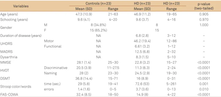
Data regarding the demographical composition of the sample, cognitive (extralinguistic) and motor performance, and duration of the disease are displayed in Table 1. HD pa-tients performed poorer than controls in all neuropsycho-logical tests.
Regarding oral language comprehension, HD patients had statistically signiicant poorer performance than con-trols in the Word Discrimination, Following Commands, Complex Ideational Material and Syntactic Processing tasks. In the Token test, there were no diferences in perfor-mance in parts 1, 2 and 3, but HD patients performed poor-er than controls in parts 4 (p=0.017), 5 (p=0.011), 6 (p=0.006) (raw data not shown) and in the total score. In oral language production, HD patients had statistically signiicant poorer
performance than controls in Verbal Agility, Non Verbal Agility, Words Repetition, Sentence Repetition and Action Naming (Table 2). In the Animal luency, Action luency tasks and in the FAS-COWA, HD patients performed poorer than controls (Tables 1 and 2).
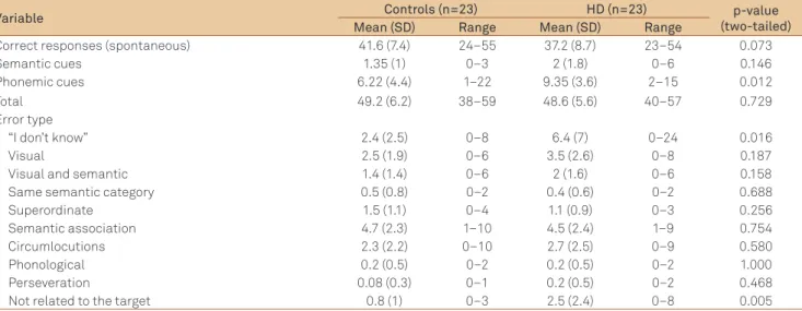
In the BNT, there were not signiicant diferences be-tween the groups in the number of spontaneous correct re-sponses. he qualitative analysis of types of errors revealed that HD patients difered from controls only in errors of the type “I don’t know” and “not related to the target” (Table 3). In written language comprehension, HD patients had lower scores than controls only in the Reading Paragraphs and Sentences task. In written language production, HD pa-tients had lower scores in the Letter Form, Letter Choice, Motor Facility and Narrative Writing tasks. In the Narrative Writing (Cookie heft Picture), HD patients performed poorer in Mechanics (p=0.002), Vocabulary (p<0.0001), Content (p<0.0001) (raw data not shown) and in the total score (Table 2).
HD patients also had lower scores than controls in the three indexes of global language performance, namely Comprehension Component, Expression Component and Language Competency Index (Table 2).
here were not any correlations between language per-formance (as measured by its three indexes of global lan-guage performance) and the UHDRS scores (motor and functional), MMSE, Stroop test, DSMT, MADRS and dura-tion of the disease. here was a positive moderate signiicant correlation between the scores in the HVOT and both the Expression Component (r=0.519, p=0.011) and the Language Competency Index (r=0.450, p=0.031).
Variables Controls (n=23) HD (n=23) HD (n=23) p-value
(two-tailed) Mean (SD) Range Mean (SD) Range
Age (years) 47.3 (10.9) 21–63 46.9 (11.2) 19–65 0.905 Schooling (years) 9.6 (4.1) 4–20 9.6 (3.7) 4–16 0.970
Gender M 8 (34.8%) 8 1.000
F 15 (65.2%) 15
Duration of disease (years) NA 6.8 (2.8) 3–12 –
UHDRS Motor NA 46.2 (19.4) 12–86 –
Functional NA 6.61 (3.2) 1–12 –
MADRS NA 12.5 (6.8) 2–32 –
Dysarthria NA 8.3 (1.5) 5–10 –
MMSE 28.1 (1.4) 25–30 22.8 (3.2) 15–27 <0.0001
HVOT Discriminative 20.5 (3.9) 11–27.5 11.3 (6.3) 2–24 <0.0001 Naming 28 (2) 23–30 24.5 (2.9) 19–30 <0.0001
DSMT 36.8 (14.4) 15–71 16 (8.9) 0–31 <0.0001
Stroop color/words time (sec.) 29 (5.8) 19–41 72.6 (53) 5–261 0.001 errors 1.4 (1.6) 0–5 3.7 (3.6) 0–13 0.010 FAS-COWA 32.4 (8.5) 18–50 14.9 (8) 4–22 <0.0001 Table 1. General characteristics of the sample: demographic data, cognitive and motor performance, and duration of the disease.
Language tests Controls (n=23) HD (n=23) p-value (two-tailed) Mean (SD) Range Mean (SD) Range
Token test 32.4 (2.2) 29–36 28.6 (5) 16–36 0.003
Action fluency 12.5 (5.3) 5–30 6.5 (4.3) 0–15 <0.0001 Animal fluency 16.5 (4.6) 10–25 9.1 (4) 3–17 <0.0001 BDAE
Word comprehension 15.3 (0.7) 14–16 14.4 (1.3) 12–16 0.004 Following commands 14.7 (0.5) 13–15 13.1 (1.8) 9–15 0.001 Complex ideational material 9.4 (2.2) 4–12 6.3 (2.8) 1–11 <0.0001 Syntactic processing 9.8 (2.4) 4–13 8.2 (2.8) 3–12 0.046 Non verbal agility 9.7 (1.7) 6–12 4.8 (2.1) 0–9 <0.0001 Verbal agility 12.8 (0.6) 14–14 10.1 (2.6) 3–13 <0.0001 Automatized sequences 3.9 (0.3) 3–4 3.9 (0.3) 3–4 1.000 Repetition of single words 14.2 (0.8) 14–15 14.6 (0.5) 13–15 0.050 Repetition of sentences 9 (0.9) 7–10 7.6 (1.5) 4–10 <0.0001 Responsive naming 19.8 (0.8) 18–22 19.6 (0.6) 18–20 0.414
Letters naming 4 (0) 4–4 4 (0) 4–4 1.000
Numbers naming 3.9 (0.2) 3–4 4 (0) 4–4 0.328
Colors naming 3.7 (0.4) 3–4 3.7 (0.5) 3–4 0.750
Action naming 9.3 (1.9) 6–12 7.9 (2.2) 3–11 0.028 Matching across cases and scripts 7.8 (0.5) 6–8 7.1 (1.6) 3–8 0.070 Number matching – fingers to Arabic numbers 2 (0) 2–2 2 (0) 2–2 1.000 Number matching – arabic numbers to dot patterns 2 (0) 2–2 1.7 (0.5) 1–2 0.005 Picture – word matching 3.6 (0.6) 2–4 3.3 (0.9) 1–4 0.351 Oral word reading 14.7 (1.2) 9–15 14.3 (2.5) 3–15 0.514 Oral sentences reading 8.6 (1.5) 6–10 7.7 (1.9) 3–10 0.079 Oral sentence comprehension 4.2 (1) 1–5 3.8 (1.3) 1–5 0.205 Reading comprehension – sentences and paragraphs 7.3 (1.5) 5–10 6.2 (1.9) 0–10 0.034 Letter form 13.5 (1.2) 9–14 10.3 (3.2) 2–14 <0.0001 Letter choice 20.6 (0.9) 17–21 18.3 (2.8) 8–21 0.001 Motor facility 13.9 (0.2) 13–14 8.8 (4.2) 0–14 <0.0001 Dictation – primer words 4 (0.5) 3–6 3.8 (0.8) 0–4 0.151 Dictation – regular phonics 2 (0.7) 1–5 1.8 (0.5) 0–2 0.233 Dictation – common irregular words 2.8 (0.7) 1–5 2.8 (0.6) 0–3 0.833 Written picture naming 2.6 (0.6) 1–4 2.6 (0.8) 0–4 0.840 Narrative writing 9.2 (1.6) 5–11 6.6 (2.2) 0–10 <0.0001 Language competency indexes
Comprehension component 79.9 (11) 60–100 51.7 (18.6) 26–90 <0.0001 Expression component 86.5 (8.4) 75–100 81.5 (7.8) 65–95 0.032 Language competency index 82.9 (8.5) 67.5–100 66.6 (12) 45.8–92.5 <0.0001 Table 2. Performance of Huntington’s disease and control groups in the Token Test, Animal fluency, Action fluency and BDAE.
BDAE: Boston Diagnostic Aphasia Examination.
BNT: Boston Naming Test; SD: standard deviation.
Variable Controls (n=23) HD (n=23) p-value
(two-tailed) Mean (SD) Range Mean (SD) Range
Correct responses (spontaneous) 41.6 (7.4) 24–55 37.2 (8.7) 23–54 0.073
Semantic cues 1.35 (1) 0–3 2 (1.8) 0–6 0.146
Phonemic cues 6.22 (4.4) 1–22 9.35 (3.6) 2–15 0.012
Total 49.2 (6.2) 38–59 48.6 (5.6) 40–57 0.729
Error type
“I don’t know” 2.4 (2.5) 0–8 6.4 (7) 0–24 0.016
Visual 2.5 (1.9) 0–6 3.5 (2.6) 0–8 0.187
Visual and semantic 1.4 (1.4) 0–6 2 (1.6) 0–6 0.158 Same semantic category 0.5 (0.8) 0–2 0.4 (0.6) 0–2 0.688
Superordinate 1.5 (1.1) 0–4 1.1 (0.9) 0–3 0.256
Semantic association 4.7 (2.3) 1–10 4.5 (2.4) 1–9 0.754 Circumlocutions 2.3 (2.2) 0–10 2.7 (2.5) 0–9 0.580
Phonological 0.2 (0.5) 0–2 0.2 (0.5) 0–2 1.000
Perseveration 0.08 (0.3) 0–1 0.2 (0.5) 0–2 0.468
DISCUSSION
Language alterations in HD are scarcely described and, according to Lepron et al.19, are primarily related to frontal
functions, such as impairment in verbal luency and syntac-tic processing. Nadeau20 states that in HD the language
disor-ders can be pervasive, due to the neurodegeneration, which progresses far beyond the head of the caudate nucleus. Our discussion addresses the alterations found in diferent lin-guistic aspects (phonetic, phonological, syntactic and se-mantic), and proposes an interpretation that takes into ac-count both linguistic and cognitive components of language. Regarding the phonetic articulatory aspects, our indings were highly predictable, given that HD largely afects frontos-triatal systems related to motor functions. Our patients were classiied as having mild hyperkinetic dysarthria according to Yorkston18, which did not compromise speech
intelligibil-ity, and did not interfere with the patients’ scoring, except in tasks that demanded verbal agility.
HD patients did not present any diiculties in tasks re-quiring preserved phonological loop, such as Repetition of Single Words and Dictation of Regular and Irregular Words. Integrity of the articulatory rehearsal processes, at least for short material, can thus be inferred. However, they did have impairment when repeating sentences. he Sentence Repetition task provides simple sentences in canonical order, being the number of syllables the only variable element.
Regarding the semantic aspects of language processing, we encountered deicits in the Word Comprehension, Following Commands and Complex Ideational Material tasks. he seman-tic system contains trace representations in nets that merge the entire subject’s knowledge about a speciic concept. he traces can be shared by several nets, thus making up catego-ries. Traces can also be distinguishing features between related items, but these are assembled in a lesser number of nets. he Word Comprehension task of the BDAE demands the identiica-tion of items within the same semantic category, thus relying on the ability to properly activate the distinguishing features.
Comprehension of Complex Ideational Material (orally pre-sented) was impaired in HD patients, indicating an inability to retain and process lengthy and integrated information. Although it is reasonable to assume that working memory impairment may play a role in these indings, it is worth noting that similar diiculties were also found in the Reading Comprehension task, where information is printed and available to the subject while performing the task. Comprehension of complex material, pre-sented either in oral or written form, depends on the interpreta-tion of its semantic and syntactic components.
Semantic loss can also be inferred by the low performance of HD patients in verbal luency tasks. Although action and letter luency greatly rely on integrity of executive functions, animal luency requires both executive functions and se-mantic knowledge integrity. Low performance in letter and
semantic luency tasks in HD was reported by Henry et al.3 in
a meta-analytic study, and a dissociation between semantic and letter luency (with a poorer performance in letter luen-cy) has also been described21. Time restriction to perform the
task may also be a factor that contributes to poor results22.
Action luency was worse in HD patients, a inding already reported by our group in a previous study23. Impaired verb
generation in HD patients was described by Péran24, and such
diiculties with action naming are hypothesized as being re-lated to frontal dysfunction25.
Moreover, action naming was the naming task that best discriminated controls and HD patients; naming tasks are said to relect an interface between language and motor ac-tion26 and the interaction between semantic, executive and
movement abnormalities, the latest two being linked to the derangement of frontostriatal circuits found in HD27.
he qualitative analysis of naming errors performed by HD patients do not allow any psycholinguistic interpretation, as the two types of errors in which HD difered from controls were “I don’t know” and “not related to the target”. HD pa-tients did not take advantage from semantic cues in the BNT, thus suggesting that visuoperceptual impairment did not signiicantly compromise their naming performance, in spite of the evidence of visuospatial impairment in the HVOT. However, visuoperceptual diiculties are more evident in the written production tasks, such as Letter Choice.
In summary, HD patients revealed deicits in situations requiring a more reined treatment of the material (e.g., vi-sual-semantic diferentiation), either when it was extensive or when its comprehension was somewhat complex, thus requiring integration of information and the realization of inferences. hey also displayed impoverishment of content in the production of semi-spontaneous language as in the nar-rative based on the Cookie heft Picture. It is worthy to note that these deicits do not characterize aphasia, but diicul-ties in high demanding tasks.
HD has been widely accepted as a disease of the basal gan-glia, more speciically the head of caudate nucleus, and sever-al studies have correlated the cognitive impairments found in HD to frontostriatal dysfunction occurring as a consequence of caudate atrophy, especially attentional deicits, working mem-ory deicits and executive dysfunction19,20,27, which are already
prominent in the early stages of the disease. Basal ganglia have been related to procedural learning and computational as-pects of linguistic processes, as those necessary for syntactic processing28. here is also evidence of a role of the striatum in
the retrieval of lexical information29. However, as the disease
progresses, difuse cortico-subcortical takes place, especially in frontal and temporal regions, and these indings correlate with increasing visuospatial, episodic memory and language disturbances20,30. Hence, it can be assumed that the
of semantic ad syntactic disturbances found in HD patients. hese multiple deicits (in lexical access, comprehension, syn-tax) quite certainly sufer the inluence of working memory, executive functions and visuospatial abilities, which were all impaired in our patients, but also relect the cortical difuse
derangement in HD18. he poor performance of HD patients
in the HVOT and the moderate positive correlation between the Expression Component (which includes the scores in the BNT) and the Language Competency Indexes and the HVOT scores favor this interpretation.
he complementary cognitive evaluation aimed to sup-port some indings previously resup-ported in HD patients, such as impaired verbal luency, verbal comprehension, syntactic production and naming abilities. Such a comprehensive eval-uation has helped us to better understand the nature of some phonological, syntactic and even semantic alterations in a broader context, for instance, that of executive dysfunction.
Limitations of this study are the small sample of HD patients and the instrument used to assess language. he BDAE was designed for the diagnosis and classiication of aphasia, and HD patients are not aphasics. Although the BDAE was well-suited for the purpose of screening the gen-eral proile of language alterations in this sample of HD pa-tients, further studies with more speciic psycholinguistic instruments are warranted.
Finally, a question that cannot be overlooked: why pa-tients sufering from HD (neither their relatives nor caregiv-ers) do not complain of language diiculties? his may be due to the fact that once the disease is diagnosed, motor (in-cluding dysarthria), psychiatric and global cognitive impair-ment turn out to be so prominent as to surpass the linguistic disturbances, hence the importance of assessing and identi-fying the linguistic deicits that might be overlooked in these patients, leading to additional impairment in quality of life.
1. Haddad MS, Cummings JL. Huntington’s Disease. In: Miguel EC, Rauch SL, Leckman JF (Eds). The Psychiatric Clinics of North America. Philadelphia: WB Saunders Company; 1997;791-807.
2. Shannon, KM. Huntington’s Disease Update. In: American Academy of Neurology 63rd. Annual Meeting Syllabi. Hawaii; 2011.
3. Henry JD, Crawford JR, Philips LH. A meta-analytic review of verbal fluency deficits in Huntington´s disease. Neuropsychology 2005;19:243-252.
4. Murray LL. Spoken language production in Huntington´s and Parkinson´s diseases. J Speech Lang Hear Res 2000;43:1350-1366.
5. Folstein SE, Leigh RJ, Parhad IM, Folstein MF. The diagnosis of Huntington’s disease. Neurology 1986;36:1279-1283.
6. Folstein MF, Folstein SE, McHugh PR. “Mini-mental state”. A practical method for grading the cognitive of patients for the clinician. J Psych Res 1975;12:189-198.
7. Smith GE, Ivnik RJ. Normative neuropsychology. In: Petersen RC (Ed). Mild Cognitive Impairment. New York: Oxford; 2003:63-88.
8. Huntington Study Group. Unified Huntington’s Disease Rating Scale: Reliability and Consistency. Mov Dis 1996;11:136-142.
9. Tumas V, Camargos ST, Jalali PS, Galesso AP, Marques Jr W. Internal consistency of a Brazilian version of the Unified Huntington’s disease rating scale. Arq Neuropsiquiatr 2004;62:977-982.
10. Smith A. Symbol Digit Modalities Test. Los Angeles: Western Psychological Services; 1982.
11. Stroop JR. Studies of interference in serial verbal reactions. J Exp Psychol 1935;18:643-662.
12. Hooper HE. The Hooper Visual Organization Test. Beverly Hills: Western Psychological Services; 1983.
13. Montgomery SA, Asberg M. A new depression scale designed to be sensitive to change. Brit J Psychiat 1979;134:382-389.
14. De Renzi E, Faglioni P. Normative data and screening power of a shortened version of the Token test. Cortex 1978;14:41-49.
15. Goodglass H, Kaplan E, Barresi, B. The Assessment of aphasia and related disorders. Philadelphia: Lippincott Williams & Wilkins; 2001.
16. Kaplan E, Goodglass H, Weintraub S. Boston Naming Test. Philadelphia: Lippincott Williams & Wilkins; 2001.
17. Hodges JR, Salmon DP, Butters N. The nature of the naming deficit in Alzheimer’s and Huntington’s disease. Brain 1991;114:1547-1558.
18. Yorkston KM, Strand EA, Kennedy MRT. Comprehensibility of dysarthric speech: implications for assessment and treatment planning. Am J Speech Lang Pathol 1996;5:55-66.
19. Lepron E, Péran P, Cardebat D, Démonet JF. A PET study of word generation in Huntington’s disease: effects of lexical competition and verb/noun category. Brain Lang 2009;110:49-60.
20. Nadeau SE. Subcortical Language Mechanisms. In: Stemmer B, Whitaker HA (Eds). Handbook of the Neuroscience of Language. London: Academic Press; 2008:329-338.
21. Taylor KI, Salmon DP, Monsch AU, Brugger P. Semantic and phonemic sequence effects in random word generation: a dissociation between Alzheimer’s and Huntington disease patients. J Int Neuropsychol Soc 2005;11:303-310.
22. Girotti F, Marano R, Soliveri P, Geminiani G, Scigliano G. Relationship between motor and cognitive disorders in Huntington´s disease. J Neurol 1988;235:454-457.
23. Azambuja MJ, Haddad MS, Radanovic M, Barbosa ER, Mansur LL. Semantic, phonologic, and verb fluency in Huntigton’s disease. Dem Neuropsychol 2007;1:381-385.
24. Péran P, Démonet JF, Pernet C, Cardebat D. Verb and noun generation tasks in Huntington’s disease. Mov Disord 2003;19:565-571.
25. Damasio AR, Tranel D. Nouns and verbs are retrieved with differentially distributes neural systems. Proc Natl Acad Sci USA 1993;90:4957-4960.
26. Pulvermüller F, Hauk O, Nikulin VV, Ilmoniemi RJ. Functional links between motor and language systems. Eur J Neurosci 2005;2:793-797.
27. Cummings JL. Movement Disorders. In: Cummings JL, Mega MS (Eds). Neuropsychiatry and Behavioral Neuroscience. New York: Oxford University Press; 2003:253-289.
28. Benítez-Burraco A. Enfermedad de Huntington: fundamentos moleculares e implicaciones para una caracterización de los mecanismos neuronales responsables del procesamiento linguístico. Rev Neurol 2009;48:75-84.
29. Teichmann M, Gaura V, Démonet JF, et al. Language processing within the striatum: evidence from a PET correlation study in Huntington´s disease. Brain 2008;131:1046-1056.
30. Montoya A, Price BH, Menear M, Lepage M. Brain imaging and cognitive dysfunctions in Huntington’s disease. J Psychiatry Neurosci 2006;31:21-29.