Arq Neuropsiquiatr 2008;66(3-B):744-746
744
Clinical / Scientiic note
BiCkerStaff’S enCephalitiS,
Guillain-Barré Syndrome and idiopathiC
intraCranial hypertenSion
Are they related conditions ?
Alfredo Damasceno
1, Marcondes C. França Jr
1, Daniel S. Pimenta
2,
Leonardo de Deus-Silva
1, Anamarli Nucci
1, Benito P. Damasceno
1enCefalite de BiCkerStaff, Síndrome de Guillain-Barré e hipertenSão intraCraniana idiopátiCa: São CondiçõeS relaCionadaS ?
School of Medicine, State University of Campinas (UNICAMP), Campinas SP, Brazil: 1Department of Neurology, 2Department of Ophthalmology.
Received 8 April 2008, received in inal form 17 June 2008. Accepted 14 July 2008.
Dr. Alfredo Damasceno – Departamento de Neurologia / FCM / UNICAMP - Caixa Postal 6111 - 13083-970 Campinas SP - Brasil. E-mail: alfredodamasceno @hotmail.com
Miller Fisher syndrome (MFS), considered a clinical variant of Guillain-Barré syndrome (GBS), is characterized by ophthalmoplegia, ataxia and arelexia1. Antecedent of
respiratory and/or gastrointestinal symptoms is frequent (up to 70%), with an average symptom-free interval of 10 days and the immune-mediated process is often associ-ated with acute phase anti-GQ1b IgG antibodies in up to 95% of all cases2. A closely related condition is
Bicker-staff’s brainstem encephalitis (BBE), which presents addi-tionally alteration of consciousness or long tract signs, but is less frequently associated with anti-GQ1b antibodies3.
Their relation and nosological position have been subject of extensive discussion in the literature. Idiopathic intra-cranial hypertension (IIH) comprises signs of increased in-tracranial pressure occurring in the absence of obvious ce-rebral pathology4. Some studies have reported cases of
intracranial hypertension associated with GBS, very few with MFS, but none with BBE.
We describe a distinct patient presenting initially with typical IIH indings but developing later signs of BBE, MFS and GBS. We also review evidence toward the possible link among MFS, BBE and GBS.
CaSe
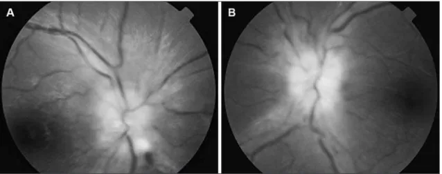
An obese 15 year-old female presented with a 5-days his-tory of frontal headache, nausea, vomiting and diplopia. Two weeks earlier she had had lu-like symptoms and diarrhea. Ex-amination disclosed normal consciousness, right sixth nerve pal-sy and bilateral papilledema (Fig 1) with normal visual acuity. Ce-rebral CT and MRI were normal. Lumbar puncture performed in the irst hospitalization day revealed an opening pressure of 100 cm H2O, and 25 mL of cerebrospinal luid (CSF) were withdrawn.
CSF protein and cell count were 15 mg/dL and two cells per mm3, respectively (Table). Despite a transient relief, the
symp-toms relapsed and acetazolamide-corticosteroid combination was begun for the hypothesis of IIH. Two days later she
Arq Neuropsiquiatr 2008;66(3-B)
745
Bickerstaff’s encephalitis, Guillain-Barré syndrome, intracranial hypertension Damasceno et al.
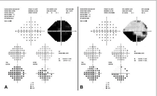
oped incomplete left third nerve palsy with affected visual acu-ity and visual ield (Fig 2A). On the next day, drowsiness was ob-served. The symptoms worsened and on the sixth day she had a Glasgow Coma Scale (GCS) score of 11, arelexia, muscle weak-ness (grade 4 on the Medical Research Council scale), urinary re-tention and mild cerebellar ataxia. Normal laboratorial investi-gation excluded metabolic encephalopathy. New CSF analysis (at the 10th hospitalization day) showed increased protein with
minimal pleocytosis. Serology for acute viral and bacterial in-fection was negative. Anti-GQ1b antibodies were not detected. Electrophysiology disclosed reduction in the amplitude of mus-cle action potential in facial nerves, right median and right ulnar nerves, besides slowed motor nerve conduction velocity in right ulnar nerve and diffusely absent F-responses and H-relex. She was diagnosed with BBE and treated with intravenous gamma-globulin (0.4g/Kg/day for 5 days). One week later she had a GCS of 15 and recovered almost completely from her ataxia, muscle weakness and urinary retention. One month later she had only a mild right sixth nerve palsy and arelexia with improved visu-al acuity and campimetry (Fig 2B). Five months later the Achil-les relex was present, though decreased.
A written informed consent to publish the case report was given by the patient and her legal responsible.
diSCuSSion
The initial clinical picture of this patient fulilled the modiied Dandy criteria for IHH. This syndrome, irst de-scribed more than 100 years ago, is particularly frequent in overweight adolescent girls and is associated with dif-ferent conditions such as medications, polycystic ovary syndrome and menstrual dysfunction4. The
pathophysi-ology of increased CSF pressure has not yet been com-pletely elucidated and different mechanisms have been
proposed, such as increased cerebral volume, CSF produc-tion rate and cerebral venous pressure, but impaired CSF outlow into the venous system does play an important role5. The course is usually benign, though sometimes it
can be complicated with visual loss4.
Our patient, however, showed an atypical evolution. Within 1-week period she developed weakness, drowsi-ness and ophthalmoplegia, features of GBS, BBE and MFS, respectively. In GBS, co-occurrence of intracranial hyper-tension has been documented, with papilledema found in about 4% of affected children, and less frequently in adults6. Although high protein content has been related
to the mechanism of pseudotumor in GBS, it does not always play a major role7, since intracranial hypertension
has also been observed in patients with only mildly or moderately elevated CSF protein content, as in our case. Other mechanisms such as an immune-mediated impair-ment of CSF outlow into the venous system may also be related. In MFS, few cases showing signs of elevated intra-cranial pressure have been documented in the literature8,
but, to our knowledge, it has never been described with BBE. This association may be under recognized since oph-thalmoscopy and CSF pressure are not routinely assessed in MFS, BBE or GBS.
Table. Cerebrospinal luid parameters through disease evolution.
CSF parameters D1 D4 D10 D25 D90
Opening pressure (cm H2O) 100 95 37 15 ND
Total protein (mg/L) 15 ND 70 88 39
Glucose (mg/L) 63 ND 99 70 54
Cell count (cells/mm3) 2 ND 5 4 5
ND, not done; D1, D4, D10, D25, and D90, days of hospitalization; numbers 1, 4, 10, 25, and 90, respectively.
Arq Neuropsiquiatr 2008;66(3-B)
746
Bickerstaff’s encephalitis, Guillain-Barré syndrome, intracranial hypertension Damasceno et al.
The classic triad of ophthalmoplegia, ataxia and are-lexia was irst described in 1956 by Fisher, who regarded it as an unusual variant of GBS resulting from peripheral nerve injury9. Interestingly, one of his patients had
drowsi-ness. One year later, Bickerstaff continued his previous work and reported eight patients with acute ophthalmo-plegia, ataxia and altered consciousness, considering this syndrome as a form of encephalitis10. Four of his patients
had arelexia. Due to the similarities of these syndromes, much begun to be debated in literature, from being or not variants of the same spectrum to their nosological position as either peripheral or central11. An important
contribution was the inding of an antibody that reacts with peripheral nerve ganglioside GQ1b in a great number of patients with MFS12 and some with BBE3. Further
ob-servations related this antibody to the presence of oph-thalmoplegia and ataxia. Greater amounts of ganglioside GQ1b have been found in cranial nerves III, IV and VI12 and
these nerves were enhanced by gadolinium on MRI in pa-tients with MFS13. A selective staining of the cerebellar
molecular layer by serum IgG antiGQ1b has also been ob-served14. Although the presence of this antibody could
be a secondary phenomenon due to destruction of neu-rons, some evidence points to a pathogenetic role, with blocking effects on the neuromuscular transmission15. The
appearance of this antibody has been related to cross re-activity with lipopolysaccharide in the bacterial coat of
Campylobacter jejuni, especially those with a particular polymorphism (Asn51) of cst-II gene16. The anti-GQ1b
an-tibodies were not detected in our patient, pointing more to the BBE side of the possible “MFS-BBE spectrum”.
Another valuable contribution came from studies with brain imaging describing abnormalities in some cases of BBE. In about 30% of the patients, high-signal lesions on T2-weighted scans were found in the brainstem, thala-mus, cerebellum and cerebrum3. A study with PET scan
also found involvement of brainstem and cerebellum17.
Similar indings have also been reported in patients diag-nosed with MFS18, even in the absence of drowsiness or
long tract signs, pointing to a central involvement in both MFS and BBE. Also supporting the spectrum hypothesis has been the evidence of concomitant central and pe-ripheral abnormalities described by Ogawara et al.19. More,
symptoms of central involvement in MFS, such as a mild somnolence, may not always be recognized.
The relation of these syndromes to GBS has been pro-posed since the irst description of Bickerstaff and Fisher, and supported by several overlapping cases3 describing the
presence of muscle weakness and albuminocytologic dis-sociation in addition to the classic triad of Fisher and/or altered consciousness or extensor plantar responses. The prognosis, however, is usually benign, especially in the “clas-sic” MFS2, and while in GBS treatment with plasmapheresis
or immunoglobulin is widely recommended1, in MFS they
are not always necessary2. In BBE or MFS/BBE overlapping
cases, this treatment has been associated with beneicial effects, but randomized controlled trials are needed to establish the value of immunotherapies or other treat-ments20. Our patient received a 5-days course of
immuno-globulin, but her improvement may be attributed either to the natural history of the disease or to the treatment.
Finally, this case shows a remarkable presentation of BBE with intracranial hypertension, a condition previously associated with GBS or MFS alone. It also contributes to the understanding of the “MFS-BBE spectrum” or acute inlammatory encephaloradiculoneuritis, which may be considered as a variant of GBS, presenting both peripheral and central involvement.
referenCeS
1. Hughes RA, Cornblath DR. Guillain-Barré syndrome. Lancet 2005;366: 1653-1666.
2. Overell JR, Willison HJ. Recent developments in Miller Fisher syndrome and related disorders. Curr Opin Neurol 2005;18:562-566.
3. Odaka M, Yuki N, Yamada M, et al. Bickerstaff’s brainstem encepha-litis: clinical features of 62 cases and a subgroup associated with Guil-lain-Barré syndrome. Brain 2003;126:2279-2290.
4. Ball AK, Clarke CE. Idiopathic intracranial hypertension. Lancet Neu-rol 2006;5:433-442.
5. Walker RW. Idiopathic intracranial hypertension: any light on the mech-anism of the raised pressure? J Neurol Neurosurg Psychiatry 2001;71:1-5. 6. Weiss GB, Bajwa ZH, Mehler MF. Co-occurrence of pseudotumor cerebri
and Guillain-Barré syndrome in an adult. Neurology 1991;41:603-604. 7. Ropper AH, Marmarou A. Mechanism of pseudotumor in
Guillain-Bar-ré syndrome. Arch Neurol 1984;41:259-261.
8. Pulitanò S, Viola L, Genovese O, et al. Miller-Fisher syndrome mimick-ing intracranial hypertension followmimick-ing head trauma. Childs Nerv Syst 2005;21:473-476.
9. Fisher M. An unusual variant of acute idiopathic polyneuritis (syn-drome of ophthalmoplegia, ataxia and areflexia). N Engl J Med 1956;255:57-65.
10. Bickerstaff ER. Brain-stem encephalitis; further observations on a grave syndrome with benign prognosis. Br Med J 1957;1:1384-1387. 11. Al-Din AN. The nosological position of the ophthalmoplegia, ataxia
and arelexia syndrome: “the spectrum hypothesis”. Acta Neurol Scand 1987;75:287-294.
12. Chiba A, Kusunoki S, Obata H, et al. Serum anti-GQ1b IgG antibody is associated with ophthalmoplegia in Miller Fisher syndrome and Guil-lain-Barré syndrome: clinical and immunohistochemical studies. Neu-rology 1993;43:1911-1917.
13. Nagaoka U, Kato T, Kurita K, et al. Cranial nerve enhancement on three-di-mensional MRI in Miller Fisher syndrome. Neurology 1996;47:1601-1602. 14. Kornberg AJ, Pestronk A, Blume GM, et al. Selective staining of the cerebel-lar molecucerebel-lar layer by serum IgG in Miller Fisher and related syndromes. Neurology 1996;47:1317-1320.
15. Lo YL, Chan LL, Pan A, Ratnagopal P. Acute ophthalmoparesis in the anti-GQ1b antibody syndrome: electrophysiological evidence of neu-romuscular transmission defect in the orbicularis oculi. J Neurol Neu-rosurg Psychiatry 2004;75:436-440.
16. Yuki N, Koga M. Bacterial infections in Guillain-Barré and Fisher syndromes. Curr Opin Neurol 2006;19:451-457.
17. Kwon HM, Hong YH, Sung JJ, et al. A case of Bickerstaff’s brainstem encephalitis; the evidence of cerebellum involvement by SPM analysis using PET. Clin Neurol Neurosurg 2006;108:418-420.
18. Urushitani M, Udaka F, Kameyama M. Miller Fisher-Guillain-Barré overlap syndrome with enhancing lesions in the spinocerebellar tracts. J Neurol Neurosurg Psychiatry 1995;58:241-243.
19. Ogawara K, Kuwabara S, Yuki N. Fisher syndrome or Bickerstaff brain-stem encephalitis? Anti-GQ1b IgG antibody syndrome involving both the peripheral and central nervous systems. Muscle Nerve 2002;26:845-849. 20. Overell JR, Hsieh ST, Odaka M, et al. Treatment for Fisher syndrome,