110
Rev Bras Oftalmol. 2015; 74 (2): 110-2
C
ASER
EPORTRevesz syndrome
Síndrome de Revesz
Dayane Cristine Issaho
1, Ana Tereza Ramos Moreira
2, Lisandro Lima Ribeiro
3,
Rommel Josué Zago
4, Christie Graf Ribeiro
21 Universidade Federal de São Paulo – São Paulo (SP), Brazil;
2,3Hospital de Clínicas da Universidade Federal do Paraná – Curitiba, (PR), Brazil;
4 Hospital de Olhos do Paraná – Curitiba (PR), Brazil.
This manuscript, or parts of it, have not been and will not be submitted elsewhere for publication
There are no conflicts of interest or financial disclosures.
Received for publication 20/05/2013 - Accepted for publication 16/11/2013
R
ESUMOA síndrome de Revesz é uma rara variante de disceratose congênita caracterizada por retinopatia exsudativa bilateral, alterações no segmento anterior ocular, retardo do crescimento intrauterino, pilificação fina e escassa, pigmentação cutânea reticular, falência da medula óssea, calcificações cerebrais, hipoplasia cerebelar e retardo neuropsicomotor. Há variações clínicas significativas entre os poucos relatos desta patologia existentes na literatura. Descrevemos o primeiro caso brasileiro de síndrome Revesz e suas características clínicas e oculares.
Descritores: Disceratose congenital/diagnóstico; Hemorragia retiniana; Anemia aplástica; Relatos de casos
A
BSTRACTRevesz syndrome is a rare variant of dyskeratosis congenita and is characterized by bilateral exudative retinopathy, alterations in the anterior ocular segment, intrauterine growth retardation, fine sparse hair, reticulate skin pigmentation, bone marrow failure, cere-bral calcification, cerebellar hypoplasia and psychomotor retardation. Few patients with this syndrome have been reported, and significant clinical variations exist among patients. This report describes the first Brazilian case of Revesz syndrome and its ocular and clinical features.
Keywords: Dyskeratosis congenital/diagnosis; Retinal hemorrhage; Anemia, aplastic; Case reports
111
I
NTRODUCTIONR
evesz syndrome is a rare variant of dyskeratosis congenital and was first described in 1992 in a Sudanese male infant who was found to have bilateral exudative retinopathy at 6 months of age. It is characterized by intrauterine growth retardation, fine sparse hair, reticulate skin pigmentation, bone marrow failure, bilateral exudative retinopathy, cerebral calcification, cerebellar hypoplasia, and psychomotor retardation(1,2).Few patients with Revesz syndrome have been reported, and the clinical features have not been fully delineated. Significant clinical variation exists among patients(1-4).
This report describes the first brazilian case of Revesz syndrome and its ocular and clinical features.
C
ASER
EPORTA 13-month-old caucasian female patient presenting with strabismus of the left eye, which had begun 2 months previously, was referred to our ophthalmology clinic. She was born at term by cesarean section with a birth weight of 3,090g. Apgar scores in the first and fifth minutes were 8 and 10, respectively. She had nonconsanguineous parents and a healthy sister.
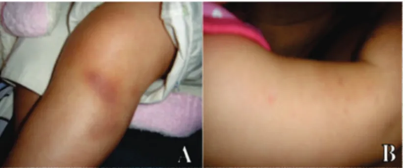
During the first examination, the child appeared to perceive an object at a distance of less than one meter with the right eye and could detect only hand movements with the left one. Neurological examination revealed developmental delay. She presented with bilateral leukocoria, and her digital ocular pressure was normal. Fundoscopy of the right eye revealed preretinal hemorrhage, coarse vitreous floaters, and no signs of disruption. The left eye exhibited a previous diffuse vitreous hemorrhage and vitreous floaters. Dermatological examination revealed petechiae in both legs and arms (figure 1).
Screening for HIV I/II, toxoplasmosis, syphilis, and cytomegalovirus was negative. Investigation revealed anemia, leukopenia, and a platelet count of 30.000/µL.
Owing to the pancytopenia, vitreous hemorrhage, and thrombocytopenic purpura, the patient was referred to a pediatric hematologist. In the subsequent months, pancytopenia was persistent. Bone marrow biopsy revealed hypoplastic marrow, consistent with myelodysplastic syndrome. Cytogenetic abnormalities of chromosome 3q were identified.
Magnetic resonance imaging of the brain revealed multiple and diffuse abnormalities in both cerebral hemispheres, consistent with calcifications or hemosiderin deposits, and cerebellar hypoplasia. The axons in the periatrial ventricular region appeared to be tenuous and this finding could be related to gliosis or terminal myelination.
Figure 1: A – Hematoma of the left leg; B – Petechiae on the right arm
During a follow-up visit, the parents complained of their child’s diminishing vision. Vitreous hemorrhage and fibrovascular proliferation were persistent in the right eye, and an examination of the left eye was impracticable owing to the retinal opacity (figure 2).
Ocular echography revealed vitreous hemorrhage and an image suggestive of retinal detachment in the right eye. In addition, the left eye exhibited hemorrhage and vitreous floaters but no sign of retinal detachment.
When the platelet count was reasonably stable, vitrectomy of the right eye was performed to correct the hemorrhage; however, the procedure was not visually successful.
The patient experienced several episodes of intestinal bleeding even after transfusion with blood products. Because of the poor prognosis, bone marrow transplantation was performed, and she was given immunosuppressive therapy. Nevertheless, even after the transplant, the child had 13 episodes of duodenal bleeding that required cauterization (figure 3).
She continued to receive supportive treatment, but we failed to control her platelet count, and the patient died at 3 years of age owing to intestinal hemorrhage.
D
ISCUSSIONIn this report, we describe a case with a rare variant of congenital dyskeratosis, Revesz syndrome. No similar cases have been described in Brazil until now.
Figure 2: Fundoscopy of the right eye showing vitreous hemorrhage and fibrovascular proliferation
Figure 3: A – Hemorrhage on the second part of the duodenum; B – intestinal cauterization
112
In 1992, Revesz et al first described this syndrome in a male infant with bilateral exudative retinopathy who developed aplastic anemia that led to his death. The child also had a history of intrauterine growth retardation, fine sparse hair, fine reticulate skin pigmentation, ataxia due to cerebellar hypoplasia, cerebral calcifications, extensor hypertonia, and progressive psychomotor retardation(1-3).
Here, we describe the ocular and clinical findings in a female patient who started to manifest symptoms within 1 year and one-month-old. Vitreous hemorrhage led to diminished vision and subsequent strabismus. We believed that the presence of petechiae and hematoma could be warning signs of the possibility of physical punishment (shaken baby syndrome); however, this notion was immediately discarded after assessing the family behavior. Thereafter, a coagulation disturbance was considered. Further examinations revealed pancytopenia, and this justified the ocular and dermatological findings.
Progressive bilateral Coats retinopathy has been described by other authors(3,4), but the hemorrhagic component in this
case impaired the visualization of retinal alterations. Our patient developed retinal detachment, similar to other cases(4,5).
The patient described herein did not present alterations in the anterior segment of the eye, such as megalocornea or buphthalmos.
Two inheritance patterns of dyskeratosis have been observed, autosomal dominant and X-linked recessive. The more common X-linked recessive form has been linked to DKC1, a gene that codes for dyskerin and is involved in telomerase function. The autosomal-dominant form of the disease has been linked to TERC, which codes for the template used by telomerase for the addition of telomeric repeat sequences(2,6). In a patient
with Revesz syndrome, Savage et al. identified a heterozygous mutation in the gene encoding TRF1-interacting nuclear factor-2 (TINFfactor-2; 604319.000factor-2), a component of the sheltering telomere protection complex(7). In our patient, cytogenetic abnormalities
of chromosome 3qwere identified, but no molecular genetics analysis was undertaken.
Regarding the neurological findings (hypoplasia and multiple cerebral calcifications), delayed neuropsychomotor development is a common feature in most patients with Revesz syndrome(3,5).
In patients with congenital dyskeratosis, the primary morbidity and mortality are due to the pancytopenia associated with this disease(1,3,5), as was observed in this case.
In conclusion, in this study, we present a case with a rare syndrome that was primarily associated with severe bone marrow aplasia and its complications, including progressive neurological abnormalities and retinal detachment.
R
EFERENCES1. Riyaz A, Riyaz N, Jayakrishnan MP, Mohamed Shiras PT, Ajith Kumar VT, Ajith BS. Revesz syndrome. Indian J Pediatr. 2007;74(9):862-3.
2. Kniffin CL. Revesz syndrome [Internet]. Baltimore: Online Men-delian Inheritance in Man. [cited 2012 Nov 25]. Available in: http://omim.org/entry/268130
3. Revesz T, Fletcher S, al-Gazali LI, DeBuse P. Bilateral retinopa-thy, aplastic anaemia, and central nervous system abnormalities: a new syndrome? J Med Genet. 1992;29(9):673-5
4. Kajtár P, Méhes K. Bilateral coats retinopathy associated with aplastic anaemia and mild dyskeratotic signs. Am J Med Genet. 1994;49(4):374-7.
5. Scheinfeld MH, Lui YW, Kolb EA, Engel HM, Gomes WA, Weidenheim KM, et al. The neuroradiological findings in a case of Revesz syndrome. Pediatr Radiol. 2007;37(11):1166-70. 6. Dokal I, Vulliamy T. Dyskeratosis congenita: its link to telomerase
and aplastic anaemia. Blood Rev. 2003;17(4):217-25. Review. 7. Savage, SA, Giri, N, Baerlocher, GM, Orr, N, Lansdorp, PM, Alter,
B. P. TINF2, a component of the shelterin telomere protection complex, is mutated in dyskeratosis congenita. Am J Hum Genet. 2008; 82(2): 501-9.
Corresponding author:
Dayane Cristine Issaho Hospital de Olhos do Paraná
Alameda Pres. Taunay, 483 – Curitiba (PR), Brazil Telephone: 55 (41) 9876-5992 - Fax: (41) 3029-4097 E-mail:[email protected]
IssahoDC, MoreiraATR, RibeiroLL ,ZagoRJ, RibeiroCG