Thiago Bruder Nascimento
, Rafaela de Fátima Ferreira Baptista
, Priscila Cristina Pereira
, Dijon Henrique Salomé
Campos
2, André Soares Leopoldo
1, Ana Paula Lima Leopoldo
2, Silvio A. Oliveira Júnior
2, Carlos Roberto Padovani
3,
Antônio Carlos Cicogna
2, Sandra Cordellini
1Departamento de Farmacologia, Instituto de Biociências - UNESP1; Departamento de Medicina Clínica - UNESP2; Departamento de Bioestatística, Instituto de Biociências - UNESP - Universidade Estadual Paulista3, São Paulo, SP - Brazil
Mailing address: Sandra Cordellini •
Rua João Carmelo, 145 - Jardim Paraíso - 18610-290 - Botucatu, SP - Brazil E-mail: [email protected], [email protected]
Manuscript received November 22, 2010; revised manuscript received January 14, 2011; accepted January 26, 2011.
Abstract
Background: Mechanisms underlying vascular abnormalities in obesity remain to be completely clarified.
Objective: L-arginine/nitric oxide pathway was evaluated on vascular response of high-fat diet-obese rats, focusing on endothelial and smooth muscle cells.
Methods: 30-day-old rats were divided in two groups: control (C) and obese (OB, high-fat diet for 30 weeks). After 30 weeks, body weight, adiposity index, blood pressure, and metabolic and endocrine profiles of the animals were recorded. Curves to noradrenaline were obtained in absence and presence of nitric oxide synthase inhibitor (L-NAME, 3x10⁻⁴M) on intact and denuded thoracic aorta from C and OB rats.
Results: Body weight, adiposity index, leptin and insulin levels were increased in OB, while blood pressure was unchanged. Obesity also produced glucose tolerance and insulin resistance. Reactivity to noradrenaline of intact aorta was similar in C and OB rats. L-NAME presence produced a similar increase in maximal responses, but a higher leftward shift of noradrenaline responses in intact aorta from C than in OB rats [EC50 (x10-7M): C=1.84 (0.83-4.07), O = 2.49 (1.41-4.38); L-NAME presence C = 0.02 (0.01-0.04)*, O = 0.21 (0.11-0.40)*†,*p < 0.05 vs respective control, †p < 0.05
vs control plus L-NAME, n = 6-7]. None of the protocols altered the reactivity to noradrenaline of denuded aortas. Conclusion: High-fat diet-induced obesity promotes metabolic and vascular alterations. The vascular alteration involved an endothelial L-arginine/NO pathway improvement was probably correlated to diet-induced hyperinsulinemia and hyperleptinemia. The greater resistance to L-NAME effects in aorta of obese rats raises concerns about the lower cardiovascular vulnerability of obese individuals in the presence of associated pathologies that impair NO-system activity. (Arq Bras Cardiol. 2011; [online].ahead print, PP.0-0)
Keywords: Obesity; rats; dietary fats; nitric oxide; endothelium, vascular.
incidence of certain forms of cancer, respiratory complications (obstructive sleep apnea), dyslipidemia, hypertension, atherosclerosis and vascular alterations1,5. Current guidelines
strongly endorse weight loss for patients who are overweight or obese. This initiative, however, do not distinguish between healthy individuals and those with chronic diseases such as heart failure and atherosclerotic coronary artery disease. Making the distinction between these populations may be important as there is evidence that in patients with chronic diseases, excess weight is paradoxically associated with a decreased risk of adverse outcomes6-8.
Several studies have shown that changes in the production and/or release of nitric oxide (NO), a major endothelial factor involved in the regulation of vascular tone9,10, are implicated
in a variety of pathophysiological responses. Although some controversy exists as to the involvement of NO-pathway on vascular disorders induced by obesity, there is evidence that impairment of NO synthesis represent a central defect triggering many of the vascular abnormalities characteristic of obesity states11,12.
Introduction
Obesity can be defined as a disease in which excess body fat has accumulated so that health may be adversely affected. It is considered to be a global epidemic and constitutes a major public health problem1,2. Although the etiology of
obesity is complex, several factors have been implicated in its development, especially hypercaloric intake3. In this context,
a variety of experimental models of obesity exist, however, dietary-induced obesity is the most relevant experimental model regarding human obesity4.
Thus, the aim of the present study was to evaluate the impact of high-fat diet-induced obesity on NO-pathway modulation of arterial tone, focusing on endothelial and smooth muscle cells. This study increases the understanding on the mechanisms of vascular alterations induced by obesity.
Methods
Animal
Thirty-day-old male Wistar rats (~150 g) were randomly assigned to one of two groups: control (C) and obese (Ob). The control group was fed a standard rat chow containing 4% fat, 42.7% carbohydrate, and 22% protein; whereas the obese animals received a high-fat diet containing 20% fat, 26.4% carbohydrate, and 20% protein. Each group was fed the diet for 30 weeks (C and Ob; n = 22). The high-fat diet was designed in our laboratory and contained powdered commercial Agroceres™ Animal Chow (Rio Claro, SP, Brazil), industrialized feed, protein supplement, vitamins and minerals. The high-fat diet was calorically rich (high-fat diet = 3.65 kcal/g versus standard diet = 2.95 kcal/g) due to the higher fat composition, made with saturated (20%) and unsaturated fatty acids (80%). All rats were housed in individual cages in an environmentally-controlled clean-air room at 23 ± 3o C with a 12-h light/dark cycle and 60 ± 5%
relative humidity.
All experiments and procedures were performed in accordance with the Guide for the Care and Use of Laboratory Animals, published by the U.S. National Institutes of Health13,
and were approved by the Botucatu Medical School Ethics Committee (UNESP, Botucatu, SP, Brazil).
Nutritional, metabolic and endocrine proiles of the animals
To analyze if dietary-induced obesity was associated with alterations in the nutritional behavior, food consumption was measured daily. Weekly calorie intake was calculated by average weekly food consumption x dietary energetic density. As obesity is defined as an excessive amount of body fat in relation to lean mass14, an adiposity index was calculated
from the sum of several fat pads. After 12-15 h fasting, animals were anesthetized with sodium pentobarbital (50 mg/kg) and euthanized by decapitation. Animals were thoracotomized and the total body fat (BF) was measured from the sum of the individual fat pad weights: visceral, epididymal and retroperitoneal. The adiposity index was calculated by the ratio: (total body fat/final body weight) x 100.
Obesity can be accompanied by metabolic and endocrine disturbances1, thus glycemic tolerance, leptinemia and
insulinemia were analyzed. Related to glycemic tolerance, after fasting for 12-15 h, rats were submitted to a glucose tolerance test (GTT). Blood samples were drawn from the tail at baseline and after administration of glucose (2 g/kg, i.p.)15,16. Blood samples were collected at 0, 15, 30, 60, 90
and 120 minutes. Glucose levels were determined using the ACCU-CHEK GO KIT glucose analyzer (Roche Diagnostic Brazil Ltda., Brazil). Glucose tolerance was analyzed from the area under curve of glycemic responses. For biochemical and
hormonal analysis, trunk blood was collected in heparinized tubes, centrifuged at 3,000 g for 15 min at 4° C. Serum leptin and insulin concentrations were determined by ELISA using commercial kits (Linco Research Inc., USA).
Homeostasis model assessment (HOMA)
HOMA, an index of insulin resistance, which employs measures of fasting plasma concentrations of glucose and insulin, was calculated according to the previously described method17.
Measurement of systolic blood pressure
Systolic blood pressure was weekly determined by using the non-invasive tail-cuff plethysmographic method (Narco Bio-Systems, Inc., Houston, TX) in conscious rats. The rats were prewarmed at 35 ± 2° C for 5 min and placed in a restrainer for blood pressure measurement. The mean of the three stable consecutive measures taken (~1 min apart) was recorded. The cuff pressure was controlled automatically and the systolic pulses detected by the pulse transducer were monitored with the audio signal. Care was taken in selecting an appropriate cuff size for the animal.
Vascular reactivity
After 30 weeks of high-fat diet or standard rat chow exposure the animals were decapitated. The descending thoracic aorta was excised and trimmed free of adhering fat and connective tissue. Two transverse rings of the same artery, each about 4 mm in length, were cut and mounted at the optimal length for isometric tension recording in organ chambers. One ring served as control, while the endothelium was mechanically removed from the other by gently rubbing the luminal surface18. The preparations were mounted in
organ baths containing 7 ml of Krebs-Henseleit solution, with composition in mM: NaCl 113.0; KCl 4.7; CaCl2 2.5; KH2PO4 1.2; MgSO4 1.1; NaHCO3 25.0; Glucose 11.0; ascorbic acid 0.11. The bathing fluid, kept at 37.0 ± 0.5o C, was saturated
with a gas mixture of 95% O2, 5% CO2. The preparations were
allowed to equilibrate for at least 1 h under a resting tension of 1.5 g, which is optimal in inducing the maximum contraction. Tension was recorded by a physiograph (Ugo Basile).
Cumulative concentration-effect curves were constructed from the response of the tissue to noradrenaline, in the absence and presence of NG-nitro-L-arginine methyl ester (L-NAME 3
x 10-4 M, inhibitor of NO synthase - NOS) (Sigma Chemical
Co., St Louis, Missouri, USA). At the end of the curves, single doses of acetylcholine (10-6 M) and sodium nitroprusside (10-4
M) were used to test the integrity of endothelial and smooth muscle layers, respectively.
Statistical analysis
Blood pressure, HOMA index and nutritional, metabolic and endocrine profileswere expressed as means ± standard deviation. Comparisons between groups were performed using the Student t-test for independent samples. The mean weekly body weight and the glucose profile of the groups
were compared by ANOVA for repeated measuresand post
The concentration of vasoactive agents producing a response that was 50% of the maximum (EC50) and maximal responses were calculated in each experiment. The EC50 values, presented as mean with 95% confidence intervals and maximum responses (g of tension), presented as mean ± SEM, were compared by ANOVA and post hoc Tukey’s test.
The level of significance was considered to be 5%.
Results
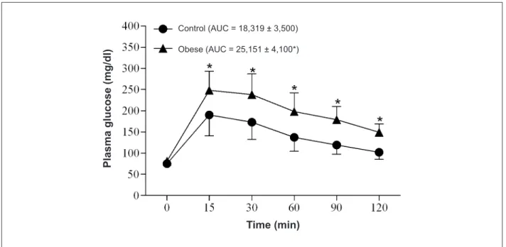
Table 1 shows nutritional, metabolic and endocrine profiles of rats.Obese rats ingested less food than control; however, calorie intake, index of obesity and final body weights at the end of 30 weeks were higher in obese than in the control group. Moreover, blood pressure did not differ between control and obese groups (Table 1). Glucose tolerance was lower in obese rats (Figure 1).
The plasma leptin (C = 2.2 ± 1.9; Ob = 7.8 ± 3.3* ng/ dl, n = 6-7), glucose levels (C = 107.0 ± 22.1; Ob = 126.0 ± 19.3* mg/dl, n = 6-7), insulin levels (C = 0.23 ± 0.08; Ob = 0.54 ± 0.07* ng/dl, n = 6-7), HOMA index (C = 18,319 ± 3,500; Ob = 25,151 ± 4,100*, n = 6-7) were significantly higher in the obese compared to the control group (*p < 0.05).
In the absence of L-NAME, the reactivity to noradrenaline of intact aorta was shown to be similar between high-fat and standard diet rats (Figure 2 and Table 2). The removal of the endothelium caused a leftward shift of the noradrenaline-aorta curves that was similar in both controls and high-fat diet obese rats (Figure 2 and Table 2). This procedure also determined a similar increase in the maximum response to noradrenaline in aortas from control and obese rats (Figure 1 and Table 2).
The presence of L-NAME produced a leftward shift of the noradrenaline curves that was lower in intact aortas from
high-fat diet obese than control rats (Figure 2 and Table 2). This inhibitor also produced an increase in the aorta maximum response to noradrenaline that was similar between control and obese groups (Figure 2 and Table 2).
Independently of L-NAME presence, the reactivity to noradrenaline did not differ among aortas without endothelium (Figure 2 and Table 2).
Discussion
In this study, high-fat diet rats developed obesity characterized by increase in final body weight and fat-pad mass. Although the obese group ingested less food, the higher
Table 1 - Effect of high-fat diet-induced obesity on the general characteristics of rats
Characteristics Control Obese
FBW (g) 498 ± 26 562 ± 36*
FI (g) 25.1 ± 1.7 21.8 ± 1.8*
CI (g kcal/day) 74.3 ± 5.4 86.1 ± 6.2*
EF (g) 6.65 ± 1.96 9.21 ± 3.80*
RF (g) 7.86 ± 2.35 11.58 ± 4.90*
VF (g) 8.31 ± 3.37 11.43 ± 4.58*
AI (%) 4.28 ± 1.65 5.96 ± 1.54*
FBP (mmHg) 130.7 ± 5.7 134.6 ± 6.9
Control rats (n = 6) received a standard diet (4% fat, 42.7% carbohydrate and 22% protein) and obese rats (n = 7) received a high-fat diet (20% fat, 26.4%
carbohydrate and 20% protein) for 30 weeks. FBW - inal body weight; FI - food intake; CI - calorie intake; EF - epididymal fat; RF - retroperitoneal fat; VF - visceral fat; AI - adiposity index; FBP - inal blood pressure. Values are expressed as mean
± SD. *p < 0.05 vs control (Student t-test for independent samples).
Figure 1 -Glucose tolerance test in control, receiving a standard diet: 4% fat, 42.7% carbohydrate and 22% protein, for 30 weeks (n = 22) and obese, submitted to 30
weeks of high-fat diet: 20% fat, 26.4% carbohydrate and 20% protein (n = 22) rats. Values are expressed as means ± standard deviation. AUC - area under the curve for glucose. *p < 0.05 control vs obese; ANOVA for repeated measures and post hoc Bonferroni test.
Pl
a
s
m
a
g
lu
c
o
s
e
(m
g
/d
l)
Time (min)
Control (AUC = 18,319 ± 3,500)
Table 2 - Maximal response and EC50% values to noradrenaline obtained in two rings, one with and the other without endothelium, in presence of absence of L-NAME (3x10⁻⁴), of the same thoracic aorta from control and obese rats
Groups Maximal response (g) EC50
With endothelium Without endothelium With endothelium (x10-7 M) Without endothelium (x10-8 M)
Control (n = 6)
Without inhibitor 2.69 ± 0.10 5.74 ± 0.40‡ 1.84 (0.83-4.07)
0.16‡ (0.06-0.42)
L-NAME 4.91 ± 0.18* 5.60 ± 0.22 0.02* (0.01-0.04)
0.14 (0.05-0.38)
Obese (n = 7)
Without inhibitor 2.35 ± 0.07 5.90 ± 0.38‡ 2.49 (1.41-4.38)
0.22‡ (0.08-0.55)
L-NAME 5.33 ± 0.18* 5.72 ± 0.28 0.21*† (0.11-0.40)
0.15‡ (0.05-0.43)
Control rats received a standard diet (4% fat, 42.7% carbohydrate and 22% protein) and obese rats received a high-fat diet (20% fat, 26.4% carbohydrate and 20%
protein) for 30 weeks. Maximal response and EC50 are expressed as mean ± SEM and mean followed by 95% conidence interval in parenthesis, respectively.*p < 0.05 vs
respective control; †p < 0.05 vs control/L-NAME; ‡p < 0.05 vs respective aorta with endothelium; ANOVA and Tukey’s test. n - number of animals.
Figure 2 -Concentration-effect curves to noradrenaline, in the absence and presence of L-NAME (3x10⁻⁴ M), obtained in two rings, one with (solid symbol) and the other
weight gain exhibited by these animals was most likely due to the increased calorie intake and feed efficiency in relation to controls. In addition, the obese rats developed metabolic disorders such as glucose intolerance, hyperinsulinemia and hyperleptinemia, characteristics commonly related to human obesity15,19,20. Insulin-resistance, defined as decreased
sensitivity and/or responsiveness to metabolic actions of insulin, is a cardinal feature of diabetes, obesity and dyslipidemia21. In
this context, HOMA index was shown to be increased in obese rats indicating insulin-resistance development in these animals as previously reported by Naderalli et al22 in diet-fed rats.
A number of experimental studies have reported a detrimental outcome of excessive high-energy diet on vascular reactivity. For example, high-fat diet impairs endothelium-dependent vasorelaxation23,24. This may, at least in part,
explain the increased risk of vascular disease in obese subject and animals. Moreover, antioxidant therapy improves endothelial function25,26,suggesting a diet-induced oxidative
stress in vasculature. In the present study, diet-fed obese rats had significant vascular alteration, particularly a marked endothelial improvement that was unveiled in the presence of L-NAME. In other words, diet-induced obesity did not alter the aorta reactivity to noradrenaline in the absence of L-NAME. However, the presence of this inhibitor produced a leftward shift of noradrenaline curves that was lower in the aorta of obese in relation to control rats. Moreover, the high-fat diet did not induce any changes in the aorta smooth muscle reactivity to noradrenaline. This conclusion could be inferred from the observation that, regardless of L-NAME presence, the reactivity of denuded aortas did not differ between control and obese rats.
As the aorta from diet-fed obese rats was more resistant to the effects of L-NAME, it can be hypothesized that the endothelial L-arginine/NO pathway is improved in the vasculature of obese rats. Moreover, this increased NO bioavailability in high-fat diet rats could represent an adaptive mechanism to counteract the reported detrimental outcome of excessive high-energy diet on vascular reactivity in obesity, mainly oxidative stress25,26.The improvement in endothelial
NO pathway could also explain the absence of changes in blood pressure of obese rats, a parameter often reported to
be increased in human and animal obesity27,28. These findings
confirm the “obesity paradox”6 and could help to understand
the mechanisms responsible for this condition.
Finally, the literature also reported that vasodilation leading to increased blood flow is a major physiological consequence of insulin-stimulated production of NO in vascular endothelium29. Moreover, leptin hormone is
able to directly activate NO production in vessels and this activation is dependent on endothelium integrity30. Thus, the
hyperinsulinemia and hyperleptinemia observed in high-fat diet animals could be partially responsible for the increased endothelial NO production and/or release in the vasculature of obese rats.
Overall, this study indicates that high-fat diet-induced obesity promotes metabolic and vascular alterations. The vascular alteration was characterized by an endothelial L-arginine/NO pathway improvement, probably correlated to diet-induced hyperinsulinemia and hyperleptinemia. Finally, the greater resistance to L-NAME effects in the aorta of obese rats in relation to controls raises concerns about the lower cardiovascular vulnerability of obese individuals in the presence of associated pathologies that impair NO-system activity.
Acknowledgments
We are grateful to FAPESP, process 07/57495-0 and 07/59747-7, for financial support.
Potential Conflict of Interest
No potential conflict of interest relevant to this article was reported.
Sources of Funding
This study was funded by FAPESP.
Study Association
This article is part of the thesis of master submitted by Dijon
Henrique Salomé Campos, from Faculdade de Medicina de
Botucatu - Unesp.
References
1. Kopelman PG. Obesity as a medical problem. Nature. 2000;404(6778):635-43.
2. O’Brien PE, Dixon JB. The extent of the problem of obesity. Am J Surg. 2002;184(6B):4S-8S.
3. Stein CJ, Colditz GA. The epidemic of obesity. J Clin Endocrinol Metab. 2004;89(6):2522-5.
4. Tschöp M, Heiman ML. Rodent obesity models: an overview. Exp Clin Endocrinol Diabetes. 2001;109(6):307-19.
5. Malnick SD, Knobler H. The medical complications of obesity. QJM. 2006;99(9):565-79.
6. Lüscher TF, Tanner FC. Endothelial regulation of vascular tone and growth. Am J Hypertens. 1993;6(7 Pt 2): 283S-93S.
7. Curtis JP, Selter JG, Wang Y, Rathore SS, Jovin IS, Jadbabaie F, et al. The obesity paradox: body mass index and outcomes in patients with heart failure. Arch Intern Med. 2005;165(1):55-61.
8. Gruberg L, Weissman NJ, Waksman R, Fuchs S, Deible R, Pinnow EE, et al. The impact of obesity on the short-term and long-term outcomes after percutaneous coronary intervention: the obesity paradox? J Am Coll Cardiol. 2002;39( 4):578-84.
9. Widlansky ME, Sesso HD, Rexrode KM, Manson JE, Gaziano MJ. Body mass index and total and cardiovascular mortality in men with a history of cardiovascular disease. Arch Intern Med. 2004;164(21):2326-32.
11. Deng G, Long Y, Yu YR, Li MR. Adiponectin directly improves endothelial dysfunction in obese rats through the AMPK-Enos Pathway. Int J Obes (Lond). 2010;34(1):165-71.
12. Martins MA, Catta-Preta M, Mandarim-de-Lacerda CA, Aguila MB, Brunini TC, Mendes-Ribeiro AC. High fat diets modulate nitric oxide biosyntesis and antioxidant defense in red blood cells from C57BL/6 mice. Arch Biochem Biophys. 2010;499(1-2):56-61.
13. Committee on Care and Use of Laboratory animals. Guide for the care and use of laboratory animals. Bethesda: Nacional Institute of Health; 1985.
14. Stunkard AJ, Wadden TA, (editors). Obesity: theory and therapy. 2nd ed. New York: Raven Press; 1993.
15. Barnes MJ, Lapanowski K, Conley A, Rafols JA, Jen KL, Dunbar JC. High fat feeding is associated with increased blood pressure, sympathetic nerve activity and hypothalamic mu opioid receptors. Brain Res Bull. 2003;61(5):511-9.
16. Fatani S, Pickavance LC, Sadler CJ, Harrold JA, Cassidy R, Wilding JPH, et al. Differencial vascular dysfunction in response to diets of differing macronutrient composition: a phenomenological study. Nutr Metab. 2007;4:15.
17. Matthews DR, Hosker JP, Rudenski AS, Naylor BA, Treacher DF, Turner RC. Homeostasis model assessment: insulin resistance and β-cell function from fasting plasma glucose and insulin concentrations in man. Diabetologia. 1985;28(7):412-9.
18. Cordellini S, Carvalho MH, Scivoletto R, Fortes ZB, Nigro D. Indirect evidence for an endothelium-derived contracting factor release in aorta of deoxycorticosterone acetate-salt hypertensive rats. J Hypertens. 1990;8(1):53-60.
19. de Moraes C, Davel AP, Rossoni LV, Antunes E, Zanesco A. Exercise training improves relaxation response and SOD-1 expression in aortic and mesenteric rings from high caloric diet-fed rats. BMC Physiol. 2008;8:12.
20. Relling DP, Esberg LB, Fang CX, Johnson WT, Murphy EJ, Carison EC, et al. High-fat diet-induced juvenile obesity leads to cardiomyocyte dysfunction
and upregulation of Foxo3a transcription factor independent of lipotoxicity and apoptosis. J Hypertens. 2006;24(3):549-61.
21. Muniyappa R, Montagnani M, Koh KK, Quon MJ. Cardiovascular actions of insulin. Endocr Rev. 2007;28(5):463-91.
22. Naderali EK, Fatani S, Williams G. Chronic withdrawal of a high-palatable obesity-inducing diet completely reverses metabolic and vascular abnormalities associated with dietary-obesity in the rat. Atherosclerosis. 2004;172(1):63-9.
23. Naderali EK, Brown MJ, Pickavance LC, Wilding JP, Doyle PJ, Williams G. Dietary obesity in the rat induces endothelial dysfunction without causing insulin resistance: a possible role for triglycerides. Clin Sci (Lond). 2001;101(5):499-506.
24. Naderali EK, Pickavance LC, Wilding JPH, Williams G. Diet-induced endothelial dysfunction in the rat is independent of the degree of increase in total body weight. Clin Sci (Lond). 2001;100(6):635-41.
25. Sato J, O’Brien T, Katusic ZS, Fu A, Nygren J, Singh R, et al. Dietary antioxidant preserves endothelium dependent vasorelaxation in overfed rats. Atherosclerosis. 2002;161(2):327-33.
26. Plotnick GD, Correti MC, Vogel RA. Effect of antioxidant vitamins on the transient impairment of endothelium-dependent brachial artery vasoactivity following a single high-fat mean. JAMA. 1997;278(20):1682-6.
27. Hall JE. The kidney, hypertension, and obesity. Hypertension. 2003;41(3 Pt 2):625-33.
28. Jones DW, Kim JS, Andrew ME, Kim SJ, Hong YP. Body mass index and blood pressure in Korean men and women: the Korean National Blood Pressure Survey. J Hypertens. 1994;12(12):1433-7.
29. Kim JA, Montagnani M, Koh KK, Quon MJ. Reciprocal relationships between insulin resistance and endothelial dysfunction: molecular and pathophysiological mechanisms. Circulation. 2006;113(15):1888-904.