Case 6/2008 - A 54 Year-Old Male with Hemiparesy and Seizures
Since Hildhood Evolving With Heart Failure 10 Years After Precordial
Pain and Normal Coronary Angiography
Gustavo Freitas Feitosa, Ricardo D`Oliveira Vieira, Paulo Jorge Moffa, Reynaldo Amato, Sérgio Rosemberg, Vera Demarchi Aiello
Heart Institute (Incor), University of São Paulo Medical School, SP - Brazil
Mailing Address: Vera D. Aiello •
InCor – Av. Dr. Enéas de Carvalho Aguiar, 44 – 05403-000 – São Paulo, SP E-mail: [email protected]
Key words
Hypothyroidism; paresis; seizures; heart failure; chest pain.
Section Editor: Alfredo José Mansur ([email protected]) Associated Editors: Desidério Favarato ([email protected]) Vera Demarchi Aiello ([email protected])
A 54-year-old man was brought to the Emergency Department with severe dyspnea. According to his medical history, he had sequelae of perinatal anoxia. The patient was experiencing difficulty speaking, motor deficit, and seizures. He also had hypothyroidism.
At age 40 he had a sudden, severe, stabbing chest pain of two hours duration. When he was first examined, in January 1993, the lungs, heart, and abdomen were normal. The patient had reduced muscle strength, right hemihypotrophy and difficulty walking.
The ECG (January 6, 1993) revealed sinus rhythm, heart rate 68 bpm, PR interval 120 msec, and QRS duration 120 msec, with initial slurring. The QRS complex was oriented posteriorly at – 40°. There was left anterior fascicular block, and ST-segment elevation in V1 and V2, interpreted as early repolarization. There was also ST-segment depression in left leads (V6) and T-wave inversion in leads 1 and aVL (Figure 1)
Laboratory findings (January 1993) were as follows: cholesterol 215 mg/dL, triglycerides 150 mg/dL, glucose 150 mg/dL, and creatinine 1.1 mg/dL.
The exercise stress test was suggestive of myocardial ischemia, and coronary angiography showed no changes in coronary arteries (January 1993). Ventriculography revealed mild hypokinesis and left ventricular hypertrophy.
The echocardiogram (May 1993) was normal (Table 1) 24-hour Holter monitoring detected 387 ventricular extrasystoles, with two nonsustained ventricular tachycardias, asymptomatic.
A brain CT scan (1994) showed hypoplastic left hemisphere and asymmetric lateral ventricles. As the patient still had severe chest pain unrelated to exertion, he was given amitriptyline.
The patient’s course remained uneventful with no further
complaints or medical visits until 2006, when he was admitted in April and June with palpitations, malaise, and dyspnea. After these episodes, he developed dyspnea with daily activities and was referred to Incor for medical review.
An echocardiogram was performed, revealing moderate left ventricular dysfunction, with ejection fraction of 41%. Coronary angiography (May 25, 2006) showed no evidence of obstructive coronary artery disease, but left ventriculography revealed moderate, diffuse hypokinesis.
On physical examination (August 29, 2006), he weighed 45.3 kg and was 1.55 m tall. His heart rate was 76 bpm and blood pressure 100x70 mm Hg. The examination of the lungs, heart, and abdomen was unremarkable.
The electrocardiogram (August 2006) showed first degree AV block, left atrial overload, left bundle branch block, left anterior fascicular block, and electrically inactive area inferoposterolateral. High resolution ECG showed standard QRS of 191.00 msec; filtered QRS of 71.00 msec (normal <100-114), low-amplitude signals below 40 µV of 3.00 msec (normal <35); and terminal 40 msec of 123.90 µV (> 20).
Laboratory findings (August 2006) were as follows: creatinine 1.32 mg/dL; glucose 89 mg/dL; hemoglobin 17.3 g/dL; hematocrit 53%, MCV 95 fl; leukocytes 5.600/mm3
(neutrophils 65%, eosinophils 1%, basophils 1%, lymphocytes 25%, and monocytes 8%); platelets 263.000/mm³; potassium 5.8 mEq/L; sodium 139 mEq/L; ANPB 1.899 pg/mL; aspartate aminotransferase 35 U/L; alanine aminotransferase 50 U/L; total cholesterol 235 mg/dL; triglycerides 123 mg/dL; HDL-cholesterol 37 mg/dL; LDL-HDL-cholesterol 173 mg/dL; potassium 5.6 mEq/L; TSH 21.4 µIU/mL; and free T4 1.2 ng/dL.
The echocardiogram (August 2006) revealed marked, diffuse LV involvement, with akinesia of the inferior wall, and moderate RV hypokinesis (Table 1).The patient was diagnosed with hypothyroidism and dilated cardiomyopathy with possible episodes of ventricular tachycardia.
Medications in use were adjusted: spironolactone was decreased to 25 mg daily and captopril was increased to 75 mg daily. The remaining drugs remained unchanged: carvedilol 6.25 mg, amiodarone 200 mg, and levothyroxine 25 µg daily.
The patient was brought to InCor Emergency Department with dyspnea accompanied by wheezing and impaired consciousness (October 26, 2006).
Figure 1 -(&*VLQXVUK\WKPLQWUDYHQWULFXODUFRQGXFWLRQGLVWXUEDQFHOHIWDQWHULRUIDVFLFXODUEORFNLQLWLDOVOXUULQJRIWKH456FRPSOH[DQGSUREDEOHOHIWYHQWULFXODU YROXPHRYHUORDG
Table 1 -Echocardiograms
May 1993 August 2006 October 2006
Left atrium (mm) 30 48 41
Right ventricle (mm) 20 28 20
Interventricular septum (mm) 8 7 10
LV posterior wall (mm) 7 7 10
LV diastolic diameter (mm) 47
-LV systolic diameter (mm) -
-/9HMHFWLRQIUDFWLRQ 22 23
Body mass index (g/m²) 90 Pulmonary artery systolic
pressure (mm Hg) - 61 41
LV motion Normal
Inferior akinesia and marked, diffuse
hypokinesis
Marked, diffuse hypokinesis
RV motion Normal Marked hypokinesis
Moderate hypokinesis
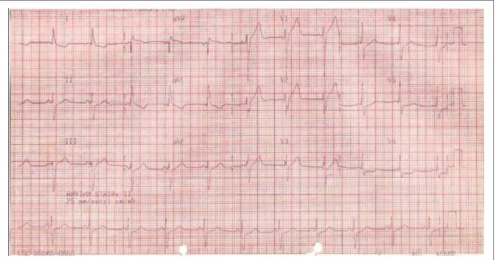
blood perfusion to the extremities, and diffuse rhonchi over both lung fields. He underwent orotracheal intubation for ventilatory support and was given continuous intravenous infusions initially of dopamine and later of norepinephrine. The electrocardiogram (October 26, 2006) displayed atrial tachycardia with a 2:1 atrioventricular block, left atrial overload, low QRS voltage in the frontal plane, electrically inactive area in the lateral and high lateral wall, prolonged QT (450 msec), plus tall and widened T waves (Figure 2).
Laboratory tests (October 26, 2006) yielded the following results: hemoglobin 16.3 g/dL; hematocrit 50%, MCV 100 fl; leukocytes 16,300/mm3 (neutrophils 82%, eosinophils 0%,
lymphocytes 9%, and monocytes 9%); creatinine 2.53 mg/dL (glomerular filtration 28 mL/min/1.73 m²); amylase 150 U/L; total bilirubin 3.18 mg/dL, direct bilirubin 1.20 mg/dL; creatine phosphokinase 168 U/L; aspartate aminotransferase 2,783 U/L; and alanine aminotransferase 2.260 U/L.
The echocardiogram (October 2006) revealed marked LV hypokinesis and moderate RV hypokinesis, in addition to enlarged left atrium with a fixed thrombus measuring 13/12 mm (Table 1).
There was no improvement in blood pressure, and the patient died of asystolic cardiac arrest a few hours after admission.
Clinical features:
Figure 2 -(&*DWULDOWDFK\FDUGLDZLWKD$9EORFNOHIWDWULDORYHUORDGDQGGLVSHUVLRQRIDWULDOUHSRODUL]DWLRQHOHFWULFDOO\LQDFWLYHDUHDLQWKHODWHUDODQGKLJKODWHUDO ZDOOVSURORQJHG47LQWHUYDODQGWDOO7ZDYHVWKDWDGGWRWKH3ZDYHVZLWKWKHLUUHSRODUL]DWLRQ
Among laboratory findings, fasting blood glucose of 150 mg/dL attracted special attention, pointing to a possible diagnosis of diabetes.
The patient underwent an exercise stress test, which was reported to be suggestive of myocardial ischemia. The criterion for positivity was not mentioned. Difficulty in walking and changes in baseline ECG make exercise stress testing less accurate as a non-invasive method to detect myocardial ischemia. In this case, pharmacological stress echocardiography and myocardial perfusion SPECT are good options. This patient underwent coronary angiography, which revealed no obstructive coronary artery disease. Ventriculography showed mild hypokinesis and left ventricular hypertrophy.
We present the case of a patient with atypical chest pain (perhaps because of the difficulty in expressing himself verbally as an anoxic sequela), ECG abnormalities, positive exercise stress test for myocardial ischemia, angiographically normal coronary arteries, and ventriculography suggestive of left ventricular hypertrophy. Changes detected by the ECG, exercise stress test and ventriculography support the hypothesis of chest pain of cardiac origin, rather than psychological, neuromuscular, pulmonary, or gastrointestinal origin.
As for the pain caused by nonatherosclerotic coronary disease, it may be associated with epicardial coronary arteries (vasospasms, myocardial bridge) or microcirculation (secondary hypertension, dilated or hypertrophic cardiomyopathy, infiltrative diseases, valvular heart disease, or idiopathic microvascular dysfunction)1.
Both the absence of a history of hypertension and normal cardiac examination make dilated cardiomyopathy, valvular hear disease, and hypertension very unlikely as the cause of pain. No systolic compression of epicardial coronary arteries (myocardial bridge) was demonstrated on coronary angiography.
The patient underwent an echocardiogram three months
after his first visit, which was normal. This excludes the diagnosis of dilated cardiomyopathy and valvular heart disease, and makes the hypothesis of hypertrophic cardiomyopathy very unlikely.
Once these possibilities were ruled out, the following conditions could explain the presence of ischemia in patients with angiographically normal coronary arteries and normal echocardiogram: coronary spasms or Prinzmetal angina, storage diseases (amyloidosis, Fabry disease), and idiopathic microvascular ischemia or syndrome X.
A 24-hour Holter monitoring revealed 387 ventricular extrasystoles (16.2/hour), with two episodes of nonsustained ventricular tachycardia (NSVT), which, according to the literature, is an independent risk factor in the presence of heart disease, whether ischemic or nonischemic2.
The patient still complained of severe chest pain unrelated to exertion and was treated with amitriptyline. At this point, two comments are necessary. First, it must be kept in mind that, despite the atypical angina, there was strong evidence of underlying heart disease (changes in ECG, exercise stress test, and ventriculography). Further etiological investigation, therefore, should be performed. Second, although tricyclic antidepressants may be used to treat chest pain of neuropsychiatric origin (perhaps this patient had this associated component), care must be taken in prescribing them to patients with evidence of ventricular arrhythmias because of their proarrhythmic effects, in these cases with possible prolongation of the QT interval.
In May 2006, an echocardiogram revealed moderate LV dysfunction with ejection fraction of 41%. Coronary angiography showed no evidence of obstructive lesions, but left ventriculography disclosed moderate, diffuse hypokinesis. What draws our attention in this case is the long latent period (12 years), after which the patient returned for medical follow-up no longer with chest pain, but rather with dyspnea, palpitations, and malaise, in addition to deterioration of ventricular function, despite normal results on coronary arteriogram.
A physical examination, in August 2006, showed body mass index of 18.8 kg/m2, heart rate of 76 bpm, and
blood pressure of 100x70 mmHg. His heart, lungs, and abdomen remained normal. The ECG showed first-degree atrioventricular (AV) block, left bundle branch block (LBBB), left anterior fascicular block, and an electrically inactive area in the inferoposterolateral wall. No late potentials were detected by high-resolution ECG. Laboratory tests showed a marked increase in B-type natriuretic peptide and TSH, in addition to moderate increase in total cholesterol and LDL-cholesterol. An echocardiogram performed at the same time revealed left ventricular dilation, akinesia of LV inferior wall and hypokinesis of the remaining walls, a marked decrease in ejection fraction, pulmonary hypertension, and right ventricular hypokinesis (Table 1).
At this time, the therapeutic regimen was changed: spironolactone was decreased to 25 mg while captopril was increased to 75 mg daily. The other medications were left unchanged: carvedilol 6.25 mg/day, amiodarone 200mg/day, and levothyroxine 25 mcg/day.
This is a patient, therefore, that 13 years ago had evidence of heart disease with “presumed” myocardial ischemia and angiographically normal coronary arteries, yet remained asymptomatic for 12 years. He progressed to ventricular dilation and dysfunction, in addition to symptoms of heart failure apparently precipitated by ventricular tachyarrhythmias.
Next, we will examine the first hypotheses formulated. Patients with Prinzmetal angina or variant angina experience chest pain almost always at rest, which is not precipitated by physical exertion or emotional stress. This condition is often associated with S-T segment elevation on ECG3, ventricular tachycardia and fibrillation, and sudden
death. These patients tend to be younger than those with atherosclerotic disease. There is also an association with tobacco use. The ECG pattern is usually characterized by episodic ST-segment elevation with pain, which can occur in any lead. Holter monitoring may detect silent ischemia, and coronary angiography may reveal “spontaneous” or provoked epicardial coronary vasospasms. The acute phase usually lasts six months, with recurrent episodes. In a series of 277 patients followed-up for 7.5 years, recurrent angina occurred in 39%, death in 3.5%, and myocardial infarction in 6.5%4. Associated
atherosclerotic disease has a significantly negative prognostic impact. After six months, recurrence tends to decrease, and those who experience remissions may benefit from therapy with calcium channel blockers. For unknown reasons, some patients may have a latent period of months or years and
then experience a recrudescence, with frequent episodes of severe ischemia, which usually respond to calcium channel blockers and nitrates.
In this specific case there was no evidence of ST-segment elevation associated with the anginal pain, nor were coronary vasospasms demonstrated on any coronary angiography. The patient’s progression to dilated cardiomyopathy may be associated with previous infarctions secondary to vasospasms, although few elements in his medical history support this hypothesis.
Most patients with idiopathic microvascular dysfunction or syndrome X are postmenopausal women5, who often present
with atypical angina. Two-thirds of patients with chest pain and normal coronary angiography have associated psychiatric disorders6. The ECG may be normal or show nonspecific
ST-T wave changes. Among patients with chest pain and angiographically normal coronary arteries, 20% have positive exercise test. These patients have a good long-term prognosis (similar to healthy individuals of the same age group, unless they are smokers or hypertensive). This diagnosis was ruled out, because this patient experienced progressive deterioration of ventricular function.
As for amyloidosis, another hypothesis initially formulated, this condition results from deposition of amyloid proteins, which, in the primary form of the disease, are made up of portions of immunoglobulin light chain (termed AL), while the secondary form (termed AA) is characterized by a different amyloid protein. Heart disease occurs in one-third of the patients, but it is virtually always present in the pathologic examination of those with this diagnosis7.
Clinical manifestations of cardiovascular involvement in amyloidosis occur in four ways that may overlap: restrictive cardiomyopathy with diastolic dysfunction and symptoms of right-sided heart failure; congestive heart failure, which tends to progress slowly, respond poorly to drug therapy and sometimes occur with angina pectoris despite angiographically normal coronary arteries; orthostatic hypotension, which is found in about 10% of the cases; and conduction disturbances and arrhythmia.
The most typical electrocardiographic change is diffuse, low-voltage QRS complex. Bundle-branch blocks and axis deviations are common. Small or absent R waves in right precordial lead and, less frequently, Q waves in inferior leads sometimes simulate areas of myocardial infarction8. Complex
ventricular arrhythmia is a frequent finding.
In advanced cases, increased thickness of ventricular walls and septum with normal-sized ventricles are often observed, as well as dilated atria. Left ventricular function may be depressed, even though it is usually surprisingly normal9. In
the early stages of cardiac amyloidosis, the echocardiogram may be “falsely” normal.
The diagnosis can be safely established by periumbilical fat biopsy. If clinical suspicion remains despite a negative result, endomyocardial biopsy may be used.
Fabry disease is an inherited, X-linked, recessive disorder caused by deficiency of the enzyme alpha-galactosidase A. In its classical form, the disease is characterized by deposition of glycosphingolipids in the kidneys, skin, and myocardium, although it can be limited to the heart. Its clinical manifestations include angina pectoris and myocardial infarction, despite angiographically normal coronary arteries. Hypertension, mitral valve prolapse, and congestive heart failure are commonly observed. Electrocardiographic changes include short PR interval, ST-segment and T-wave abnormalities, atrioventricular block, left ventricular overload, and QRS widening10. The echocardiogram often reveals
increased left ventricular wall thickness, which was not the case in this patient, making this diagnosis unlikely. The diagnosis was confirmed by endomyocardial biopsy.
Another hypothesis that has not yet been mentioned but deserves to be highlighted is myocarditis. Considering the patient’s progression to left ventricular dysfunction, the diagnosis would be that of subacute viral myocarditis.
Clinically, these patients may have a condition suggestive of viral infection. The portal of entry for the virus is the upper respiratory or gastrointestinal tract, and the infection precedes by days or weeks the onset of cardiac symptoms, such as heart failure, embolism, or arrhythmia. The initial viral infection may go unnoticed11.
Electrocardiographic changes may include nonspecific ST-T wave changes and atrial or ventricular arrhythmias. Atrial, ventricular or intraventricular conduction delays can also occur. Echocardiography may demonstrate increased left ventricular thickness, more evident in fulminant myocarditis12.
The diagnosis is based on endomyocardial biopsy. The type of pain experienced by this patient at the time of his initial presentation is not characteristic of this disease. All the same, this diagnosis should be considered, because after the initial symptom of chest pain he developed myocardial failure with ventricular dilation.
It is important to point out that nuclear magnetic resonance would have been of great value in establishing the diagnosis.
In October 2006, the patient was readmitted with impaired consciousness, wheezing and hemodynamic shock. He was intubated and treated with vasoactive drugs. Of note, liver and kidney enzyme levels were elevated: AST 2783 U/L, ALT 2.260 U/L, total bilirubin 3.18 mg/dlL, direct bilirubin 1.2 mg/ dL, and creatinine 2,53 mg/dL. Electrocardiographic findings included sinus rhythm with heart rate of 65 bpm, left atrial overload, low QRS voltage in the frontal plane, electrically inactive area in the lateral wall, prolonged QT, and tall and widened T waves.
This electrocardiogram no longer showed left bundle branch block, as mentioned earlier. In addition, the low QRS voltage in the frontal plane may be due to infiltrative diseases or advanced dilated cardiomyopathy itself. The inactive area observed laterally corresponded to a region that was already electrocardiographically altered 12 years ago. The QT prolongation may be attributed to hypothyroidism.
An echocardiogram was performed, and results were similar to those obtained previously, except for the presence
of a mobile thrombus in the left atrium.
A few hours after admission, the patient had an asystolic cardiorespiratory arrest.
Considering all the above, we concluded that the cause of death was cardiogenic shock secondary to dilated cardiomyopahy.
Gustavo Freitas Feitosa, MD, and Ricardo D`Oliveira Vieira, MD
Diagnostic hypothesis
The primary etiological hypothesis is viral myocarditis that progressed to dilated cardiomyopathy.
Dr. Gustavo Freitas Feitosa e Dr. Ricardo D`Oliveira Vieira
Comments on electrocardiograms
The electrocardiogram obtained on January 6, 2003, showed sinus rhythm, heart rate of 68 bpm, PR interval of 120 msec, QRS duration of 120 msec, QRS axis directed superiorly (-50°) and posteriorly, left anterior fascicular block, and T-wave inversion in leads I, aVL and V6.
The electrocardiogram obtained on October 26, 2006, showed atrial tachycardia with a 2:1 AV block; heart rate of 65 bpm; left atrial overload in leads V3 to V6; a deep, downward deflection comparing the atrial regularization of the first P wave (dispersion of atrial repolarization); low voltage QRS complex in the frontal plane in leads V5 and V6; wide R waves from V1 to V3, corresponding to an electrically inactive area in the
inferoposterolateral region; prolonged QT interval; and tall T waves that add to the P waves with their repolarization.
Paulo Jorge Moffa, MD
Comments on chronic coronary artery diseases
This clinical case, the diagnostic possibilities of which were reported by the medical resident and suggest the presence of cardiomyopathy, illustrate how difficult it is to establish a diagnosis based on unreliable information provided by a patient with motor deficit and speech difficulty. For the diagnosis of chest pain, the patient underwent two invasive procedures performed three years apart, which yielded normal angiographic findings.
Reynaldo Amato, MD
Autopsy studies
Postmortem examination of the heart revealed an extensive, healed infarction involving the entire ventricular septum and left ventricular anterior wall, in addition to the paraseptal portion of the right ventricular anterosuperior wall (Figure 3). In addition, small mural thrombi were found in both ventricles. The left atrium was dilated and had a large thrombus (3.5 x 2.5 cm) attached to its septal surface (Figure 4). No recent myocardial infarctions were found on histological examination.
Figure 3 -6KRUWD[LVFURVVVHFWLRQRIWKHKHDUWGHSLFWLQJH[WHQVLYH¿EURVLVSUHYLRXVLQIDUFWLRQDUHDHQFORVHGE\GRWWHGOLQHVLQYROYLQJ/9DQWHULRUZDOOWKHHQWLUH YHQWULFXODUVHSWXPDQGWKHSDUDVHSWDOSRUWLRQRI59DQWHULRUZDOO7KHUHDUHDOVRFDYLWDU\WKURPELDVWHULVNV$6DQWHURVXSHULRU3,SRVWHURLQIHULRU/OHIW'ULJKW
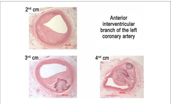
Figure 5 -3KRWRPLFURJUDSKRIVHJPHQWVRIWKHDQWHULRULQWHUYHQWULFXODUEUDQFKRIWKHOHIWFRURQDU\DUWHU\VKRZLQJFDOFL¿HG¿EURXVSODTXHV+HPDWR[\OLQHRVLQVWDLQLQJ
with approximately two weeks’ duration. On histological examination, epicardial coronary arteries were obstructed by predominantly fibrocalcific atheromatous plaques, with no inflamed walls and little lipid accumulation. The greater degree of obstruction was found in the fourth centimeter of the anterior interventricular branch of the left coronary artery (70%) (Figure 5)
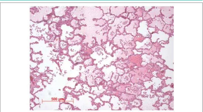
The remaining segments showed stenosis ranging from 20% to 50%. In the distal right coronary artery there was a recent, not totally occlusive thromboembolism. The lungs showed evidence of chronic passive congestion and alveolar edema (acute edema – Figure 6), which ultimately led to the patient’s death.
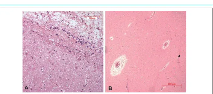
The brain weighed 1140 g. The left hemisphere showed considerable volume loss compared to the right, but with no apparent atrophy. Vertico-frontal sections of the brain confirmed the presence of hypotrophy without evidence of clastic lesions (Figure 7). The most distinctive finding on microscopic examination was a moderate decrease in neuronal cells of the isocortex in all areas studied. This decrease seemed to affect all layers indistinctively. Associated with this finding, there was reactive astrocytic gliosis.
A significant subpial Chaslin’s gliosis was also found, accompanied by a great number of amylaceous bodies, which were also present in the subcortical white matter. Dilation of Virchow-Robin spaces without reaction in surrounding tissues was present in basal ganglia and white matter. No changes were found in the hippocampus (Figure 8).
Pathologic diagnoses
Ischemic heart disease characterized by extensive, healed transmural infarction involving the septum and anterior wall. Coronary atherosclerosis. Massive left atrial thrombosis. Acute pulmonary edema. Cerebral changes consistent with chronic hypoxia.
Vera Demarchi Aiello, MD, PhD, Sérgio Rosemberg, MD, PhD
&RPPHQWVRQFDUGLRYDVFXODUÀQGLQJV
Histopathological studies have unequivocally shown the presence of ischemic heart disease. Although coronary obstructive lesions were not critical, they cannot be considered negligible either, since there were calcified atherosclerotic plaques. The primary hypothesis for the development of myocardial infarction is coronary vasospasm. It is highly unlikely that thrombosis or thromboembolism caused the previous infarction, since we found no evidence of thrombus formation in the coronary tree.
Regarding the discrepancy between data from coronary angiography (no obstructions) and that from histopathological examination, well-known studies have demonstrated that positive remodeling (increased external diameter), in the presence of atherosclerotic plaque, may result in little or no luminal obstruction on coronary angiography13-15.
Figure 6 -3KRWRPLFURJUDSKRIWKHOXQJVKRZLQJDOYHRODUVSDFHV¿OOHGE\DPRUSKRXVHRVLQRSKLOLFPDWHULDODFXWHHGHPDLQDGGLWLRQWRKHDUWIDLOXUHFHOOV+HPDWR[\OLQ HRVLQVWDLQLQJ
Figure 8 -3KRWRPLFURJUDSKRIWKHEUDLQ,Q$WKHFHUHEUDOFRUWH[VKRZVDVXESLDODUHDZLWKHRVLQRSKLOLF¿EHUVUXQQLQJSDUDOOHOWRWKHVXUIDFHWKHVRFDOOHG&KDVOLQ¶V JOLRVLVLQDGGLWLRQWRDP\ODFHRXVERGLHVURXQGEDVRSKLOLFVWUXFWXUHV,Q%GLODWLRQRI9LUFKRZ5RELQVSDFHVLVVHHQEULJKWSHULYDVFXODUDUHDV+HPDWR[\OLQHRVLQ [DQG[UHVSHFWLYHO\
&RPPHQWVRQQHXURSDWKRORJLFDOÀQGLQJV
In the absence of a reliable pre-, peri- or postnatal history, neuropathological findings were interpreted randomly. Both the type of volume reduction and histological findings do not support an hypoxic-ischemic etiology during that period (peri-natal), which would be characterized by ulegyria, that is, scarred cerebral gyri with almost completely neuronal loss, fibrous gliosis and cavitation, or by multicystic encephalomacia, which, as the name suggests, is characterized by multiple cysts usually in the white matter.
The abnormal findings indeed support the existence of a
hypoxic process, but a more chronic, progressive process. One possibility that may be considered is the so-called HHE syndrome (hemiconvulsions, hemiplegia, epilepsy). In this condition, children under the age of two, for any reason (fever, for instance), develop a unilateral status epilepticus. The consequence is neuronal death in the contralateral hemisphere, with gliosis and an epileptical scar, leading to chronic epilepsy.
Neuropathological findings (diffuse neuronal loss, nonfibrous reactive gliosis, and Chaslin’s gliosis) are fully consistent with the hypotheses formulated.
Sérgio Rosemberg, MD, PhD
References
1. Yang EH, Lerman A. Angina with normal angiogram. Herz. 2005;30:1-25. 2. Ruberman W, Weinblatt E, Goldberg J, Frank C, Shapiro S. Ventricular
premature beats and mortality after myocardial infarction. N Engl J Med. 1977;297:750-6.
3. Prinzmetal M, Kennamer R, Merliss R, et al: Angina pectoris. I. A variant form of angina pectoris: Preliminary report. Am J Med. 195;27:375-88 4. Bory M, Pierron F, Panagides D, et al: Coronary artery spasm in patients with
normal or near normal coronary arteries. Long-term follow-up of 227 patients. Eur Heart J. 1996:17:1015-21.
5. Cannon RO III: Chest pain and the sensitive heart. Eur J Gastroenterol Hepatol. 1995;7:1161-71.
6. Potts SG, Bass CM: Psychological morbidity in patients with chest pain and normal or near-normal coronary arteries: A long-term follow-up study. Psychol Med. 1995;25:339-47,
7. Trikas A, Rallidis L, Hawkins P, et al: Comparison of usefulness between exercise capacity and echocardiographic indexes of left ventricular function in cardiac amyloidosis. Am J Cardiol.1999;84:1049-54.
8. Gertz MA, Lacy MQ, Dispenzieri A: Amyloidosis. Hematol Oncol Clin North
Am. 1999;13:1211-33.
9. Cacoub P, Axler O, De Zuttere D, et al: Amyloidosis and cardiac involvement. Ann Med Interne (Paris) 2000;151:611-7.
10. Linhart A, Magage S, Palecek T, et al: Cardiac involvement in Fabry disease. Acta Paediatr Suppl. 2002;91:15-20.
11. Imazio M, Trinchero R. Myopericarditis: etilogy, management and prognosis. Int J Cardiol. 2008;127:17-26.
12. Felker GM, Boehmer JP, Hruban RH, et al: Echocardiographic findings in fulminant and acute myocarditis. J Am Coll Cardiol. 2000;36:227-32. 13. Stiel GM, Stiel LSG, Schofer J, Donath K, Mathey DG. Impact of compensatory
enlargement of atherosclerotic coronary arteries on angiographic assessment of coronary artery disease. Circulation.1989;80:1603–9
14. Mintz GS, Popma JJ, Pichard AD et al. Limitations of angiography in the assessment of plaque distribution in coronary artery disease. Circulation 1996;93:924–31.
![Figure 2 - (&*DWULDOWDFK\FDUGLDZLWKD$9EORFNOHIWDWULDORYHUORDGDQGGLVSHUVLRQRIDWULDOUHSRODUL]DWLRQHOHFWULFDOO\LQDFWLYHDUHDLQWKHODWHUDODQGKLJKODWHUDO ZDOOVSURORQJHG47LQWHUYDODQGWDOO7ZDYHVWKDWDGGWRWKH3ZDYHVZLWKWKHLUUHSRODUL]DWLRQ](https://thumb-eu.123doks.com/thumbv2/123dok_br/15558214.600198/3.892.64.770.160.524/fdugldzlwkd-eorfnohiwdwuldoryhuordgdqgglvshuvlrqridwuldouhsrodul-dwlrqhohfwulfdoo-lqdfwlyhduhdlqwkhodwhudodqgkljkodwhudo-zdoovsurorqjhg-lqwhuydodqgwdoo-zdyhvwkdwdggwrwkh-zdyhvzlwkwkhluuhsrodul.webp)