C. Şengül et al. Let ventricular microistulizaion 299
Dicle Tıp Derg / Dicle Med J www.diclemedj.org Cilt / Vol 39, No 2, 299-301
Yazışma Adresi /Correspondence: Dr. Cihan Şengül
Universal Çamlıca Germany Hospital, İstanbul, Turkey Email: [email protected] Copyright © Dicle Tıp Dergisi 2012, Her hakkı saklıdır / All rights reserved
Dicle Tıp Dergisi / 2012; 39 (2): 299-301
Dicle Medical Journal doi: 10.5798/diclemedj.0921.2012.02.0146
CASE REPORT / OLGU SUNUMU
Left ventricular microistulization: A rare cause of ischemia in a patient with normal
coronary arteries
Sol ventriküler mikroistülizasyon: koroner arterleri normal olan bir hastada iskeminin nadir
bir nedeni
Cihan Şengül, Ayşegül Sünbül, Ender Semiz, İsmet Dindar Universal Çamlıca Germany Hospital, Division of Cardiology, İstanbul, Turkey
Geliş Tarihi / Received: 10.10.2011, Kabul Tarihi / Accepted: 11.02.2012
ÖZET
Fizik egzersiz esnasında göğüs ağrısı oluşan 71 yaşın -da bayan hasta kardiyoloji bölümüne başvurdu. Miyokard perfüzyon sintigraisinde inferior ve anteroapikal seg -mentlerde hipoperüzyon saptandı. Selektif koroner anji -yograide sol ön inen arter ve sağ koroner arterden köken alan ve önemli koroner arter darlığı yapmadan sol ven -triküle boşalan çoklu korono-kameral istüller saptandı. Koroner arter istülleri bir koroner arter ile bir kalp odacığı veya büyük damar arasındaki anormal bağlantılar olarak tanımlanır. Sol ventrikülde sonlanan korono-kameral is -tüller nadirdir. Küçük is-tüller genellikle hemodinamik açı -dan tehlike yaratmaz. Fakat daha büyük ve çoklu istüller koroner çalma fenomenine atfedilen miyokard iskemisine neden olabilirler. Kameral istüller çok nadir görüldüğü için nasıl en iyi şekilde tedavi edilecekleri belirsizdir. Bu olgu -da 50 mg/gün metoprolol ile anti-iskemik te-davi yapıldı ve hiçbir girişim yapılmaksızın hasta altı ay olaysız takip edildi.
Anahtar sözcükler: Anjiyograi, iskemi, istül ABSTRACT
A 71-year-old woman with chest pain occurring on physi -cal exercise was admitted to cardiology department. Myocardial perfusion scintigraphy revealed inferior and anteroapical segment hypoperfusion. Selective coronary angiography revealed multiple coronary-cameral istulas originating from the left anterior descending artery and the right coronary artery and emptying into the left ventri -cle without any signiicant coronary artery stenosis. Coro -nary artery istulas are deined as abnormal communica -tions between a coronary artery and a cardiac chamber or major vessel. Coronary-cameral istulas terminating in the left ventricle are uncommon. Small istulas usually do not cause any hemodynamic compromise. However, the larg -er and multiple istulas may cause myocardial ischemia ascribed to a coronary steal phenomenon. The best way to manage cameral istulae is uncertain largely due to the rarity of the condition. In the present case, anti-ischemic medications with metoprolol 50 mg/day provided an un -eventful follow-up of six months without any intervention. Key words: Angiography, ischemia, istulae
INTRODUCTION
Coronary-cameral istulas (CCFs) are deined as ab-normal communications between a coronary artery and a cardiac chamber or major vessel, such as the vena cava, right or left ventricle, pulmonary vein, or pulmonary artery. Most patients with coronary ar-tery istula are asymptomatic thus they are discov-ered incidentally during angiographic evaluation for coronary vascular diseases. They may present with symptoms of angina caused by coronary steal.
CASE REPORT
C. Şengül et al. Let ventricular microistulizaion 300
Dicle Tıp Derg / Dicle Med J www.diclemedj.org Cilt / Vol 39, No 2, 299-301
anterior descending coronary artery and the right coronary artery and emptying into the left ventricle without evidence of coronary stenosis (Figure 1, Figure 2). Anti-ischemic medications (metoprolol 50 mg/day) provided an uneventful follow-up of six months.
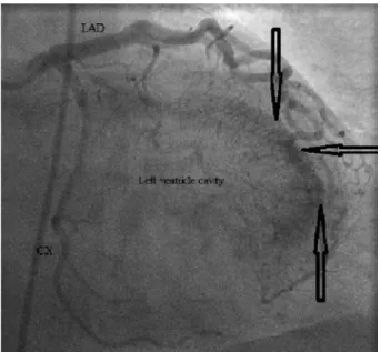
Figure 1. Right anterior oblique view showing the
multiple microistulas draining into the left ventricle. Arrows show opaciication of the left ventricle by is -tulas
Figure 2. Left anterior oblique view showing micro -istulae originating from the right coronary artery (RCA). Arrow shows opaciication of the left ven -tricle
DISCUSSION
Coronary-cameral istulas are thought to arise as a persistence of sinusoidal connections between the lumens of the primitive tubular heart that supply myocardial blood low in the early embryologic peri-od. Another explanation may be faulty development of the distal branches of the coronary artery
recti-form vascular network.1 The routine angiographic
detection of a CCF is rare, occurring in an estimated 0.2% of patients who undergo catheterization.2 In
children, the diagnosis of coronary artery istula can often be made with transthoracic 2-dimensional and color-low Doppler echocardiography. However, in adults cardiac catheterization with coronary angiog-raphy which shows the size, anatomy, number, orig-ination, and termination site of the istulas remains the gold standard for the diagnosis of CCFs.3 Al-though asymptomatic in most cases, CCFs may pro-duce symptoms such as angina pectoris, myocardial infarction, congestive heart failure, rhythm distur-bances, subacute bacterial endocarditis,
thrombo-embolism, and sudden death.3,4 Magnetic resonance
imaging, and multidetector computed tomography can also be used to evaluate the CCFs. Small is-tulae usually do not cause any hemodynamic
com-promise.4 However, the larger and multiple istulae
may cause ischemia by coronary steal phenomenon leading to myocardial ischemia.5 The best way to manage cameral istulae is not well-known largely due to the rarity of the condition. Once a CCF is detected, the management should be established in-cluding antibiotic prophylaxis and in case of aneu-rysmal dilatation of istula-related coronary artery or istulous vessel antiplatelet regimen is recom-mended. Permanent occlusion of istulas by surgi-cal ligation is addressed when CCFs are presented with multiple connections, tortuous course, acute angulations, complex anatomy, distal localization, large istula with high istulous low, side branch at risk, and complicated with aneurysmal dilatation. Percutanous therapeutic transcatheter embolisation or graft stent implantatios may be another option for the selected cases. Factors favoring percutanous intervention are: proximal location, older patients, and absence of concomitant cardiac disorders neces-sitating surgery.6 Small caliber and multiple istulas
like in the present case are unlikely to be amenable to surgical or percutanous intervention and can be
C. Şengül et al. Let ventricular microistulizaion 301
Dicle Tıp Derg / Dicle Med J www.diclemedj.org Cilt / Vol 39, No 2, 299-301 REFERENCES
1. Luo L, Kebede S, Wu S, Stouffer GA. Coronary artery istu-lae. Am J Med Sci 2006;332(2):79-84.
2. Iadanza A, del Pasqua A, Fineschi M, Pierli C. Three-vessel left-ventricular microistulization syndrome: a rare case of angina. Int J Cardiol 2004;96(1):109-11.
3. Lessick J, Kumar G, Beyar R, Lorber A, Engel A. Anomalous origin of a posterior descending artery from the right pul-monary artery: report of a rare case diagnosed by multide-tector computed tomography angiography. J Comput Assist Tomogr 2004;28(6):857-9.
4. Brooks CH, Bates PD. Coronary artery-left ventricular istula with angina pectoris. Am Heart J 1983;106(2):404–6. 5. Yilmaz H, Basarici I, Demir I. A rare cause of ischemia:
congenital coronary-cameral istula: case report. Turkiye Klinikleri J Cardiovasc Sci 2006;18(2):158-61.
6. Said SA, van der Werf T. Dutch survey of congenital coro-nary artery istulas in adults: corocoro-nary artery-left ventricu-lar multiple micro-istulas multi-center observational sur-vey in the Netherlands. Int J Cardiol 2006;110(1):33-9. 7. Sheikhzadeh A, Stierle U, Langbehn AF, Thoran P, Diederich