UNIVERSIDADE FEDERAL DE OURO PRETO
NÚCLEO DE PESQUISA EM CIÊNCIAS BIOLÓGICAS PROGRAMA DE PÓS-GRADUAÇÃO EM CIÊNCIAS BIOLÓGICASProduction and expression of inflammation
and angiogenic parameters triggered by
different genetic population of
Trypanosoma cruzi
DEENA SHRESTHA
SUPERVISOR: Prof. Dr. ANDRE TALVANI
CO-SUPERVISOR: Prof. Dr. MARIA TEREZINHA BAHIA
OURO PRETO 2014
Tese apresentada ao programa de
Pos-Graduação em Ciências Biológicas da
Universidade Federal de Ouro Preto,
como parte integrante dos requisitos
para a obtenção do titulo de Doutor em
Ciências
Biológicas,
área
de
concentração:
Imunobiologia
de
Catalogação: sisbin@sisbin.ufop.br
S561p Shrestha, Deena
Production and expression of inflammation and angiogenic parameters triggered by different genetic population of Trypanosoma cruzi [manuscrito] / Shrestha Deena. - 2014.
101f.: il., grafs.; tabs.
Orientador: Prof. Dr. André Talvani.
Co-orientadora: Profª. Drª. Maria Terezinha Bahia.
Tese (Doutorado) - Universidade Federal de Ouro Preto. Instituto de Ciências Exatas e Biológicas. Núcleo de Pesquisas em Ciências Biológicas. Programa de Pós-graduação em Ciências Biológicas.
Área de concentração: Imunobiologia de Protozoários.
1. Trypanossoma Cruzi - Teses. 2. Chagas, Doença de - Teses. 3. Inflamação
- Teses. 4. Cardiopatias - Teses. I. Talvani, André. II. Bahia, Maria Terezinha. III. Universidade Federal de Ouro Preto. IV. Título.
iii This work was developed in the Laboratory of Chagas Disease / Núcleo de Pesquisas em Ciências Biológicas at Universidade Federal de Ouro Preto under the supervision of Prof. Dr. Andre Talvani with the financial support from International Society for Infectious Diseases (ISID), Fundação de Amparo à Pesquisa do Estado de Minas Gerais (FAPEMIG), Coordenação de
Aperfeiçoamento de Pessoal de Nível Superior
v
In biology, nothing is clear, everything is too complicated, everything is a mess,
and just when you think you understand something, you peel off a layer and find
deeper complications beneath. Nature is anything but simple.
I would like to express my appreciation and heartily gratitude to my supervisor Prof. Andre Talvani for his continual guidance, constructive suggestions and whole-hearted cooperation throughout this research work. I would also like to thank him for giving me this PHD opportunity and helping me to live in Brazil.
My sincere gratitude and admiration goes to Prof. Luis Carlos Crocco Afonso who opened
a door for me to come to Brazil and has helped me in many ways. I am also thankful to Prof.
Maria Terezinha Bahia for her guidance and suggestions for this work.
I also would like to thank Prof Sandra, Prof. Laser, Prof. William from UFOP and Prof.
Geovani, Prof Enio, Prof Silvia from UFMG and others professors who have helped and supported me in my research work.
My thanks go to all my friends of Laboratory of Chagas Disease, Laboratory of
Immunoparasitology and NUPEB especially to Ivo, Magna, Bia, Guilherme, Carol, Arlete, Ludmilla for making my work easier. I would also like to appreciate Nicole, Paula, Libia, Vivian, Ana Luisa, Ana Salome, Alvaro and all others from the lab who are unnamed here.
I would also like to acknowledge NUPEB, UFOP and all funding agencies; TWAS,
CNPQ, ISID, FAPEMIG, CAPES making my work possible.
I am indebt to my dear friends Leandro (shushu), Hellem, Joao, Fabiano. I must admire
their family as well. They always made me feel part of their family.
I am deeply grateful to all people who helped me without realizing and remained unnamed but contributed greatly to my work.
I would like to express my deepest thanks to Ba, Ma, Ritesh and Anju for constant support,
cooperation and encouraging concern. I am equally beholden to Ridyan for his innocence
that makes me happy when I see him and my caring brother Sudin who always have patience
for me.
My profoundest acknowledgement goes to my husband, Bijay, for his unfaltering patience,
unswerving support and loan of paradise in my work as well as in my life. My
acknowledgement and debt would be incomplete without my parents, Mom and Dad who
RESUMO
vii RESUMO
A cardiopatia induzida pela infecção pelo Trypanosoma cruzi aprensenta a inflamação como
sua principal característica imunopatológica. Differente células inflamatórias contribuem para a produção de mediatores inflamatorios e regulatórios promotores diretos ou indiretos do processo denominado angiogênese inflamatória. As células cardíacas de mamíferos apresentam-se como importantes alvos do T.cruzi e, consequentemente, do próprio sistema
imune do hospedeiro que objetiva a eliminação desses parasitos. Esse processo inflamatório culmina em uma destruição celular, em fibrose, hipóxia e alterações funcionais ao coração. A hipóxia e a produção de mediadores inflamatórios são estímulo bem definidos para o início da angiogênese. Assim, o objetivo desse estudo é estudar a participação do T.cruzi na
produção de fatores inflamatórios e angiogênicos em animais C57BL/6 infectados com diferentes cepas desse parasito. Demonstrou-se a formação de novos vasos sanguíneos na matriz da esponja culminando no 14º dia pos-implante dorsal. Demonstrou-se, ainda, alta produção de mediadores inflamatórios no plasma de animais infectados com as cepas Y e
Colombiana do T.cruzi. Esses animais também apresentaram elevados níveis plasmáticos de
VEGF. Além disso, observou-se que a cepa Colombiana do T.cruzi foi capaz de induzir
elevado infiltrado leucocitário e parasitos no tecido cardíaco. Possivelmente, devido ao alto parasitismo induzido pela cepa Colombiando do parasito, houve baixa produção (VEGF, ANG-1 e ANG-2) e expressão (VEGF) de fatores angiogênicos no tecido cardíaco. De forma
interessante, animais infectados pela cepa VL-10 do T.cruzi demonstraram reduzida
parasitemia, produção de citocinas inflamatórias e quimiocinas, bem como de infiltrado celular no tecido cardíaco. Além disso, o Benznidazol, a droga de escolha para o tratamento da doença de Chagas com alta citotoxicidade, foi capaz de manter os parâmetros angiogênicos similares aos animais não infectados. Também, o Enalapril e a Sinvastatina (fármacos utilizados para o tratamento de doenças cardioavasculares) foram capazes de reduzir citocinas inflamatóras em camundongos infectados, mas não a produção e a expressão de VEGF que mostrou-se similar aos animais infectados não tratados. Em suma, este estudo apresenta mediadores da angiogênese locais (coração) e sistêmicos durante a infecção pelo
T.cruzi, sendo esses mediadores definidos pela genética e pela quantidade de parasitos
ABSTRACT
Inflammation is a key immune-pathophysiological characteristic of cardiomyopathy induced by Trypanosoma cruzi. Different inflammatory cells can contribute to the release of pro- and
anti-inflammatory mediators which can directly or indirectly promote inflammatory
angiogenesis. Mammalian cardiac cells are important targets to the protozoan T. cruzi driving
an immune system that attempts to eliminate parasites. This inflammation culminates in cellular destruction, fibrosis, hypoxia and heart disturbances. Hypoxia and releasing of inflammatory mediators are well-defined stimuli to initiate angiogenesis. Therefore, we
aimed to study the participation of T. cruzi in the production of inflammatory and
angiogenesis mediators in C57BL/6 mice infected with different strains of this parasite. We found that in sponge matrix from infected mice, formation of new blood vessels were abrogated on day 14. Our data also demonstrated higher production of inflammatory
mediators in plasma from mice infected with Y and Colombian strains of T.cruzi. These mice
also demonstrated increase plasma VEGF. In addition, Colombian strain induced higher leukocyte infiltration and tissue parasites in cardiac tissue. However, possibly by the Colombian strain-associated higher tissue parasites, there were low production (VEGF, ANG-1 and ANG-2) and mRNA expression (VEGF) of angiogenic factors in cardiac tissue. It is very interesting that VL-10 infected C57BL/6 mice demonstrated decreased circulating parasite, production of inflammatory cytokines and chemokines, cellular infiltration in cardiac tissue. Furthermore, Benznidazole, a drug of choice for the treatment of Chagas disease, although exhibits numerous side effects, we observed that treament with this drug maintained angiogenic parameters similar to uninfected mice. Enalapril and Simvastatin (used for the treatment of cardiovascular diasease) treatment to infected mice diminished inflammatory cytokines but not VEGF expression and production which was similar to untreated infected mice. In summary, this study demonstrates angiogenesis mediators in local
tissue (heart) and systemically during T. cruzi infection may be defined by the genetic and
TABLE OF CONTENTS
ix TABLE OF CONTENTS
TABLE OF CONTENTS
ACKNOWLDGEMENT ... vi
RESUMO ...vii
ABSTRACT ... viii
TABLE OF CONTENTS ... ix
LIST OF FIGURES ... xi
LIST OF TABLES ... xiii
LIST OF ABBREVIATIONS ... xiv
1. INTRODUCTION ... 16
1.1 Angiogenesis ... 17
1.2 Process of angiogenesis ... 17
1.3 Inflammation and its association with angiogenesis ... 19
1.4 Trypanosoma cruzi infection in human and experimental model ... 22
1.5 Pharmacologies ... 27
JUSTIFICATION ... 29
2. OBJECTIVES ... 31
3.1 General Objective ... 32
3.2 Specific Objectives ... 32
4. MATERIALS AND METHODS ... 33
4.1 Strains of Trypanosoma cruzi ... 34
4.2 Mice ... 34
4.3 Trypanosoma cruzi infection and drug administration in experimental models ... 34
4.4 Heart mass measurement ... 35
4.5 Enzyme linked immunosorbent assay (ELISA) ... 35
4.6 Histopathology ... 36
4.7 mRNA expression of angiogenic factors by quantitative PCR ... 37
4.7.1 Extraction of RNA ... 37
4.7.3 Reverse trancriptase ... 37
4.7.4 Amplification ... 38
4.8 Preparation of sponge disc and implantation ... 39
4.9 Quantification of angiogenesis by hemoglobin measurement ... 39
4.10 Immunohistochemistry analysis ... 40
4.11 Statistics ... 40
4.12 Experimental Design ... 41
5. RESULTS ... 42
5.1 Study of inflammation and angiogenenic parameters in sponge implanted animals ... 43
5.1.1 Evaluation of inflammatory chemokine and cytokine levels in sponge supernatant and plasma of infected and uninfected mice ... 43
5.2 Inflammation and angiogenesis by different strains of Trypanosoma. cruzi .. 49
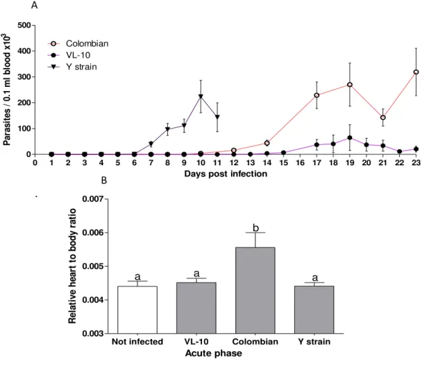
5.2.1 Parasitemia curve and relative weight of heart to body ratio ... 49
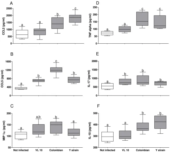
5.2.2 Profile of inflammatory mediators in plasma in C57BL/6 mice infected with different strains of parasite in acute phase ... 51
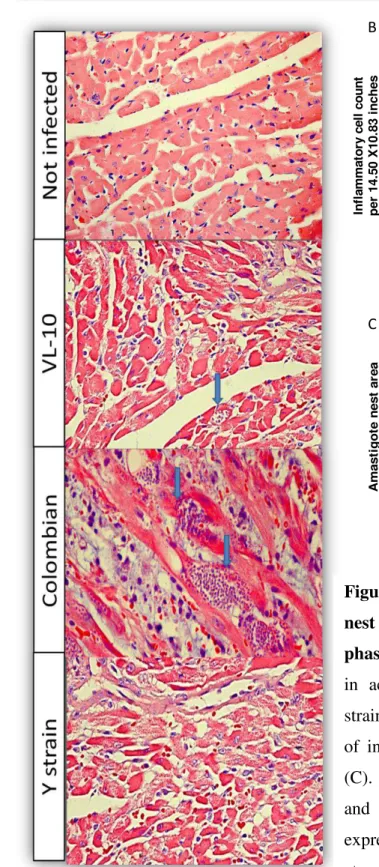
5.2.3 Inflammatory infiltrate and parasitism in cardiac tissue ... 53
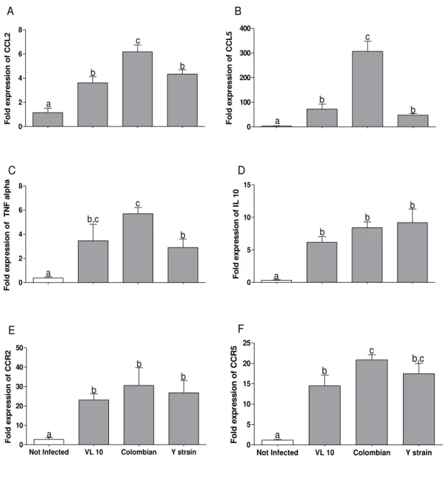
5.2.4 Expression of angiogenic mediators in cardiac tissue infected by VL-10, Colombian and Y strains of Trypanosoma cruzi. ... 56
5.2.5 Quantification of blood vessel by immunohistochemistry ... 57
5.3 Pharmacological effects of different drugs on inflammation and angiogenesis ... 59
5.4 Tabulated summary of results ... 66
6. DISCUSSION... 68
7. SUMMARY ... 77
8. CONCLUSION ... 80
9. PERSPECTIVES ... 82
10.REFERENCES ... 84
LIST OF FIGURES
xi LIST OF FIGURES
Figure I Diagrammatic representation of Typanosoma cruzi antigen activated
inflammation mediated angiogenesis in sponge model ... 24
Figure.II The model of our proposed idea ... 25
Figure III Hypothesis of angiogenesis modulation in experimental Trypanosoma cruzi
infection ... 73
Figure 1 Evaluation of inflammatory mediators in sponge implants obtained from
infected and uninfected C57BL/6 mice ... 43
Figure 2 High chemokine and cytokines production in plasma from infected C57BL/6
mice ... 44
Figure 3 Hemoglobin and VEGF content was reduced in sponge from Trypanosoma
cruzi infected C57BL/6 mice ... 45
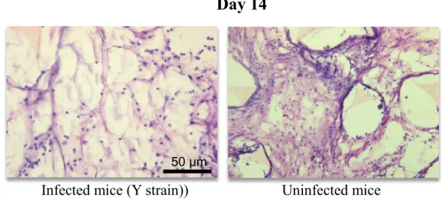
Figure 4 H & E staining of sponge sections of 14 days from infected (Y strain of
T. cruzi) and uninfected mice ... 46
Figure 5 Decreased vascularization in sponge implants in infected C57BL/6 mice
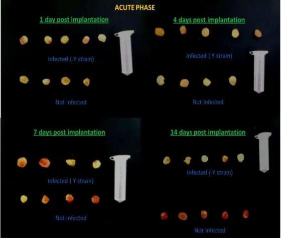
on day 14 ... 47
Figure 6 Parasitemia curve and relative weight of heart to body ratio from C57BL⁄6
mice infected with different strains of T. cruzi ... 49
Figure 7 Increased production of inflammatory mediators in Colombian strain
infected C57BL⁄6 mice in acute phase of infection ... 51
Figure 8 Vascular endothelial growth factor (VEGF) in plasma from C57BL⁄6
mice infected with different strain of Trypanosoma cruzi in acute
phase of infection ... 52
Figure 9 Relative expression of CCL2, CCL5, TNF-alpha, IL-10, CCR2 and
CCR5 in heart tissue from C7BL/6 mice in acute phase of infection ... 53
Figure 10 Inflammatory cell count and amastigote nest area in heart from C7BL/6
mice during acute phase of infection ... 54
Figure 11 Concentration and relative expression of vascular endothelial growth
factor (VEGF) mRNA in heart tissue from C7BL/6 mice in acute phase of infection ... 55
Figure 12 Relative mRNA expression of Angiopoeitin 1 and 2 and Thrombospondin
Figure 13 Quantification of blood vessel in heart section from infected and uninfected C57BL/6 mice in acute phase of infection ... 57
Figure 14 Parasitaemia curve and weight of heart to body ratio in C57BL/6 mice
under different pharmacological treatments ... 59
Figure15 Production of inflammatory mediators in plasma in C57BL/6 mice under
different pharmacological treatments ... 60
Figure 16 Concentration of inflammatory and regulatory cytokines in plasma in
C57BL/6 under different pharmacological treatments ... 61
Figure 17 Inflammatory cell count and amastigote nest area in heart from C57BL/6
mice under different pharmacological treatments ... 62
Figure 18 Concentration of vascular endothelial growth factor in plasma of C57BL/6
mice under different pharmacological treatments ... 63
Figure 19 Relative expression of vascular endothelial growth factor mRNA in heart
LIST OF TABLES
xiii LIST OF TABLES
Table 1: List of primers (sense and antisense). Primer sequences were determined
based on nucleotide obtained from GeneBank Database ... 38
Table 2: Inflammation and angiogenesis parameter using sponge model on14 day (Y
strain of Trypanosoma cruzi ) ... 65
Table 3: Inflammatory mediators and VEGF in plasma from mice with acute phase of
Trypanosoma cruzi infection ... 65
Table 4: Table 4: Inflammatory cell infiltration and angiogenic parameters in heart
from mice with acute phase of Trypanosoma cruzi infection ... 65
Table 5: Pharmacological effects on study of inflammation and angiogenesis in mice
LIST OF ABBREVIATION
ACE Angiotensin converting enzyme
ANG Angiopoeitin
ANOVA Analysis of variance
bFGF Basic fibroblast growth factor
CCR2 C-C chemokine receptor type 2
CCR5 C-C chemokine receptor type 5
CD4 Cluster of differentiation 4
CD8 Cluster of differentiation 8
cDNA Complementary deoxyribonucleic acid
CRT Calreticulin
Ct Threshold cycle
Cols Colaboradores
DAB Diaminobenzidine
dATP Deoxyadenosine triphosphate
DC Dendritic cell
dCTP Deoxycytosine triphosphate
dGTP Deoxyguanosine triphosphate
DNA Deoxyribonucleeic acid
dTTP Deoxythiosine triphosphate
DTU Discrete typing unit
EC Endothelial cells
EDTA Ethylenediaminetetraacetic acid
ELISA Enzyme Linked Immunosorbent Assay
Ephrs Ephrin receptors
GPI Glycosylphosphatidylinositol
H & E Hematoxyllin and Eosin
Hb Hemoglobin
HGFH Hepatocyte growth factor
HRP Horse Radishperoxide
HTAB Hexa-1,6-bis-decyltrimethylammonium bromide
IFN γ Interferon gamma
IgG Immunoglobulin
IL-1 Interleukin-1
IL-10 Interleukin-10
IL-12 Interleukin-12
IL-17 Interleukin-17
IL-4 Interleukin-4
M1 Classically activated macrophage
M2 Alternatively activated macrophage
MAPK Mitogen- activated protein of kinase
MCP-1 Monocyte chemotactic protein-1
MIP 1 Macrophage Inflammatory protein 1
MMP Matrix metalloproteinase
mRNA Messenger ribonucleic acid
LIST OF ABBREVIATIONS
xv
NF Nuclear factor
NK Natural killer cell
NOD Nucleotide-binding oligomerization domain
OD Optical Density
PAMPs Pathogen associated membrane proteins
PBS Phosphate buffer saline
PCR Polymerase chain reaction
PECAM Platelet endothelial cell adhesion molecule
PlGF Placental Growth Factor
PMN Polymorphonuclear leukocytes
qPCR Quantitative Polymerase Chain Reaction
RANTES Regulated and normal T cell expressed and
secreted
RNA Ribonucleic acid
TGF-β Transforming growth factor β
Th-1 Type I helper T cell
Th-17 T helper 17 cell
Th-2 Type II helper T cell
TIE Tyrosine kinase
TIMP Tissue inhibitors of metalloproteinase
TLR Toll like receptor
TMB Tetramethylbenzidine
TNF alpha Tumor necrosis factor alpha
TSP Thrombospondin
VEGF Vascular Endothelial Growth Factor
VEGFR Vascular Endothelial Growth Factor Receptor
INTRODUCTION
17 1. INTRODUCTION
1.1 Angiogenesis:
Angiogenesis, the formation of new capillary network from a preexisting vasculature, is a tightly regulated multistep process which maintains the balance between production and release of pro-angiogenic and anti-angiogenic factors (Sunderkotter et al., 1991). The process involves degradation of basement membrane, proliferation of endothelial cells, survival, and migration towards angiogenic stimulus, elongation and anastomosis
(Costa et al., 2007). Judah Folkman (1933-2008) first stated that tumor growth was directly
dependent on angiogenesis (Folkman, 1997). Angiogenesis occurs physiologically such as in embryo development, during wound healing and in several pathological conditions including atherosclerosis, proliferative retinopathies, tumor growth and chronic inflammation with abnormal proliferation of blood vessels (Carmeliet, 2000).
1.2 Process of angiogenesis:
Under normal conditions endothelial cells remain in the state of quiescence, however, when stimulated, the endothelial cells become active and initiate a cascade of events that culminate in the formation of new vessels (Laurent et al., 2007, Mariscalco et al., 2011).
There are several pro- and anti-angiogenic components and signaling pathways that have been described in angiogenesis mechanism. Vascular endothelial growth factor (VEGF) and its receptors (VEGFRs), basic fibroblast growth factor (bFGF) Angiopoietin-Tie,
Ephrin-EphRs, and Delta-Notch are major factors that play direct role in angiogenesis (Longatto et
al., 2010). VEGF-A, the best characterized and important angiogenic factor, stimulates proliferation and migration of endothelial cells. VEGF-B and VEGF-C also play a key role in
angiogenesis observed specially in extracellular matrix degraded area (Lohela et al., 2009).
Placental growth factor (PIGF), a VEGF homolog, stimulates angiogenesis in a variety of conditions in vivo and is a key molecule in regulating angiogenic switch in pathological
conditions (Luttun et al., 2002). Another signaling system, endothelium specific tyrosine kinase (Tie-2) receptor is a ligand for angiopoietins (Ang)-1 and -2, and presents an essential role in vessel growth, maturity, integrity, maintenance, and stabilization (Fagiani & Christofori, 2012). As a key regulator for vascular maintenance and stabilization, it plays critical role in tumor angiogenesis. Ang1 is constitutively expressed in many organs, whereas Ang-2 is predominantly expressed by activating endothelial cells at the site of vascular
remodeling (Bachet al., 2007; Galeet al., 2002; Huanget al., 2011). Ang-2 is pro angiogenic
molecule destabilizing the vessel to make it responsive to angiogenic growth factors such as VEGF (i.e., functions as a ‘trigger’ of remodeling) (Peterset al., 2004; Viscontiet al., 2002) whereas Ang-1 promotes vascular stabilization and counteracts VEGF-induced angiogenesis
(Fukuharaet al., 2009). Gene knockouts have shown that Ang-1 and Tie-2 play crucial roles
in late vascular development. Therefore, vascular maturation appears to be controlled by the precise balance between Ang-1 versus Ang-2 (Thurston & Daly, 2012).
Thrombospondins (TSP)-1 and -2 are matricellular endogenous proteins that inhibit angiogenesis impairing endothelial migration, proliferation, survival and induction of endothelial cell apoptosis and suppressing the activity and availability of VEGF (Armstrong & Bornstein, 2003). TSP-1 and -2 antagonize the endothelial cell migration which is important to the formation of sprouting capillaries in CD-36 dependent or independent
fashion (Dawsonet al., 1997; Yamauchiet al., 2007; Oganesianet al., 2008).
INTRODUCTION
19
angiogenesis and increase VEGF release (Page-McCaw et al., 2007; Rundhaug, 2005).
Opposing to MMPs, tissue inhibitors of metalloproteinases (TIMPs) abrogate angiogenic factor-induced endothelial cell proliferation and angiogenesis in MMP inhibition dependent
or independent manner (Seoet al., 2003). Therefore, together with these circulating proteins,
enzymes and receptors contribute to the equilibrium of angiogenesis process.
1.3 Inflammation and its association with angiogenesis:
Association between angiogenesis and inflammation can be evidenced by increased vascular permeability, monocyte/macrophage and neutrophil recruitment at angiogenic sites (Barcelos et al., 2004). Inflammation is the response of immune system triggered by the invading pathogens or damaged/injured tissue. It is a multifactorial process that regulates vascular responses, cellular (migration and activation of leucocytes) and systemic reactions aiming the homeostasis of the organism. The inflammatory response is eventually linked to the repair process which is accomplished by formation of new blood vessels. Therefore, angiogenesis is an essential process in the progression of many diseases aiding inflammatory response. Newly formed blood vessels are involved in the maintenance of the inflammatory state by transporting inflammatory cells to the site of inflammation and by supplying nutrients and oxygen to the inflamed tissue (Jackson et al., 1997).
The inflammatory reactions can be triggered by various stimuli, infections, trauma, physical and chemical agents, necrosis, foreign bodies and immune responses. Pathologists tend to classify acute and chronic inflammation in accordance with the morphological characteristics. The acute inflammatory process begins when cells sense the injury and release chemical mediators like histamines, proteases and cytokines promoting exudation of fluid, plasma proteins (edema) and migration of leukocytes, primarily the neutrophils (Rao et al., 2007). Chronic inflammation is associated with the infiltration of lymphocytes and macrophages, proliferation of blood vessels, tissue necrosis and fibrosis. Inflammation ends when the stimulus is removed and secreted mediators are destroyed or dispersed. There are also active anti-inflammatory mechanisms which modulate the response and prevent from tissue exacerbation. During the inflammatory process, chronic phase is usually observed with healing attempts by the replacement of damaged tissue by connective tissue leading to fibrosis of tissues (Wynn & Ramalingam, 2012).
slowly rolling along the vascular endothelium and then transiently adhere to the endothelium. When recruited by chemokines, the leukocytes increase the avidity of its links with endothelial adhesion molecules and then pass between adjacent endothelial cells, a phenomenon called transmigration or diapedesis. These leukocytes in the interstitium migrate toward to harmful stimuli by chemotaxis (McIntyre et al., 2003, Kumar et al., 2010). Polymorphonuclear neutrophils are the predominant cells exuded during the first 24 hours after the initiation of the inflammatory process, with short half- life (Charo & Ransohoff, 2006). Another type of inflammatory cells such as monocytes migrate into the inflammatory site by chemotaxis, where they differentiate into dendritic cells and macrophages. Monocytes start migrating from the vessels 18 and 24 hours after initiation of diapedesis, these cells accumulate and becomes predominant cells after 48 hours (Visser et al., 2006; Rao et al., 2007). The macrophages produce a variety of cytokines and growth factors responsible for a wide variety of responses in many cell types including endothelial cells, epithelial cells and mesenchymal cells. Macrophages release biologically active substances that result in tissue remodeling and recruitment of additional leukocytes such as B, CD4+ and CD8+ lymphocytes, antigen-specific, that will amplify the immune response (Visser et al., 2006). Among these biological substances, reactive oxygen and nitrogen species, proteases, cytokines, chemokines, coagulation factors and arachidonic acid metabolites plus growth factors such as platelet derived growth factor (PDGF), fibroblast growth factor (FGF) and transforming growth factor (TGF)-beta, fibrinogenic cytokines and angiogenesis factors can be responsible for formation of new vessels (Maruotti et al., 2013). The inflammatory response is regulated by a balance between pro and anti-inflammatory factors that coexist in the injured site (Trace, 2002). The imbalance of these factors results in increased production of proteases, proteoglycans, mediators, prostaglandins lipid and that concomitantly, enhance inflammatory process (Mrowietz & Boehncke, 2006). As the inflammation involves the migration and extravasation of immune cells through the microcirculation, the endothelium plays a fundamental role in this process and as well as in angiogenesis too.
INTRODUCTION
21
Interlukin-1 (IL-1). It has been shown to be a strong promoter of angiogenesis by modulating
endothelial cells (Naldini & Carraro, 2005). The importance of IL-1 signaling in the host has been demonstrated by the dramatic reduction of inflammatory and angiogenic responses in
matrigel plugs implanted in IL-1 receptor type I knockout mice (Carmiet al., 2009).
Interleukin-17 (IL-17): an inflammatory cytokine, expressed predominantly by Th17 cells,
induces angiogenesis, cell migration, and cell invasion (Duet al., 2012; Moranet al., 2011).
Tumor Necrosis Factor alpha (TNF-alpha): It also enhances the expression of VEGF
receptors and VEGF protein (Balkwill & Mantovani, 2001). Using subcutaneous implants of genetically deficient TNFR1, it was shown that there is decrease in angiogenesis (blood
vessel formation) and VEGF levels (Barceloset al., 2005).
Chemokines: Several studies have shown that the CXC chemokine carries important role in
angiogenesis associated with inflammation, repair and tumor (Belperioet al., 2000; Mooreet
al., 1998; Szekanecz et al., 1998). It has been shown that CXC chemokines that contain the
ELR motif (ELR +) are potent angiogenesis inducer in vivo. In contrast, the CXC without
ELR motif (ELR-) are potent angiostatic factors (Strieter et al., 1995). It has been
demonstrated that KC/CXCL1that binds to the CXCR receptor, monocyte chemotactic protein-1 (MCP-1)/CCL2, macrophage inflammatory protein (MIP)-1alpha /CCL3 and regulated on activation, normal T Cell Expressed and Secreted (RANTES)/CCL5 are
involved in formation of new vessels (Addisonet al., 2000; Stamatovicet al., 2006; Suffeeet
al., 2012).
IL-12 and IFN-gamma: Whereas pro-inflammatory and Th1 cytokines, such as IL-12 and
IFN-γ behave as negative regulators of angiogenesis through direct and indirect effects on endothelial, tumor and immune cells (Naldini & Carraro, 2005).
Not only cytokines and chemokines, depending upon the type of phenotypes, inflammatory cells and its products may contribute to the fate either as anti- or pro-
angiogenenic. For example, alternatively activated macrophages (M2),
polymorphonuclear neutrophils phenotype (PMN) 2, decidual-like nature killer (NK) cell, alternatively activated dendritic cells (DC) possess angiogenesis properties, whereas classically activated macrophages (M1), PMN1 phenotype of neutrophil, activated NK cells
Although inflammation is an important defense mechanism present in the host system during infection, prolonged inflammation can result into adverse condition as described in various diseases. Inflammation induced angiogenesis, well referred as double edge sword, may either further exaggerate tissue damage due to excess recruitment of inflammatory leucocytes or may ameliorate the affected sites by providing necessary oxygen as well as nutrients along with the inflammatory cells to combat the injury leading to wound healing. During infection, angiogenesis can therefore be essential host mechanism to improve the disease conditions or may also be exploited by the parasites to guarantee their survival in the host (Ribatti et al., 2008).
1.4 Trypanosoma cruzi infection in human and experimental model:
Trypanosoma cruzi infection is responsible for the acute and chronic inflammation
driving cardiomyocytes towards a progressive damage with consequent fibrosis and loss of functionality. Approximately 7-8 million of people in the world are infected with this parasite (WHO, 2013). T. cruzi is transmitted to humans through the bite wound of its triatomine
insect host, the reduviid bug. Once within the body, T. cruzi is able to enter a variety of host
cells where it differentiates into mammalian proliferative forms, the amastigotes. Within the cell, amastigotes divide and then differentiate into blood forms trypomastigotes. The parasitized cell ruptures, releasing trypomastigotes, which may infect adjacent cells or be disseminated through the blood and infect cells at other locations (de Souza et al., 2010)
Clinical symptoms of human and experimental T. cruzi infection in animal model
consist of both acute and chronic phases. In the acute phase of the human infection, the signs and symptoms are either absent or mild and nonspecific. For these reasons, on the basis of
clinical symptoms, most individuals are not diagnosed with T. cruzi infection until they are in
the chronic phase of the disease. In human chagas disease, heart may be inflamed and exhibits mild enlargement during acute infection (Moncayo & Ortiz Yanine, 2006). Initially, cardiac inflammation is focal and lesions coalesce over time, followed by congestion, edema, and infiltration of the heart by mononuclear cells, mast cells and neutrophils are observed. Additionally, apart from cardiomyopathy, the disease may affect several other systems like digestive and neurologic (Py, 2011). It has been shown that parasitism occurs mostly in muscle fibers but may also be present in macrophages and ganglionic Schwann cells.
Non-parasitized muscle fibers can also show distinct lesions (Wong et al., 1992). There are two
INTRODUCTION
23 individuals, or entry into the chronic phase of infection. When present, the acute myocarditis
resolves after several weeks and the heart regains its functions (Mayaet al., 2010)
Over the next 10 to 30 years, only focal myocarditis of mild degree is observed in the majority of cases and most individuals remain in this state for all life. However, thirty percent of infected people develop a chronic, progressive, fibrosing cardiomyopathy of variable degree. The infiltrate in chronic Chagas heart disease consists of lymphocytes (T cells predominate and CD8+ lymphocytes are two to three times more abundant than are CD4+ cells) and to a lesser extent, macrophages, eosinophils, plasma cells, neutrophils and mast cells (Brener & Gazzinelli, 1997; dos Santos et al., 2001). The most part of this leukocyte recruitment into the heart tissue is driven by chemokine and chemokine receptors
and, their over expression is clearly associated with heart dysfunction (Talvani et al., 2000;
Talvani et al., 2004a; Talvani et al., 2004b). The perpetuation of parasites in leukocyte is rarely found in the heart but parasite DNA can be detected in some inflammatory lesions
(Tarleton et al., 1997). Inflammatory cell infiltrates can lead to destruction of myocardium,
epicardium and endocardium which is followed by replacement with connective tissue (fibrosis) and therefore responsible for decrease in heart contractility, reduced cardiac muscle
mass, and loss of cardiac innervations (Dutraet al., 2005; Machadoet al., 2005)
Trypanosoma cruzi possesses very fascinating mechanisms for its survival in the
host cell and tissue. In T. cruzi infection, host immune system maintains to achieve balance
between inflammatory and anti-inflammatory response In the acute phase, there is a huge inflammatory response to clear the infection whereas in the chronic phase, it is rather controlled in such a way that immune response is targeted to maintain the parasite load and inflammatory response low so as to keep infection under control and to prevent excessive
tissue damage (Garciaet al., 2010; Maya et al., 2010). Absence or decrease in inflammatory
response may lead to parasite multiplication and subsequently to the death of animals, in contrast to this, enormous inflammation may lead to tissue damage and again leading to death of the animals (Aliberti et al., 2001; Golgher & Gazzinelli, 2004; Talvani & Teixeira, 2011; Teixeiraet al., 2002).
producing other cytokines (TNF-alpha, IL-12) and chemokines (CCL2/MCP1, RANTES/CCL5). These cytokines, chemokines and their receptors have a determinant role in
the course of the inflammatory response during T. cruzi infection because the action of these
mediators leads to leukocyte recruitment to infected tissues (Paiva et al., 2009). IL-12 stimulates the Natural Killer (NK) cells to produce more IFN-gamma and recruit lymphocytes, leading indirectly to produce more TNF-alpha, IL-12, chemokines and free radicals, a positive feedback system (Talvani & Teixeira 2011). This amplifying system can employ excessive amount of inflammatory cells and can be harmful for the host tissue. Hence, the increase of inflammatory markers is regulated with the production of cytokines such as IL -10 and IL-4 (Brenner & Gazzinelli, 1997, Aliberti et al., 2001; De Oliveira, 2007; Talvani et al., 2009).
It is known that membrane proteins of T. cruzi activate Toll like receptors
(TLR)-1,2, 4 , 5, 6 or 9 present in macrophages and dendritic cells which in turn trigger the activation of nuclear factor kB (NF-kB ) and mitogen- activated protein of kinase (MAPK) that lead to the synthesis of pro-inflammatory cytokines and chemokines (Campos and Gazzinelli, 2004; Bafica et al., 2006; Koga et al., 2006; McGettrinck & O'Neill, 2010; Nagajyothi et al.; 2012; Higashikuni et al., 2013) . Furthermore, other receptors, such as mannose receptors and nucleotide-binding oligomerization domain (NOD)-like receptor family and as retinoic acid-inducible gene I have also recently been shown to induce activation of inflammatory mediators. (Garrido et al., 2011, Silva et al., 2010). Different parasite proteins and glycoproteins like glycosylphosphatidylinositol (GPI), GPI-mucin, GPI
transialidase, TC52, cruzipain and nucleic acid of T. cruzi bind with these receptors present
on cells of the monocytic lineage and participate to activate these cells to produce nitric oxide (NO) and secrete inflammatory mediators (Fernandez-Gomez et al., 1998; Saavedra et al.,
1999; Gazzinelli et al., 1999; Coelho et al., 2002, Gao & Pereira, 2001; Giordanengo et al.,
2002; Shoda et al., 2001 ).
Hence, inflammation and angiogenesis are pivotal processes and interrelated in the progression of many diseases under inflammatory conditions, studying angiogenesis process in T cruzi infection can be a new and promising area of investigation. Because of prolong
inflammation, there is impairment of cardiomyocytes leading to aggressive and progressive damage in the heart and is one of the main reasons for congestive heart failure in Chagas disease (Hidron et al., 2010). Recently, our group has found that T. cruzi antigens are
INTRODUCTION
25
suggesting T. cruzi might induce inflammatory mediators leading to angiogenesis. The result
of this experiment is represented by the following diagram:
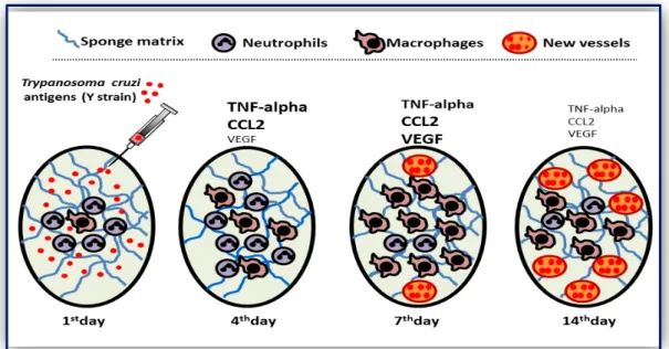
Figure I: Diagrammatic representation of Typanosoma cruzi antigen activated
inflammation mediated angiogenesis in sponge model. After injecting T. cruzi antigen in
sponge implant, there was increase in inflammatory cytokines (CCL2 and TNF-alpha) on day 4 and increase in VEGF as well as macrophage recruitment on day 7 consequently increasing the number of new blood vessels day 14. Increase in size of word denotes the increase production of soluble mediators and vice-versa.
However, studies have shown that calcium-binding proteins (Calreticulin - CRT) from T. cruzi can exert anti-angiogenesis and anti-tumor activity in the host and protect
against ongoing neoplasic damage (Ferreira et al., 2004; Ferreiraet al., 2005; Ramirez et al.,
2011; Ramirez et al., 2012). Lopez and cols showed in ex-vivo angiogenesis assay, T. cruzi
CRT completely abrogated capillary growth (Lopez et al., 2010). It has also been described that an augment of Thrombospondin-1 protein by calreticulin enhances cellular infection (Johnson et al., 2012). But, several infections have been reported to induce angiogenesis,
known as infection induced angiogenesis through the regulation of TLRs (Grote et al.,
2011).Interestingly, recent findings have shown link between Toll Like Receptor (TLR)
stimulation and angiogenesis during inflammation (Cho et al., 2007; Grote et al., 2010;
Jagavelu et al., 2010; Pollet et al., 2003; Varoga et al., 2006) and previous studies have
demonstrated the role of several membrane proteins and glycoproteins such as GPI of T. cruzi
bring about inflammation via TLR (Baficaet al., 2006; Campos & Gazzinelli, 2004; Kogaet
al., 2006; McGettrick & O'Neill, 2010). Similarly, different inflammatory chemokines and
cytokines such as TNF-alpha, IFN-γ, MCP-1/CCL2, RANTES/CCL5, MIP-1α/CCL3, IL-10,
cruzi infection, are involved in immunopathology of Chagas disease (Paiva et al., 2009;
Araujo-Jorgeet al., 2008; Silvaet al., 1991; Talvaniet al., 2004b; Talvani & Teixeira, 2011).
Therefore, it is plausible that there is presence of angiogenesis may be present during T. cruzi
infection. Angiogenesis is further related with the MMPs (that stimulates angiogenesis) and
increases VEGF release (Rundhaug, 2005; Zheng et al., 2006). It has been evidenced in T.
cruzi infection, there is increased activities of cardiac matrix MMP-2 and MMP-9 (Gutierrez
et al., 2008) that can reinforce angiogenesis in T. cruzi infection. Therefore, with all these
studies and evidences, we hypothesize the possible role of T. cruzi in angiogenesis which is
represented by following figure:
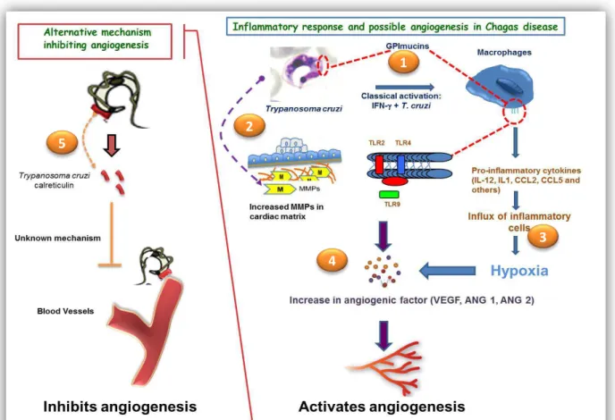
Figure II: The model of our proposed idea
1. Different antigens (PAMPs) of T. cruzi will be recognized by different inflammatory
cells, for instance macrophages, through its TLRs releasing various proinflammatory chemokines and cytokines.
2. Parasites can also lead to production/release of MMPs in cardiac matrix.
3. Inflammatory mediators recruit more inflammatory cells at site of injury and cause hypoxic condition.
1
2
3
INTRODUCTION
27 4. All these different factors can induce direct or indirect production of angiogenic
proteins which can activate angiogenesis process, formation of new vessels.
5. On other hand, calreticulin can inhibit angiogenesis but the process of inhibition for
the formation of new vessels is still unknown.
1.5 Pharmacologies:
Chemotherapies to treat T. cruzi infection are still a challenge even though various
drugs have been discovered and are in use. Current standard chemotherapies are unsatisfactory because of their side effects and are inefficient to cure in chronic phase. Because of persistent parasite, in T. cruzi infection, inflammation is a characteristic feature
which is responsible for the destruction of tissue and leading to the loss of tissue function. Therefore it is very important to develop chemotherapy strategies that not only control parasite but also inflammation so that inflammation related tissue remodeling (eg. angiogenesis) can be ignored. Another strategy that can help T. cruzi infected patient is to use
drugs that improve functionality of the organ in combination with standard anti- T. cruzi
drugs.
(i) Benznidazole, standard drug, is considered beneficial in controlling parasite load and
disease progression. But during chronic Chagas cardiopathy in the symptomatic individuals, Benznidazole has nominal effect in heart and patient. Hence, treatment with Benznidazole along with combination with other anti-trypanosomal drugs and/or drugs used in cardiovascular diseases might be effective to improve heart condition in chronic symptomatic patients (Coura & Borges-Pereira, 2011). Our laboratory has observed that pharmacological
therapies such as Enalapril and Simvastatin have benefited hearts in animals infected with T.
cruzi (de Paulaet al., 2010 Meloet al., 201; Silva et al., 2012).
(ii) Simvastatins, inhibitors of 3-hydroxy-3-methyl-glutaryl-coenzyme A reductase, are
statins that reduce lipid and present cardiovascular protective properties (Kapur & Musunuru, 2008). Simvastatin has pleiotropic properties with anti-inflammatory effects and can
stimulate angiogenesis (Zhu et al., 2008). It has been demonstrated that Simvastatin therapy
has ameliorated cardiac remodeling in animals infected with T. cruzi (Meloet al., 2011; Silva
et al., 2012)
(iii) Enalapril, an inhibitor of angiotensin converting enzyme (ACE), is generally used in the
treatment of cardiovascular diseases (e.g. heart failure and coronary artery diseases) (Bowling
can have anti-T. cruzi and anti-inflammatory activities reducing inflammatory cells during
acute phase of experimental T. cruzi infection in mice model (de Paulaet al., 2010).
It is generally accepted, that inflammatory angiogenesis is ongoing process during
development of Chagas disease. In experimental cardiomyopathy induced by T. cruzi
parasites, little is known about the expression and the role of many different pro- and
anti-angiogenesis stimuli. Understanding anti-angiogenesis and their mediators in experimental T.
cruzi may help to better understand host parasite interaction and as well as focusing on
30 2. JUSTIFICATION
Angiogenesis and inflammation are always cross-linked as both processes are
well interconnected. T. cruzi infection is characterized by intense inflammation in both acute
and chronic stages of the infection. Therefore it is likely possible that persistence of inflammation in infected individuals may promote angiogenesis. New blood vessel formation helps functioning of tissues and organs. It can, however, also augment inflammation, as increased formation of new blood vessels may further induce recruitment of inflammatory cells which may follow with increased production of cytokines and chemokines. Most of the fatalities of the disease seen in T. cruzi infection are mainly by organ failure, especially
cardiac malfunctioning. Likewise in experimental model for example, in mice, the severity and/or mortality of the disease is extremely high if not treated. The death is usually caused by high inflammatory response and high parasitemia.
Recently, there are few evidence highlighted in the literature that calreticulin may exhibit angiogenic and tumor effects. But the mechanism of this protein for
anti-angiogenesis is still not known in T. cruzi infection although there are few papers that have
shown increased thrombospondin 1, an anti-angiogenic factor, by this protein. Based on our previous studies using sponge model, there is an indication of increase in angiogenesis in the sponge implanted with total antigen of T. cruzi (Y strain) in mice.
It is yet not clear whether angiogenesis is friend or foe in T. cruzi infection. The
process may be one of the mechanisms that parasite may induce in the host in order to sustain within it or on the other hand, a possible host response against infection whilst it may improve cardiac functioning in infected host. Angiogenesis is ongoing process and plays significant role in wound healing and chronic inflammation; our study on an inflammation induced angiogenesis in T. cruzi infection would be of great value with regards to the
understanding pathophysiological characteristics of the disease condition. There are very few
information available on whether T.cruzi induces or inhibits angiogenesis in vivo. Therefore,
our study will be helpful to evaluate unique properties of different T. cruzi strains to evolve T.
cruzi infection associated with inflammation and angiogenesis. Furthermore, study of these
intertwined processes will help to improve understanding of mechanisms related to
3. OBJECTIVES
3.1General objective:
To determine inflammation and angiogenesis parameters in Trypanosoma cruzi infection
locally and systemically using three different strains of the parasite and analyze possible interference of pharmacological drugs in inflammation and angiogenic mediators.
3.2 Specific objectives:
(i) Evaluate inflammatory angiogenesis in a polyester-polyurethane sponge discs in
T. cruzi (Y strain) infected mice.
(ii) Quantify inflammatory mediators and VEGF in plasma from C57BL/6 mice
infected with Y, VL-10 and Colombian strain of T. cruzi in acute phase of
infection
(iii) Determine inflammatory cell infiltration and area of amastigote nest in heart from
C57Bl/6 mice infected with Y, VL-10 and Colombian strain of T. cruzi in acute
phase of infection.
(iv) Determine mRNA levels of pro- and anti-angiogenic factors by real time PCR in
heart from C57BL/6 mice infected with Y, VL-10 and Colombian strain of T.
cruzi in acute of infection.
(v) Monitor blood vessels using anti-CD31 antibody by immunohistochemistry in
heart during acute experimental T. cruzi infection.
(vi) Analyse the interference of anti-T. cruzi therapy (Benznidazole) and heart disease
34 4. MATERIALS AND METHODS
4.1 Strains of Trypanosoma cruzi
VL-10 strain is considered as a resistant to Benznidazole (Caldas et al., 2008; Filardi &
Brener, 1987) and according to the new nomenclature (Zingaleset al., 2009) it is classified as
discrete typing unit II (DTU II) (Morenoet al., 2010)
Y strain is considered partially sensitive to Benznidazole and was classified as T. cruzi II
(Zingaleset al., 2009)
Colombian strain, resistant to Benznidazole, was isolated from Colombia and was typed as
Biodeme Type III and recently classified T. cruzi I, as referred by Momem (Camandarobaet
al., 2001; Zingaleset al., 2009)
All these strains were maintained by successive passages in Swiss mice in the Animal Care Facility of Universidade Federal de Ouro Preto (UFOP), Ouro Preto, Minas Gerais, Brazil.
4.2 Animals
C57BL/6 male mice of age 8-10 weeks were used for the purpose of our study. Mice were housed and maintained at the animal central facility in the (UFOP). All animal experiments and procedures were approved by the Institution’s Committee on the ethical handling of laboratory animals (CEUA Protocol No 043/2010).
4.3 Trypanosoma cruzi infection and drug administration in experimental models
In the first step in order to show the process of inflammatory angiogenesis after infection, polyester polyurethane sponge discs were implanted subcutaneously on neck pouch of animals (n=5) followed by intraperitoneal infection with Y strain (100 trypomastigotes) of
T. cruzi. In second step, animals (n=10) were infected with 100 trypomastigotes forms of T.
cruzi using following strains: (i) Y (ii) VL-10 and (iii) Colombian. In final stage, to verify
MATERIALS AND METHODS
35 Parasitemia was determined daily by analysing tail bleed samples (5 μl) on the optic microscopy (Brener., 1962) and mortality was evaluated during the experiment. Animals were euthanized in acute phase (12 days of infection for Y strain and 24 days of infection for Colombian and VL-10 strain). For sponge implanted groups, animals were euthanized on day 1, day 4, day 7 and day 14 according to the protocol described by (Andrade et al., 1987). Blood was collected for immune assay. Half part of hearts were fixed with 10% formalin to perform immunohistochemistry and histology and another half part of hearts were designated to study immunological, morphological and angiogenic parameters. Sponges were collected for biochemical, immunological and histological analysis.
4.4 Heart mass measurement
Ex vivo hearts were carefully excised and gently blotted on absorbent paper to remove
blood before wet weight measurement. The relative heart weight was calculated using heart weight / mouse body weight and used to evaluate cardiac mass measurement at the time of
euthanasia with each group infected with different strains of T. cruzi (n=10/group).
4.5 Enzyme-linked immunosorbent assay (ELISA)
Plasma from all experimental groups of animals and supernatants from polyester polyurethane sponge disc (described latter) was collected and maintained at -80oC. In parallel, fragment of 10mg of cardiac tissue from each animal was homogenized in cold
RIPA buffer with protease inhibitor and supernatant was collected and also stored at -80oC.
Later, these biological samples were defrosted and were used to measure soluble biological
markers (TNF-alpha, CCL2, CCL5, VEGF, IL-10, IL-17 and MIP-1α). Sandwich ELISA was
performed following manual from the manufacturer (Peprotech and R&D systems). 96 well plates were first coated adding 100ul of monoclonal antibodies anti TNF-alpha (Peprotech), anti CCL2 ( R & D Systems), anti CCL5 (Peprotech), anti VEGF (Peprotech), anti CCL3 (Peprotech), anti IL-10 (Peprotech) and anti IL-17 (Peprotech) and were incubated overnight at room temperature. Then, plates were washed four times with wash buffer (PBS and 0.05% TWEEN) followed by blocking step to avoid nonspecific binding. Blocking step was performed by adding block buffer (PBS and 1% BSA; pH 7.4) and incubated at room temperature for 1 hour. It was then washed once again as before. Standards (100μl/well) in mentioned dilutions and samples were added (25 μL/well), followed by incubation at room
36 biotinylated detection antibody diluted in reagent diluent (PBS, 1% BSA and 0.05% TWEEN for R&D system, PBS, 0.1% BSA and 0.05% TWEEN for Peprotech; pH 7.4) was added and incubated for 2 hours at room temperature. Plates were then washed once again. For the R & D system, streptavidin-horseradish peroxidase in 1:200 dilution was added (100 μL/well), and the plates were incubated for 20 min at room temperature. Plates were again washed and then 100 μL/well of the substrate solution (1:1 mixtue of hydrogen peroxide and tetramethylbenzidine- Sigma Chemical Co.) was added, and plates were incubated in dark for 30 min at room temperature. Reaction was terminated adding 50 μL/well 1 M H2SO4 solution. Plates were read at 450nm in ELISA plate reader (SoftMaxPR040). For Peprotech, after detection antibody step, avidin horseradish peroxidase in1:2000 dilution was added (100μl/ml) and plate was incubated for 30 min. Plates were washed and then 100μl of the substrate solution (ABTS liquid substrate Solution, Sigma Cat#A3219) was added. Plates were incubated at room temperature for color development and the color development was read at 405nm wavelength in ELISA plate reader. The plates were monitored at 5 min intervals for approximately 30 min and then, reading at 20 min were taken for our study.
4.6 Histopathology analysis
To analyze and quantify inflammatory infiltrates and amastigote nest areas, fragments of cardiac tissue were stained by hematoxylin and eosin (HE) staining. Hearts and polyester polyurethane sponge discs were fixed for 24 hours in 10% buffered formalin (conventional histology). Tissues were embedded in paraffin/paraplast, sectioned, stained with Hematoxylin-Eosin staining and examined by light microscopy. Two sections were taken from each heart including both atria and ventricle. Each section was examined for evidence
of mononuclear and polynuclear cellular infiltration and T. cruzi nests. Nucleus of each cell
in the section was quantified from 20 images whereas amastigote nest area was quantified from 35 images taken at an objective lens of 40X by Microcamera Leica DM 5000 B (Leica Application Suite, version 2.4.0R1). Quantification of infiltration was analyzed by using Leica Qwin V3 program and amastigote nest area was quantified by Image J 1.45s, NIH, USA (www.imagej.nih.gov/ij)
MATERIALS AND METHODS
37 4.7.1 Extraction of RNA
Extraction of RNA was done according to instructions provided by RNA-SV total RNA isolation system kit-promega. Fragment of heart approximately 40 mg from each of infected and control mice (studied under different conditions) was macerated and homogenized in 175μl of RNA lysis buffer. This lysate was transferred into new tube and 350μl of RNA dilution buffer was added. This mixture was homogenized, incubated at 70oC for 3 min and then centrifuged for 10 min at 13,000×g for selective precipitation of cell
proteins. Supernatant that contained RNA was transferred carefully into a new tube. 200μl of
95% ethanol was added to precipitate RNA. This solution was transferred into a spin column assembly and centrifuged at 12,000 x g for 1 min. In this step, RNA will be attached on silica membrane layer on the spin basket. The liquid obtained in the collection tube was discarded and RNA was washed using 600μl of RNA wash solution. After centrifugation, 50ul of DNase mix (40μl of yellow core buffer, 5 μl of 0.09M MnCl2and 5 μl of DNAse I enzyme) was added in column to digest contaminated genomic DNA. After incubation of 15 min at
20-25oC, reaction was stopped using 200μl of DNA stop solution and centrifuged at 14,000 x
g for 1 min. It was then followed by washing process using RNA wash solution to purify RNA, eliminating contaminants, proteins and cellular impurities. Finally, total RNA was eluted through membrane with the addition of nuclease free water.
4.7.2 Quantification of RNA-determination of yield and quality
Quantification of RNA was performed in NanoVue Plustm (GE Healthcare). Pure RNA will exhibit an A260/A280 ratio of 2.0. However, it should be noted that, due to the variations between individual starting materials and in performing the procedure, the
expected range of A260/A280 ratios for RNA will be 1.7–2.1 and for A260/A230 ratio will
be 1.8–2.2.
4.7.3 Reverse transcriptase
Reverse transcription was done according to the instruction of GeneAmp RNA PCR
kit- Applied Biosystems. Mastermix was prepared containing 2μl of 10X RT buffer, 0.8μl of
25X deoxynucleotides mix (dATP, dGTP, dCTP and dTTP), 1 μl of RNase inhibitor, 1μl of
multiScribe reverse transcriptase, 2μl of 10X random primers, sample in concentration of 0.035μg/ μl and DEPC water in sufficient quantity to make the final volume 20μl. Then, the
38
min, 85 C 5 min and rest at 4 C. After this process cDNA was ready for PCR and maintained
at -80oC.
4.7.4 Amplification
Relative quantitative gene expression for Real time PCR was done in ABI 7300 (applied Biosystem), utilizing SYBER® Green Master mix for detection of amplicons. Messenger RNA expression of CCL2, CCL5 and their receptors CCR2, CCR5, TNF-alpha, IL -10, VEGF, ANG-1, ANG-2, TSP-1 and TSP-2 was analyzed by quantitative real time PCR using the primers as shown in table 1. For each reaction, mix containing 5μl of SYBER®Green Master Mix, 0.7μl of each sense and anti-sense primer (10pmol/μl), 2μl of cDNA and ultra-pure water in sufficient quantity to make a volume of 10 μl were used.
Conditions for thermocycler was as following: 50oC for 20 min, 95oC for 10 min, 40 cycles
of 94oC for 30 sec, 59.6oC for 30 sec and 72oC for 1 min. PCR amplification was performed
in duplicate wells using standardized conditions. Samples containing the highest amount of each target will be used to create standard curves of the transcribed cDNA. In the same run, the threshold cycle (Ct) values of the samples (20-fold diluted cDNA) will be measured and relative expression levels were determined. PCR amplifications will be performed in duplicate wells using standardized conditions. Ct values obtained from duplicate were used to calculate the expression of target gene after normalizing by reference gene (β actin gene). Then, was verified difference of Ct observed between test samples and Ct obtained from the amplification of pool of cDNA from mice which were not infected and same pool was used in all experiments. Then it was checked how many times the test samples expressed mRNA expression in relation to that normally present in uninfected mice using formula 2-∆∆Ct.
MATERIALS AND METHODS
39
Primer Primer sequences
Angiopoeitin 1 5’ GGGGGAGGTTGGACAGTAA 3’ 5’ CATCAGCTCAATCCTCAGC 3’
Angiopoeitin 2 5’ GATCTTCCTCCAGCCCCTAC 3’ 5’ TTTGTGCTGCTGTCTGGTTC 3’
Vascular Endothelial
Growth factor A 5’ AAAAACGAAAGCGCAAGAAA 3’ 5’ TTTCTCCGCTCTGAACAAGG 3’
Thrombospondin 1 5’ GAAGCAACAAGTGGTGTCAGT 3’ 5’ ACAGTCTATGTAGAGTTGAGCCC 3’
Thrombospondin 2 5’ CCTCAACTACTGGGTAGAAGGC 3’ 5’ TGACACTGTCGATAAGATCGCA 3’
CCL2 5’ AACTGCATCTGGCTGAGC 3’ 5’ CAGCACCAGCCAACTCTC 3’
TNF alpha 5’ TGAGTGACCAAGGGACAGAACC 3’ 5’ AGCCAGGAGGGAGAACAG 3’
CCL5 5’ ACCCTCTATCCTAGCTCATC 3’ 5’ CGTGTTTGTCACTCGAAG 3’
CCR2 5’ CCTGTCCACTAATGCGTTTC 3’ 5’ GCAAAGCCAGACCACAATG 3’
CCR5 5’ CCCTGTCATCTATGCCTTTG 3’ 5’ GCTTGCACGATCAGGATTG 3’
IL-10 5’ ACTACCAAAGCCACAAGG 3’ 5’ AAGAGCAGGCAGCATAG 3’
4.8 Preparation of sponge disc and implantation
Polyester-polyurethane sponge discs 5mm thick and 8mm diameter (Vitafoam Ltd, Manchester, UK) were used as the matrix for fibrovascular tissue growth. The sponge discs were soaked overnight in 70% v/v ethanol and sterilized by boiling in distilled water for 15 min before the implantation surgery as previously described (Andrade et al., 1987; Barcelos et al., 2004). Animals were anesthetized injecting intraperitoneally with Xilazine and Ketamine (8mg/kg and 60mg/kg respectively). Dorsal hair of mice was then shaved and skin was wiped with 70% ethanol. Sponge discs were aseptically implanted into a subcutaneous
neck pouch. Animals were then infected with Y strain (100 parasites) of T. cruzi. Animals not
infected were used as control groups.
4.9 Quantification of angiogenesis by hemoglobin measurement
40 13000rpm for 40 min. The supernatant was filtered through a 0.22mm Millipore filter. Hemoglobin concentration of samples was determined by measuring absorbance at 540nm using an ELISA plate reader and was compared against a standard curve of hemoglobin (Barceloset al., 2004).
4.10 Immunohistochemistry analysis
Consecutive 4μm sections were cut and mounted on gelatinized slides. Tissue
sections were deparaffinized and rehydrated in graded ethanol. The CD-31 antigens were retrieved using solution retrieval (DAKO ) at pH 6.0 and heated for 20 minutes in water bath at 98°C. The slides were immersed in 3% hydrogen peroxide for 15 minutes to ensure endogenous peroxidase blocking. Then Slides were incubated in 2% bovine serum albumin (Sigma-Aldrich) for 30 mins at room temperature to reduce non-specific protein binding. The histological sections were incubated in a humid chamber for 1 hour at room temperature with a primary rat monoclonal IgG2a antibody for PECAM 1 (Santa cruz Biotechnology) in 1:50 dilution. Antigen amplification was done with the Advance HRP link (DAKO) for 30 minutes at room temperature. For all slides, 3, 3′-diaminobenzidine (DAKO) was used as a chromogen. Sections were counterstained with Mayer's hematoxylin, dehydrated, and mounted. Negative controls were obtained by omitting the primary antibody.Numbers of blood vessels were counted from 10 photos taken from each heart. A grid with 100 intersections was used and blood vessels at the intersection were counted using program Corel DRAW version 11.633 windows.
4.11 Statistics
MATERIALS AND METHODS
41 4.12 Experimental Design
20
Experiment 1: Sponge implantation
experiment
1 4
DAY
Sponge implantation &
Infection (T. cruzi, Y strain)
0 14
ANIMALS EUTHANIZED on 1, 4, 7 and 14 day Sponge, Heart & Blood collected
7
Experiment 3: Pharmacological therapies intervention
experiment
Heart, Blood collected Infection with
T. cruzi, Colombian strain DAY
Treatment with Simvastatin,Enalapril and Beznidazole
Animals euthanized
0 1 24
1. Immunoaasay 4. qPCR
2. Biochemical analysis 5. Immunohistochemistry
3. Morphometric analysis
Post euthanasia techniques
Experiment 2: Different strains
of T. cruzi
infection
experiment
DAY
Heart, Blood collected Y strain infected
animals euthanized
VL 10 & Colombian infected animals euthanized
Infection with T.cruzi
Y, VL-10, colombian strain
24 12
RESULTS
43 5. RESULTS
Previously, our group has experiment on sponge implant using antigen from
Trypanosoma cruzi (Y strain) in Swiss mice. We found that sponge implant of 14 days from
mice which were injected with the antigen, demonstrated increased number of blood vessels than sponge with the PBS as described in figure I (da Silva et al unpublished). Therefore, our first step was to use live parasite and observe the response in sponge implant. Sponges were
implanted subcutaneously in C57BL/6 mice and 100 trypomastigote forms of T. cruzi (Y
strain) were inoculated in peritoneum. Inflammatory and angiogenic parameters were assessed on day 1, 4, 7 and 14 after sponge implantation in infected and control groups. In the second step, we focused our study in heart in C57BL/6 mice infected with different strains
(Colombian, Y and VL-10 strains) of T. cruzi to study inflammation and angiogenesis in the
acute phase of infection. In the final step, we used different pharmacological drugs (Simvastatin, Enalapril and Benznidazole) to study the interferences of these drugs in
inflammation and angiogenesis in mice infected with Colombian strain of T. cruzi.
5.1. Study of inflammation and angiogenic parameters in sponge implanted animals
5.1.1 Evaluation of inflammatory chemokine and cytokine levels in sponge supernatant and plasma of infected and uninfected mice:
C57BL/6 mice were implanted with sponge and then infected with Y strain of T.
cruzi. The implants were well tolerated by all animals. No signs of infection at the implant
44
1 4 7 14
100 120 140 160
Not infected Infected (Y strain)
Days after sponge implantation and infection
IL -1 0 ( p g /m l) s p o n g e s u p e rn a ta n t
1 4 7 14
0 100 200 300 *** * C C L 5 ( p g /m l) s p o n g e s u p e rn a ta n t
Figure 1. Evaluation of inflammatory mediators in sponge implants obtained from infected and uninfected C57BL/6 mice. Implanted sponges were excised from C57BL/6 mice on day 1, 4, 7 and 14. CCL2, (A), CCL5 (B), TNF-alpha (C) and IL-10 (D) were measured in sponge supernatant by ELISA. Data are shown as a mean of 5 animals. *,** &*** denote that difference in the production is significant at P<0.05.
B
D A
C
1 4 7 14
100 120 140 160 180 200 C C L 2 ( p g /m l) s p o n g e s u p e rn a ta n t
1 4 7 14
400 450 500 550 600 650 **
Days after sponge implantation and infection