UNIVERSIDADE FEDERAL DE OURO PRETO
NÚCLEO DE PESQUISA EM CIÊNCIAS BIOLÓGICAS
PROGRAMA DE PÓS-GRADUAÇÃO EM CIÊNCIAS BIOLÓGICAS
ADENOSINE PRODUCTION VIA CD39/CD73
PATHWAY PROMOTES
Leishmania amazonensis
SURVIVAL IN MACROPHAGES
BIJAY BAJRACHARYA
OURO PRETO
UNIVERSIDADE FEDERAL DE OURO PRETO
NÚCLEO DE PESQUISA EM CIÊNCIAS BIOLÓGICAS
PROGRAMA DE PÓS-GRADUAÇÃO EM CIÊNCIAS BIOLÓGICAS
ADENOSINE PRODUCTION VIA CD39/CD73
PATHWAY PROMOTES
Leishmania amazonensis
SURVIVAL IN MACROPHAGES
BIJAY BAJRACHARYA
SUPERVISOR: Prof. Dr. Luis Carlos Crocco Afonso
OURO PRETO
2014
Tese apresentada ao programa de
Pos-Graduação em Ciências Biológicas da
Universidade Federal de Ouro Preto,
como parte integrante dos requisitos
para a obtenção do titulo de Doutor em
Ciências
Biológicas,
área
de
concentração:
Imunobiologia
de
Catalogação: sisbin@sisbin.ufop.br
B594a Bijay, Bajracharya
Adenosine production via CD39/CD73 pathway promotes Leishmania
amazonensis survival in macrophages [manuscrito] / Bajracharya Bijay -
2014.
95f.: il. color.; graf.; tab.
Orientador: Prof. Dr. Luís Carlos Crocco Afonso.
Tese (Doutorado) - Universidade Federal de Ouro Preto. Instituto de Ciências Exatas e Biológicas. Núcleo de Pesquisas em Ciências Biológicas. Programa de Pós-Graduação em Ciências Biológicas.
Área de concentração: Imunobiologia de Protozoários.
1. Células Killer - Teses. 2. Adenosina trifosfato - Teses. 3. Células imunocompetentes - Teses. 4. Leishmaniose - Teses. I. Afonso, Luis Carlos Crocco. II. Universidade Federal de Ouro Preto. III. Título.
ACKNOWLEDGMENT
First and the foremost, I would like to express my heartfelt gratitude to my supervisor Prof. Dr. Luís Carlos Crocco Afonso for continuous support of my PhD study and research, for his patience, motivation, enthusiasm and immense knowledge. His guidance throughout this period of time helped me to grow both professionally and scientifically. It was his belief and his optimistic vision that I and my wife are here today accomplishing our dreams. He, not only as a professor, but also as a friend, assisted me in solving problems both in the lab and off the lab. I would not have imagined living a good life or doing science in Brazil without his help. I owe him a debt of gratitude for all he had done for me.
I am equally grateful to Prof. Dr. Ricardo Gonçalves for supervising me in my work. It was his contribution that helped me to solve some of the experimental problems that I was struggling initially with. Without his efforts, this present work would not have had this shape. I am also indebt to Prof. Dr. Andre Talvani, who not only accepted my wife as her doctorate student but also gave us all support, friendship, love, and care. He and his family were so generous all the time that we felt like we are in our own home. I would also like to offer my special gratitude to Prof. Leda for being so welcoming and nice all the time and for being such an amazing person. Her energy is exceptional. She owes my respect.
I am so obliged to the funding agencies; CNPq, TWAS, CAPES, FAPEMIG and UFOP for bearing all the expenses of my living and studying in Brazil. Without their financial support, it would not have been possible to complete my doctorate.
I would also like to express thanks to all professors from NUPEB for allowing me to use their equipments and reagents whenever necessary. Particularly, I am grateful to the assistance given by Prof. William, Prof. Riva and Prof. Ieso in some of my experiments. Many thanks to Secretary sector of NUPEB, people from animal facility and from RU for their outstanding dedication, efforts and performance which somehow assisted me in living and doing things properly at the university.
the lab but also did a lot to settle down in Ouro Preto. I also thank Mr. Marcorelio for technical assistance, his friendship, generosity and positive thoughts about life.
I am thankful to my fellow lab mates Hellem, Tiago, Rodrigo, Pauline, Amanda, Renata, Miriam, Jiame, Pedro, Marina, Leandro, Rodringinho, Luana, Glauber, Marcia, Hebert, Priscila (too many to list here but you know who you are!) for the healthy discussions, assistance in the experiments and giving me company in the lab and out of the lab. I thank you all for the patience, friendship and cooperation throughout my stay. I would also like to appreciate Prof. Sandra, Prof. Cristiane, Prof. Denise and Prof. Simone for their generosity and suggestions. I also equally thank to people from LDC and friends from other laboratories for offering their help whenever possible.
I am grateful to my dearest friends; Hellem, Joao and Fabiano for friendship, love, attention and brotherhood. Without you all, life would not have been so fun filled in Brazil. I also thank to their families for treating me and my wife as their own children. My appreciation also goes to all my beloved friends from Nepal who constantly motivated me although millions of miles away.
My deepest appreciation goes to my wife, Mrs. Deena Shrestha, for her support, love, positive energy and cheering me up all the times. She never complained me of coming home late and even when I stayed whole night in the lab. I appreciate for her patience and trusting me all the time.
RESUMO
ABSTRACT
Cutaneous leishmaniasis (CL) caused by L. amazonensis is characterized by intense immune-suppression and uncontrolled parasite multiplication in experimental models and is usually severe in humans ranging from cutaneous to diffuse cutaneous leishmaniasis. There are no precise mechanisms known how L. amazonensis modulates immune response so that
macrophages (MФ) infected with L. amazonensis are refractory to activation by effector T cells. Here, we investigated the possible regulatory mechanism that Leishmania can likely induce in
TABLE OF CONTENTS
ACKNOWLEDGMENT...v
RESUMO ... vii
ABSTRACT ... ix
TABLE OF CONTENTS ...x
LIST OF FIGURES ... xii
LIST OF ABBREVIATIONS ... xiii
1. INTRODUCTION ... 15
1.1 Leishmaniasis ... 16
1.2 Life cycle of leishmaniasis ... 16
1.3 Host parasite interaction and immunomodulation during leishmaniasis... 17
1.4 Macrophage and Leishmania interaction ... 19
1.5 Leishmania amazonensis ... 20
1.6 Virulence factors of Leishmania and role of ectonucleotidases during infection ... 20
1.7 Purinergic signaling and immune responses ... 21
1.8 Hypoxia inducing factor and its role in immune response ...25
2. JUSTIFICATION ...29
3. OBJECTIVES ... 31
3.1 General objective ...32
3.2 Specific objectives ...32
4. MATERIALS AND METHODS ... 33
4.1 Animals ...34
4.2 Preparation of parasites ...34
4.3 CFSE labeling ...34
4.4 Harvest peritoneal macrophages ...35
4.5 In vitro infection of macrophages ...35
4.6 CD39 and CD73 inhibition experiments...36
4.7 Study of role of A2a and A2b receptors in vitro ...36
4.9 Effects of HIF-1α on the expression of CD39 and CD73 in MФ in vitro
infection ...37
4.10 Flow cytometry ...37
4.11 Cytokine assays ...38
4.12 Nitric oxide measurement ...38
4.13 Relative mRNA of expression of adenosine receptors by real time PCR ...39
4.14 Statistical analysis ...40
5. RESULTS ... 41
5.1 Resident macrophages are characterized by high CD39 and CD73 expression ...42
5.2 Resident macrophages down regulate CD73 expression in vitro ...43
5.3 L. amazonensis increases CD73 expression in rested macrophages ...45
5.4 L. amazonensis increases CD73 expression in rested macrophages during 48hrs of infection ...47
5.5 L. amazonensis does not affect cytokine and NO production ...48
5.6 Infected macrophages express high CD39 and CD73 in in ex-vivo studies ...49
5.7 CD39 and CD73 activity determine parasite survival in infected macrophages ...51
5.8 Evaluation of cytokine and NO production from treated macrophages ...53
5.9 Inhibition of A2a and A2b adenosine receptors reduces L. amazonensis survival in rested macrophages ...54
5.10 Evaluation of cytokine and NO production from treated macrophages ...56
5.11 Effects of HIF-1α inhibitor on growth of L. amazonensis ...57
5.12 Inhibition of HIF-1α and its role in L. amazonensis infection ...57
5.13 Role of HIF-1α on CD39 and CD73 expression in resident macrophages during L. amazonensis infection ...59
5.14 Expression of purine receptors in L. amazonensis infected macrophages ...60
6. SUMMARY OF RESULTS ...63
7. DISCUSSION ...65
8. CONCLUSION ...75
9. PERSPECTIVES ...77
LIST OF FIGURES
INTRODUCTION
Figure I Life cycle of Leishmania 17
Figure II Cell surface-located catabolism of extracellular nucleotides and potential
activation of receptors for nucleotides (P2 receptors) and adenosine
(P1 receptors) 23
Figure III Model of coordinated nucleotide metabolism and nucleotide signaling in
hypoxia and inflammation 27
RESULTS
Figure 1: Peritoneal resident macrophages are major cell populations that express both
CD39 and CD73 in ex-vivo 43
Figure 2: Resident macrophages down regulate CD73 expression in vitro 44
Figure 3: L. amazonensis upregulates CD73 expression in rested resident
macrophages 46
Figure 4: L. amazonensis keeps CD73 expression high in infected macrophages
during 48hrs of infection 47
Figure 5: L amazonensis does not alter cytokine and NO production 48
Figure 6: L. amazonensis infected macrophages show high CD73 expression in
in ex-vivo studies 50
Figure 7: CD39 and CD73 activity determine survival of L. amazonensis 52
Figure 8: Inhibition of CD39 and CD73 activity does not alter cytokine and NO
production by infected macrophages 53
Figure 9: Survival of L. amazonensis depends on A2a and A2b receptors 55
Figure10: Inhibition of A2a and A2b receptors does not alter cytokine and NO
production by infected macrophages 56
Figure11: Growth curve of L. amazonensis in presence of FM19G11 57
Figure12: HIF-1α inhibition in resident macrophages alters parasite internalization and
survival 58
Figure13: Inhibition of HIF-1α does not alter expression of CD39 and CD73 expression
in macrophages during infection with L amazonensis 60
Figure14: Relative expression of mRNA of A2a and A2b receptors in resident
macrophages 61
LIST OF ABBREVIATIONS
AIDS acquired immune deficiency syndrome
ANOVA analysis of variance
ATP adenosine triphosphate
BSA bovine serum albumin
cAMP cyclic adenosine monophosphate
cDNA complementary DNA
CFSE carboxyfluorescein diacetate sccinimidyl ester
CL cutaneous leishmaniasis
CREB cyclic responsive element binding protein
Ct threshold cycle
DC dendritic cell
DIDS 4,4′-Diisothiocyanatostilbene-2,2′-disulfonic acid disodium
salt
DMEM dulbecco’s modified eagle medium
DMSO dimethyl sulfoxide
dNTPs deoxynucleotide triphosphate
Ecto-NTPDase ectonucleoside tri-phosophodihydrolases
ELISA enzyme linked immunosorbent assay
FBS fetal bovine serum
Fig figure
FM19G11 2-oxo-2-(p-tolyl)
ethyl]3-[(2,4-dinitrobenzoyl)amino]benzoate,3-[(2,4-
initrobenzoyl)amino]- benzoic acid 2-(4-methylphenyl)-2-oxoethyl ester
G gauge
Gp63 glycoprotein63
GPCRs G-protein coupled receptors
HIF-1 hypoxia-inducible factor 1
IFN-γ interferon-gamma
IL-1 Interleukin-1
IL-10 interleukin-10
IL-12 interleukin-12
IL-13 Interleukin-13
IL-1β interleukin-1β
IL-4 Interleukin-4
IL-6 interleukin-6
InsP3 inositol triphosphate
LPG lipophosphoglycan
LPS lipopolysaccharide
MFI mean fluorescence intensity
MHC-II Major histocompatibility complex class II
mRNA messenger ribonucleic acid
MRS 1724 4-[((4-Cyanophenyl) carbamoylmethyl)
MRS MRS 1724
MФ macrophages
NO nitric oxide
P parasite
PAMPs pathogen associated membrane patterns
PBS phosphate buffer saline
PCR polymerase chain reaction
PHDs prolyl hydroxylases
RNA ribonucleic acid
ROS reactive oxygen species
SD standard deviation
SP1 specific protein 1 transcription factor
TGF-β transforming growth factor-Beta
Th1 type 1 helper T-cell
Th2 type 2 helper T-cell
TNF-α tumor necrosis factor- alpha
Treg regulatory T cells
UFOP Universidade Federal de Ouro Preto (Federal University of
Ouro Preto)
VL visceral leishmaniasis
ZM ZM241385
ZM241385 4(-2-[7-amino-2-{2-furyl}{1,2,4}triazolo{2,3-a}{1,3,5}
triazin-5-yl-amino]ethyl
1. INTRODUCTION 1.1 Leishmaniasis
Leishmaniasis is a vector born protozoan infection and is a potentially fatal disease threatening about 350 million people in more than 80 countries around the world. In addition, 12 million people are currently at the risk of this infection, with the estimated 1-2 million of new cases occurring every year (Bern et.al., 2008). Today, this disease presents a huge global challenge to clinicians due to the development of drug resistant forms and occurrence of co-infections with various other diseases such as AIDS making it hard to treat. Leishmaniasis in humans, which presents remarkably different pathological responses, is caused by more than 20 different species of Leishmania. The disease can have diverse clinical manifestations which may be cutaneous, mucocutaneous or visceral. Cutaneous leishmaniasis (CL) is the most common form which accounts for more than 50% of new cases, and is usually characterized by the formation of self-resolving skin ulcers at the bite site mainly on exposed parts of the body whereas visceral leishmaniasis (VL) is the most severe form in which vital organs of the body are affected. It has been reported that approximately 20,000-30,000 deaths occur annually and yet this disease is categorized as neglected tropical disease (http://www.who.int/leishmaniasis/burden/en/) (http://www.cdc.gov/parasites/leishmaniasis/).
1.2 Life cycle of Leishmania
Fig I. Life cycle of Leishmania: Infective forms of Leishmania inoculated by female sandflies are taken up by the phagocytic cells mainly MФ via complemented mediated phagocytosis. Inside MФ, promastigotes multiply within phagolysomes and then transform into amastigotes. Once they multiply sufficiently, these parasites are released from MФ which are again taken up by the female sandflies completing a cycle. Adapted from (Sacks & Noben-Trauth, 2002)
1.3 Host parasite interaction and immunomodulation during leishmaniasis
Several immune cells participate in immunopathogenesis of leishmaniasis. The outcome of disease is determined by the interaction and activation of these immune cells and how the parasites modulate the immune response (Nylen & Gautam, 2010).
inoculation of L. major in mice (Goncalves et.al., 2011). By the use of two-photon intra-vital microscopy, it is confirmed that neutrophils are the primary cells that infiltrate at the site of inoculation of L. major in the dermis of C57Bl/6 (Peters et.al., 2008). Neutrophils act as intermediate hosts where these parasites live shortly. Following ingestion, parasites induce apoptosis in neutrophils by the virtue of which MФ that are recruited at the site of infection engulf neutrophils together with the parasites. They are popularly regarded as Trojan horses allowing the parasites to silently get inside MФ (Laufs et.al., 2002; Zandbergen G.et.al., 2004)
Dendritic cells (DC) are professional antigen presenting cells and are distributed throughout the body. The interaction between Leishmania and dendritic cells depends on several factors including parasite species and type of DC, their maturation and activation. Function of DC in leishmaniasis is mainly known for its ability to induce naïve T cell polarization as Leishmania infected MФ cannot secret IL-12 and thus cannot produce antigen specific Th1 response (Kima et.al., 1996)). It has been described that dermal migratory dendritic populations are the principal antigen presenting cells that migrate to draining lymph nodes for antigen presentation to T lymphocytes (Moll et.al., 1993; Ritter et.al., 2004). Other studies also indicate that the origin of DC in lymph nodes, as early as 24hrs post infection by L. major, can stimulate T cell proliferation (Iezzi et.al., 2006). In L. major infection, DC from BALB/c mice shows upregulation of IL-4 receptor expression but downregulates IL-12p40 production indicating that this parasites can inhibit DC Th1 polarization functions (Moll et.al., 2002). L amazonensis can modulate several functions of DC by modulating the expression of MHC-II, CD80, CD86 and CD40 as well as IL-10 and IL-12 production (Favali et.al., 2007; Xin et.al., 2008). Together with MФ, infected DC can determine the outcome of the disease in resistant and susceptible mice (Liu & Uzonna, 2012).
Th1 and Th2 paradigm has been explained in relation to resistance and susceptibility of towards L. major infection where the disease is determined based on dominance of either of Th1or Th2 cytokines (Sacks & Noben-Trauth, 2002). However, this observation is not always true such as in L. amazonensis infection where a mixed Th1/Th2 response can be observed (Afonso & Scott, 1993; Ji et.al., 2002). In the case of leishmaniasis, Treg cells suppress effector T cell functions through production of IL-10 (Rai et.al., 2012). IL-10 is a potent inhibitor of IFN-γ production and has been shown to be a key cytokine that favors the persistence of Leishmania in the skin lesions (Belkaid et.al., 2001)
1.4 Macrophage and Leishmania interaction:
MФ are innate immune cells that have an indispensable role in early pathogen immune responses, and at the same time, they play important role in the healing of excessive inflammation (Stempin et.al., 2010). Although several immune cells including neutrophils, dendritic cells, and monocytes can migrate to the site of infection and interact with Leishmania,
MФ are the key immune cells in leishmaniasis serving as the primary host cells as well as the major effector cells (Mauel, 1990). Parasites first attach to MФ via complement receptors CR1, CR3 (Mac-1), fibronectin receptor, and the mannose-fucose receptor (MR) present on the surface of MФ (Kane & Mosser, 2000). Once attached, surface molecules such as lipophosphoglycan (LPG), GP3, and Proteophosphoglycans can enhance phagocytosis of the parasites (Naderer et.al., 2008; Yao et.al., 2003).
For a productive infection to occur, Leishmania must establish themselves in MФ since
MФ contain very potent antimicrobial functions, Leishmania need to avoid MФ activation and antigen presentation to T cells. For this, the parasites use several strategies of immune evasion; for instance alteration of host complement system (Brittingham & Mosser, 1996), protection against leishmanicidal products such as impairment of oxidative burst (Buchmuller-Rouiller & Mauel, 1987; Chan et.al., 1989; Passwell et.al., 1994; Zandbergen G et.al., 2004), cytokine modulation (Cummings et.al., 2010), inhibition of antigen presentation (Kaye et.al., 1994; Pinheiro et.al., 2004), and alteration of T cell differentiation and function (Alexander & Bryson, 2005). Understanding the possible mechanisms how these parasites avoid activation of macrophages is a key question in leishmaniasis.
1.5 Leishmania amazonensis
L. amazonensis, a member of the Leishmania mexicana complex, constitutes important group of parasites which causes severe infections with diverse clinical forms in humans (Almeida et.al., 1996). The disease caused by this parasite is characterized by uncontrolled parasite replication and profound immunosuppression (Afonso & Scott, 1993; Ji et.al., 2002; Ji et.al., 2003; Jones et.al., 2002). This parasite has been shown to alter the host cell defense mechanisms in several ways such as inhibition of antigen presentation and inhibition of reactive oxygen species (ROS) and NO production (Gomes et.al., 2003; Horta et.al., 2012; Meier et.al., 2003). Studies conducted in experimental cutaneous leishmaniasis mouse models demonstrate that most inbred mouse strains, which are resistant to L. major infection, are susceptible to L. amazonensis representing different modes of pathogen induced regulatory mechanisms for immune down modulation (Afonso & Scott, 1993; Ji et.al., 2002).
1.6 Virulence factors of Leishmania and role of ectonucleotidases during infection.
5'-nucleotidase) that have ability to cleave extracellular adenosine triphosphate (ATP) into adenosine has been reported in the parasites as a potential regulator of immune response in leishmaniasis (Sansom et.al., 2008). These enzymes are specifically used to obtain purine nucleosides from the extracellular environment in order to compensate for the lack of de novo synthesis (Marr et.al., 1978). Adenosine, the end product from ATP hydrolysis by ecto-enzymes, is a very strong immunomodulatory molecule which can suppress immune responses (Ohta & Sitkovsky, 2001). Our laboratory has discovered that the level of ectonucleotidase activity can determine the severity of the infections. L. amazonensis shows higher ectonucleotidases activity comparing with other species of Leishmania and usually presents severe infections in experimental models (de Almeida Marques-da-Silva et.al., 2008; de Souzaet.al., 2010; Maioli et.al., 2004). In addition, it has also been proposed that the level of ectonucleotidase activity may also determine the clinical manifestations of the disease in humans. Clinical isolates having high ectonucleotidase activities have been found to inhibit DC activation as well as NO production by MФ in vitro (Leite et.al., 2012).
1.7 Purinergic signaling and immune response
number of other immune cells such as the effector T cell functions. Lymphocytes proliferation and activation are also driven by the presence of extracellular ATP (Bours et.al., 2006)
Adenosine, the final product of ATP hydrolysis, is a potent immunosuppressive molecule. The extracellular concentration of adenosine is usually low, however, its concentration increases during ATP hydrolysis by ectonucleotidases or by suppression of adenosine kinase (Deussen, 2000; Pastor-Anglada et.al., 2001). During excessive inflammation and infection, both activated MФ and neutrophils produce adenosine (Deussen, 2000; Hasko & Cronstein, 2013; Pastor-Anglada et.al., 2001). Adenosine can inhibit recruitment of neutrophils to vascular endothelium (Cronstein et.al., 1983) and also reduces phagocytosis as well as oxidative toxic metabolites such as ROS in activated neutrophils and MФ (Cronstein et.al., 1983; Hasko & Pacher, 2012; Taylor et.al., 2005). It is observed that in presence of adenosine, maturation and differentiation of monocytes are considerably decreased when MФ are treated with extracellular adenosine thereby leading to reduce pro-inflammatory cytokine production. Adenosine has been found to inhibit IL-12 and TNF alpha production from MФ. Recently, studies have indicated that adenosine can transform into alternatively activated MФ (Hasko & Pacher, 2012). Furthermore, it has also been shown that accumulation of extracellular adenosine, driven by ATP hydrolysis, is important to induce LPS stimulated MФ into regulatory cells (Cohen et.al., 2013). Regulatory MФ are characterized by low production of IL-12 and TNF and increase IL-10 and TGF-β that can favor for parasitic survival and growth (Mosser, 2003; Noel et.al., 2004).
Adenosine specifically binds to P1 receptors (Figure II). The P1 class has four types of G protein-coupled adenosine receptors A1, A2a, A2b and A3. A1, A2a, and A3 receptors can be activated by low concentration of adenosine; whereas A2b has low affinity towards adenosine (Hasko.,et.al., 2007). MФ can express all four adenosine receptors depending on the activation state (Kobie et.al., 2006). A1 and A3 have proinflammatory and A2a and A2b have anti-inflammatory activities (Abbracchio et.al., 2009; Fredholm et.al., 2001). A2 receptors are associated with delayed maturation, differentiation of phagocytic cells as well as decreased activation and reduced phagocytic activities (Xaus et.al., 1999). A2 receptors also inhibit production of IL-12 production by DC and interfere with the differentiation of naïve T cells into the Th1 phenotypes (Haag et.al., 2007). Adenosine receptors have also been studied in association with tumors. The progression of the disease is delayed when A2a receptor is blocked (Ryzhov et.al., 2008). In addition, adenosine receptor activation can allow for the expansion of T cells lacking effector functions in presence of excessive adenosine (Ernst et.al., 2010)
Figure II: Cell surface-located catabolism of extracellular nucleotides and potential activation of receptors for nucleotides (P2 receptors) and adenosine (P1 receptors). Taken from (Robson et.al., 2006)
a number of cells but its expression varies differently according to maturation and activation state (Zimmermann, 1992). It has been demonstrated that CD73 is the main source of adenosine production and depletion of this enzyme reduces adenosine production while it increases leukocyte adhesion in endothelial barriers (Grunewald & Ridley, 2010). A group of studies have demonstrated that in CD73-/- mice, leukocyte infiltration is reduced in inflammatory situations (Mills et.al., 2008; Reutershan et.al., 2009; Takedachi et.al., 2008). CD73 mediated adenosine production participates in specific immunosuppressive functions through the inhibition of TNF-alpha, IL-1, IL-6, and IL-12 synthesis (Bours et.al., 2006; Desrosiers et.al., 2007; Hasko et.al., 2008).
CD39 and CD73 work together to produce an adenosine rich environment. Expression of these enzymes on the surfaces of immune cells largely determines the pathophysiological conditions. It has been noted that immune suppression such as inhibition of proinflammatory cytokines release (Imai et.al., 2000) or DC inactivation (Borsellino et.al., 2007) occurs via combined actions of CD39 and CD73 expressed on the activated Treg cells (Borsellino et.al., 2007; Deaglio et.al., 2007). Adenosine is able to hamper lymphocyte migration in to draining lymph nodes (Takedachi et.al., 2008) as well as hinders TCR-mediated T cell proliferation through stimulation of adenosine receptors (Huang et.al., 1997; Koshiba et.al., 1997). Furthermore, Th17 cells expressing CD39 and CD73 also participate in adenosine derived immunomodulations (Longhi et.al., 2014).
Classically activated MФ have less ectonucleotidase activity whereas alternatively activated MФ show increase capacity to ATP hydrolysis (Hasko & Pacher, 2012; Zanin et.al., 2012). It has been found that MФ expressing CD39 are able to self-regulate LPS induced activation by producing adenosine via CD39 activities. Therefore, the immunomodulation by adenosine production via the actions of CD39/CD73 can convert classically activated MФ into regulatory type macrophages (Cohen et.al., 2013). The effects of ectonucleotidases on the immune response can be one of the processes of immune suppression that are likely to be exploited by Leishmania to ensure their survival inside the host MФ.
produces adenosine as a virulence factor to escape from the immune response (Thammavongsa et.al., 2009). Similarly, Streptococcuss anguinis survives from immune patrol through the immunosuppressive actions of adenosine (Fan et.al., 2012). Fungus such as Candida parapsilosis inhibits phagocytosis by the production of adenosine (Russo-Abrahao et.al., 2011). AIDS progression has been found to be associated with CD39/CD73 activities. CD39 expression on Treg cells exhibits high immunosuppressive activities via A2a receptors in HIV positive patients (Nikolova et.al., 2011). In parasites, such as in Leishmania, Toxoplasma gondii, T. cruzi, the disease outcome is believed to be associated with ectonucleotidase activities of the parasites as well as of the host enzymes. In addition, the role of these enzymes has been also described in autoimmune diseases for instance, rheumatic arthritis or inflammatory bowel diseases and in the progression of tumor. Adenosine release by Treg has also been shown to increase progression of cancer (Antonioli et.al., 2013)
1.8 Hypoxia inducing factor and its role in immune response
During excessive inflammation, extracellular ATP release and its degradation can be governed by several factors. Hypoxia is a common phenomenon at the foci of severe inflammation and is characterized by low local tissue oxygen levels due to congestion of leukocyte trafficking termed as inflammatory hypoxia (Karhausen et.al., 2005). Hypoxia-inducible factor 1 (HIF-1), a transcription factor, is induced in hypoxic tissues (Pugh & Ratcliff e, 2003; Semenza, 2003). It is a heterodimeric protein that consists of HIF-1α and HIF-1β subunits (Wang et.al., 1995). The overall activity of this molecule is determined by intracellular HIF-1α levels. HIF-1β is constitutively expressed while HIF-1α is tightly regulated by level of oxygen tensions in the tissues. Under normal physiological conditions, prolyl hydroxylases (PHDs) together with ubiquitin cascades degrade HIF-1 α (Ivan et.al., 2001). In contrary, deficiency of oxygen in hypoxic condition halts the functions of PHDs so that HIF-1 α is no longer further degraded and is accumulated inside the hypoxic cells. However, there is also evidence saying that HIF-1α can be induced under non-hypoxic conditions (Richard et.al., 2000). It has been reported that HIF-1α can be stablized in normoxic tissues and is important regulator of tissue homeostasis (Stroka et.al., 2001).
(Cramer et.al., 2003). HIF-1α–null phagocytes, for example, cannot effectively kill bacteria. HIF-1α also regulates generation of ATP molecules at the inflamed tissues thereby stimulating aggregation, motility, invasiveness, and bactericidal activity of myeloid cells (Cramer et.al., 2003; Peyssonnaux et.al., 2005). It may be advantageous for some parasites such as Leishmania to regulate HIF-1α in order for to survive within their host cells.
Interestingly, a number of adaptive immune cells are also influenced by the presence of HIF-1α (Sitkovsky & Lukashev, 2005). Existing evidence supports that increased production of HIF-1α in T cells can shift type 1 helper T-cell (Th1) phenotype, which is essential for MФ activation and cytotoxic T cells proliferation, to a type 2 helper T-cell (Th2) phenotype (Ben-Shoshan et.al., 2009) which, via release of IL-10 and TGF-β, can suppress IFN-γ, TNF-alpha and IL-12 production. HIF-1α also upregulates differentiation and proliferation capacity of Treg (Ben-Shoshan et.al., 2008) as well as enhances extracellular adenosine production (Deaglio et.al., 2007) which prevents tissues from aggressive effector T activities to maintain homeostasis (Sitkovsky, 2009).
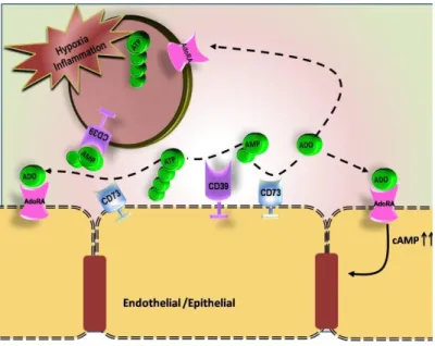
As illustrated in figure III, ATP degradation is enhanced by HIF-1α driven increased enzymatic conversion into adenosine (Colgan et.al., 2006; Eltzschig et.al., 2003; Eltzschig et.al., 2009). HIF-1α can stimulate release of extracellular adenosine (Synnestvedt et.al., 2002) and at the same time can inhibit the process of its uptake into the intracellular compartment as well as its intracellular metabolism (Morote-Garcia et.al., 2009) preventing cells from generating effective innate immune responses (Ohta & Sitkovsky, 2001; Sitkovsky et.al., 2004). HIF-1α can also influence adenosine receptors thereby leading to enhanced adenosine receptor signaling (Eckle et.al., 2008; Kong et.al., 2006) and elevating intracellular cAMP and CREB activation (Stiles, 1992). HIF-1α induced adenosine receptors, such as A2b, have shown to produce increased intracellular cAMP levels by human DCs inducing Th2 cell polarization (Yang et.al., 2010).
conditions. Therefore, the effects of these enzymes can be altered by HIF-1α levels during inflammation.
Figure III. Model of coordinated nucleotide metabolism and nucleotide signaling in hypoxia and inflammation (Colgan et.al., 2006)
Studies done in cutaneous lesions in experimental models infected with Leishmania amazonensis have shown that Leishmania can induce HIF-1α and HIF-1β (Arrais-Silva et.al., 2005) during chronic phase of infection. However, recently it becomes clear that the cells do not need to be hypoxic for HIF-1α expression in Leishmania infected HIF-1α (Degrossoli et.al., 2007). It is suggested that induction of HIF-1α (relative to hypoxia) could affect the microbial activities and protein expression of MФ yielding different phenotypes from that of the normoxic counterparts so that this phenomenon may take part in modulating immune responses in pathological conditions (Degrossoli & Giorgio, 2007). It may be possible that Leishmania can alter immune functions of immune cells such as mononuclear phagocytes in Leishmania infection with the regulation of HIF-1α regardless whether cells are hypoxic or not.
2. JUSTIFICATION
CL associated with L. amazonensis infection is usually severe in experimental mice as well as in humans. It often causes diffuse cutaneous leishmaniasis or in rare cases, VL in South American countries (Almeida et.al., 1996). The severity of the disease is further complicated due to failure in several treatment regimens and emerging co-infections (Chkravarty & Sundar, 2010). The underlying mechanisms for the pathogenesis of non-healing lesions are, however, still obscure. L. amazonensis can dampen both innate as well as adaptive immune responses at the site of infection where conditions can further be worsened by drastic changes in tissue metabolism, for example, increased nucleotide metabolism (Cramer & Johnson, 2003) and with the up-regulation of HIF-1α (Degrossoli et.al., 2007). The possibility that L. amazonensis may exploit host properties (such as host ectonucleotidases in this context) has not yet been explored in the early stages of their interaction with MФ. Production of adenosine may also regulate MФ activation state by triggering A2a and A2b receptors. These receptors can further be influenced by HIF-1α.
3. OBJECTIVES
3.1 GENERAL OBJECTIVE:
Determine the effects of L. amazonensis infection on the expression of CD39 (ecto-NTPDase), CD73 (ecto-5’ectonucleotidases) and the involvement of adenosine receptors in the parasitic survival in resident MФ
3.2 SPECIFIC OBJECTIVES:
1. Determine the expression of CD39 and CD73 in resting resident MФ
2. Determine the role of CD39 and CD73 in survival of L. amazonensis
3. Determine the role of A2a and A2b receptors in survival of L. amazonensis
4. Determine the effects of HIF-1α on the expression of CD39 and CD73 as well as in parasite survival and infection in MФ in vitro
5. Investigate mechanisms involved in parasite survival in resident MФ
4.1 Animal:
C57BL/6 mice (8-12 weeks, both male and female) were used for the purpose of our study. Mice were housed and maintained at the central animal facility in the Universidade Federal de Ouro Preto (UFOP). All animal experiments and procedures were approved by the institution’s committee on ethical handling of laboratory animals. (Protocol 2012/56)
4.2 Preparation of parasites:
Leishmania amazonensis (IFLA/BR/1967/PH8) were cultivated at the initial concentration of 1x10⁵ at 250C in grace’s base (Sigma Aldrich Inc, St.Louis, MO, USA) supplemented with 10% inactivated fetal bovine serum (FBS) (SFB-LGC Biotecnologia, Cotia, SP, Brasil), 2mM L-glutamine (GIBCO BRL-Life Technologies, Grand Island, NY, MO, EUA), 100U/ml penicillin G (USB Corporation, Cleveland, OH, USA), pH 6.5. Five days old stationary phase promastigotes were used for metacyclic isolation and purification as described in protocol by Spath e Beverely (Spath & Beverely, 2001) and adapted from Marques-da-Silva (de almeida Marques- da- Silva et.al. 2008). Parasites were washed twice with phosphate buffered saline (PBS), and centrifuged at a speed of 1540 x g, 40C for 10 min. Parasites were resuspended in
dulbecco’s modified eagle medium (DMEM) (Sigma Aldrich Inc) pH 7.2, and 2ml of parasitic suspension was distributed in tubes. This was followed by Ficoll® (Amersham Biosciences do Brasil, Sao Paulo, Sp, Brasil) gradient by adding 2ml of 10% Ficoll. This preparation was centrifuged at a speed of 1070 x g, 250C, for 15 min. After centrifugation, supernatant was collected in another tube without disturbing the pellet. Supernatant, rich in metacyclics, was further washed twice with PBS to remove any remaining contamination from Ficoll and other ingredients. Parasites were always kept in ice in an interval between proceeding steps.
4.3 CFSE labeling:
in DMEM supplement medium/10% FBS and kept in ice until use. CFSE tagged parasites were always protected from direct light.
4.4 Harvest peritoneal macrophages:
Animals were euthanized and the abdomen was gently massaged and peritoneal lavage was collected by using ice cold PBS with 16G needle and peritoneal lavage was collected by injecting 10ml of ice cold PBS with 16G needle (Zhang, X et.al., 2008). Cells were centrifuged at a speed of 210 x g, 40C for 10 min and were suspended in DMEM supplemented with 10% FBS pH 7.2. Viability of the cells was confirmed by using Trypan blue (Sigma-Aldrich). Cells (5x10⁵ cells) were distributed in vitro medium Dulbecco’s modified eagle´s medium (DMEM -Sigma) supplemented with/10% FBS, 100 U/ml penicillin G, 2mM glutamine, 25mM HEPES (Sigma), 1.2mM sodium bicarbonate (Vetec Quimica Fina Ltd) and 50M 2-mercaptoethanol (Pharmacia Biotech). Freshly harvested MФ were pooled before treatment or any further incubation. Once harvested, MФ were either analyzed in ex-vivo or cultivated in vitro culture medium. In vitroexperiments, MФ were rested for 24, 48 and 72hrs and subsequently incubated at 330C/5% CO2 without any external stimulus. The shift in temperature was made considering
the fact that L. amazonensis is sensitive to 370C (Ref). Therefore, in all experiments, resident cell population was rested for 72hrs at 370C/5% CO2 prior to any further treatment and analysis.
Further incubation was done at 330C/5%CO2 for any longer period of time. For flow cytometry
experiment, 6 mice were utilized whereas for in vitro infection in plates, 10 mice were used for each experiment.
4.5 In vitro infection of macrophages:
For analysis by flow cytometry, 72hrs rested resident cells were infected with metacyclics forms of L. amazonensis labeled with CFSE in a ratio of 1:3 in DMEM supplemented medium. In parallel, another group was treated with LPS obtained from E. coli (Sigma Aldrich Inc) at the concentration of 5µg /ml and mixed well. The cells were then incubated at 330C/5% CO2 for 24hrs or 48hrs. The expression of CD39 and CD73 was then
analyzed by flow cytometry.
resident cells were removed by washing two times with PBS before infection or further treatment. New fresh medium was added to the rested resident MФ and then infected with metacyclic forms of L. amazonensis in a ratio of 1:3. Cells were incubated at 330C/5% CO2 for 3
hours and excess parasites were then removed by washing twice with PBS. Cells were further incubated at 330C/5% CO2 for 24hours or 48hours.
4.6 CD39 and CD73 inhibition experiments
Inhibitors of CD39 and CD73, DIDS (4,4′-Diisothiocyanatostilbene-2,2′-disulfonic acid disodium salt hydrate-Sigma) and αβ MAD (α,β-Methyleneadenosine 5′-diphosphate sodium salt-Sigma) were added at a concentration of 200µM after 3hrs of initial infection and were then kept throughout the infection study. These inhibitors were dissolved in PBS.
4.7 Study of role of A2a and A2b receptors in vitro
Resting MФ were treated with MRS 1754 8-[4-[((4-Cyanophenyl) carbamoylmethyl) oxy] phenyl]-1,3-di(n-propyl) xanthine hydrate (Sigma-Aldrich), inhibitor for A2b receptor and ZM241385(4(-2-[7-amino-2-{2-furyl}{1,2,4}triazolo{2,3-a}{1,3,5}triazin-5-yl-amino]ethyl) Phenol (Sigma-Aldrich), for A2a receptor, at the concentration of 5µM at the time of infection and left throughout the incubation time period. These antagonists were prepared in DMSO and therefore control groups were treated with DMSO not exceeding 1% in final volume.
In all conditions, coverslips were removed 3hrs, 24hrs and 48hrs post infection from macrophage culture plates. Coverslips were then fixed in methanol for 10 min (Vetec Fine Chemistry), dried and stained using the kit Panótico Rápido (Laborclin, Pinhais, PR, Brazil) following manufacturer’s instructions. Coverslips were analyzed using an Olympus BX50 optical microscope (Olympus, Center Valley, PA, USA). A minimum of 200 macrophages per coverslip was examined and number of uninfected, infected and amastigotes in infected macrophages were recorded.
4.8 In vivo analysis of CD39 and CD73 expression in macrophages:
Mice were euthanized after 24hrs inoculation. Peritoneal lavage by ice cold PBS was performed and cells were harvested and studied in in ex-vivo for the expression of CD39 and CD73 in
resident MФ.
4.9 Effects of HIF-1α on the expression of CD39 and CD73 in MФ in vitro infection:
In in vitro infection, 72hrs rested cell population was treated with the FM19G11 (2-oxo-2-(p-tolyl)ethyl] 3-[(2,4-dinitrobenzoyl)amino]benzoate, 3-[(2,4- initrobenzoyl)amino]- benzoic acid 2-(4-methylphenyl)-2-oxoethyl ester) (Sigma-Aldrich), an inhibitor of HIF-1α, at a concentration of 100nM and 200nM. The inhibitor was added at the time of infection together with the parasites and was left there throughout the infection. This inhibitor was dissolved in DMSO. Control groups were always treated with DMSO. The expression of CD39 and CD73, in presence of FM19G11, was evaluated by flow cytometry after 24hrs of incubation with parasites.
Effects of the inhibitor were initially evaluated by cultivating L. amazonensis with the inhibitor and plotting a growth curve. L. amazonensis was cultivated in Grace´s medium/10% FBS. Inhibitor to HIF-1α in two different concentrations of 100nM or 200nM was added at the time of cultivation. Cultures were incubated at 250C for 6 days and growth curve was plotted. In a control group, DMSO was added.
4.10 Flow cytometry:
4. 11 Cytokine assays:
TNF-alpha, and IL-10 cell culture supernatants were determined by ELISA kits (Mouse TNF-alpha DuoSet catalogue DY410, Mouse IL-10 Duoset catalogue DY417E from R&D system). Assays were performed according to manufacturer´s instructions. Briefly, flat-bottom 96-well microtiter plates (Nunc) were coated with 100 μL/well of TNF-alpha and IL-10 specific monoclonal antibodies (0.2 µg/mL and 2.0 µg/mL, respectively) for 18hrs at 4o C and then washed with PBS buffer (pH 7.4) containing 0.05% Tween 20 (wash buffer). Nonspecific binding sites were blocked with 300 µL/well of 1% BSA in PBS. Plates were rinsed with wash buffer, and 100 µL of samples and standards were added followed by incubation for 2hrs at room temperature. Seven fold serial dilution of standards of each these cytokines was prepared. Plates were then washed and 100 µL of the appropriate TNF-alpha (50 ng/mL) and IL-10 (100 ng/mL) biotinylated detection antibodies diluted in blocking buffer containing 0.05% Tween 20 were added for 1hr per well at room temperature. Plates were, then, washed and streptavidin-horseradish peroxidase (0.1 µg/mL) added for 30min of incubation at room temperature. Finally, plates were washed and 100 µL of the substrate solution (1:1-mixture H2O2 and
Tetramethylbenzidine) was added per well and, after 30 min of a dark incubation at room temperature, the reaction was stopped by 50 µL/well of 1M H2SO4 solution. Plates were read at
450 nm with wavelength (Microplate Reader, model 680, BioRad). All samples were assayed in triplicate using DuoSet® ELISA Development System, R&D Systems®, Minneapolis, MN systems.
4.12 Nitric oxide measurement:
4.13 Relative mRNA expression of adenosine receptors by real time PCR:
RNA was extracted from cultivated cells by treating with brazol (1ml for 5-10 x 106 cells). Cells were incubated with brazol for 5 min at 15-300C. Cells were then centrifuged at 12,000 x g/2-80C/15min. Supernatant was then collected in another tube. 0.2ml of chloroform was added to each 1ml of brazol used to lyse the cells and then vortexed vigorously for 15sec. It was incubated on ice for 2 min. It was then centrifuged at 12,000 x g/2-80C/15min. After centrifugation, top layer which contains RNA was collected. RNA was then treated with 0.5ml of isopropyl alcohol and incubated it for 10 min at 15-300C. It was then centrifuged at 12,000 x g/2-8oC/10 min. Supernatant was removed and precipitated RNA was washed with 75% of alcohol followed by centrifugation at 7500 x g/2-80C/5 min. Precipitate was then dissolved in RNAse free water and was quantitated in spectrophotometer. Pure RNA will exhibit an A260/A280 ratio of 2.0.
cDNA was prepared using high capacity cDNA reverse transcription kit (Applied Biosystems). Mastermix was prepared containing 2μl of 10X RT buffer, 0.8μl of 25X deoxynucleotides mix (dNTPs), 1μl of RNase inhibitor, and 1μl of multiScribe reverse transcriptase, 2μl of 10 X random primers and 3.2 μl DEPC water and mixed with 10μl of sample (0.025µ/µl) to make the final volume 20μl. Then, the tubes were submitted in thermocycler with following temperatures: 25oC 10 min, 37oC 120 min, 85oC 5 min and rest at 4oC. After this process cDNA was ready for PCR and maintained at -20oC.
Finally, it was then checked how many times the test samples expressed mRNA expression in relation to that uninfected group using formula 2-∆∆Ct.
Melting curve analysis was also performed during amplification to determine any non-specific amplification. Data were normalized to the expression levels of reference gene (β actin). Primer sequences for the target gene were illustrated below.
Primer Sequences
Gene Forward sequence Reverse sequence
β- actin ACTGCTCTGGCTCCTAGCAC ACATCTGCTGGAAGGTGGAC
A2a GGCTCCTCGGTGTACATCAT GTTCTGCAGGTTGCTGTTGA
A2b TTCTTTGGGTGTGTCCTTCC CCTGGAGTGGTCCATCAGTT
4.14 Statistical analysis:
5. RESULTS:
Expression of CD39 and CD73 in resident MФ has been illustrated here in vitro.
Regulation of CD39 and CD73 expression by rested resident MФ in in vitro culture medium has also been described. The effects of L. amazonensis on CD39 and CD73 expression have been
reported for the first time here in resident MФ and at the same time, the role of these molecules in parasite survival and infection has been discussed. Adenosine production via CD39 and CD73 enzyme activity binding to A2a and A2b receptors and its significance in parasite growth and survival has been shown. A possible link between HIF-1α and CD39 and CD73 has also been studied during L. amazonensis infection. The regulation of A2a and A2b receptors by HIF-1α during L. amazonensis infection at transcript levels was studied after 24hrs of incubation.
5.1 Resident macrophages are characterized by high CD39 and CD73 expression
In order to determine the effects of L. amazonensis on CD39 and CD73 expression in
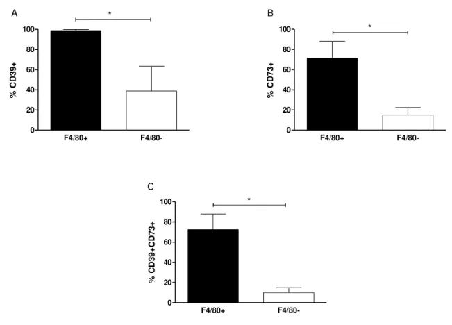
MФ, we first analyzed expression of these enzymes in resident cell population based on F4/80 marker. When analyzed in flow cytometry, whole F4/80+ population (MФ) expressed CD39
(Fig. 1A) indicating CD39 as a predominant ectonucleotidase enzyme in MФ while 39% of non
-macrophage groups presented this enzyme. CD73 expression was equally higher in MФ than
non-MФ (Fig. 1B). In total, MФ represented the major cell population expressing both ecto
Figure 1: Peritoneal resident macrophages are major cell populations that express both CD39 and CD73 in ex-vivo. Resident MФ were harvested and pooled from peritoneum of naïve C57BL/6 mice. Cells were labeled with
anti-murine F4/80, anti-CD39 and anti-CD73 antibodies and then were analyzed by flow cytometry. In the figure,
the percentage of cells expressing A. CD39 and B. CD73 and C. CD39CD73 is shown for F4/80+ and F4/80- cells.
This result is the mean±S.D of at least 3 independent experiments. *p<0.05 means the statistically different using Paired two tailed Student´s t- test.
5.2. Resident macrophages down regulate CD73 expression in vitro
Before studying the role of CD39 and CD73 in Leishmania infection, whole peritoneal cell population was cultivated in vitro medium in order to determine if these surface enzymes can self-regulate in in vitro conditions. As shown in Fig. 2, although the percentage of MФ remained constant during incubation period (Fig. 2B), these cells surprisingly down regulated CD73 while their CD39 expression remained unaltered (2C–2F). CD73 expression gradually decreased over the incubation period suggesting that the culture conditions may have an influence in the expression of CD73 in vitro. On the other hand, it may also be possible that the expression of CD73 could have been increased by the harvesting procedure and then slowly returned to the basal level.
Figure 2: Resident macrophages down regulate CD73 expression in vitro. Resident MФ were harvested from
naïve C57BL/6 mice. Total peritoneal cell population was counted and viability of the cells was determined by
Trypan blue. Freshly harvested cells were rested for 24, 48 and 72hrs prior to further incubation at 33oC/5%CO
2. Six
mice were used for each experiment and cells were pooled for the analysis. Fig.2A shows size and granularity for
total peritoneal population. In figure 2B, F4/80+ cells were first gated and then cells expressing C. CD39 and D.
CD73 in F4/80+ population from 24, 48 and 72hrs rested MФ were over laid in histograms. The percentage of cells
expressing E. CD39 and F. CD73 is represented in bar diagrams for MФ. This result is representative of 3 independent experiments. *p<0.05 indicates statistically different using Paired two tailed Student’s test
5.3. L amazonensis increases CD73 expression in rested macrophages
Once it was determined that CD73 expression could be down regulated by the rested MФ, our next approach was to observe if the infection by L. amazonensis can further influence
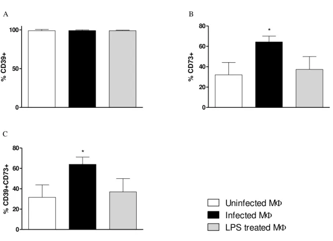
in CD39 and CD73 expression in MФ. Seventy two hours rested MФ were incubated with CFSE labeled metacyclics of L. amazonensis. After 24hrs of infection, it was found that although the percentage of CD39+ cells did not alter (Fig. 3A), there was a significant increase in the percentage of cells expressing CD73 (Fig. 3B). LPS treatment did not affect the expression of CD39 or CD73, suggesting that activation does not alter CD73 expression (Fig 3A and 3B). Figure 3C demonstrated that the combined expression of CD39 and CD73 was higher amongst
Figure 3: L. amazonensis upregulates CD73 expression in rested resident macrophages. Resident cells were
collected and pooled from naïve C57BL/6 mice and rested for 72hrs prior to infection. They were infected with
CFSE tagged metacyclics of L. amazonensis and additionally another group was treated with 5µg/ml of LPS. Cells
were then further incubated for 24hrs at 330C/5% CO2 and flow cytometry was performed. The percentage of cells
expressing A. CD39 B. CD73 and C. CD39CD73 in uninfected, infected or LPS treated MФ was analyzed by flow
cytometry. This result is the mean±SD of at least 3 independent experiments* p<0.05 indicates statistically
significant between infected and control groups. One-way analysis of variance (ANOVA) followed by a posttest Bonferroni test.
Uninfected M Infected M LPS treated M
Fig 5.4: L. amazonensis increases CD73 expression in rested macrophages during 48hrs of infection
Furthermore, seventy two hour rested MФ were incubated with CFSE labeled metacyclics of L. amazonensis and infection was prolonged to 48 hours of incubation. We observed that the percentage of cells expressing CD73 was higher in infected groups than in control groups (Fig 4A-C).
Figure 4: L. amazonensis keeps CD73 expression high in infected macrophages during 48hrs of infection. Seventy two hour rested MФ were prepared and treated with parasite or LPS as described in materials and methods.
Cells were incubated at 330C/5% CO2 for 48hrs infection. In given figure, the percentage of MФ expressing A.
CD39 and B. CD73 was analyzed for uninfected, infected and LPS treated MФ by flow cytometry. Similarly, in
figure C, the combined expression of CD39CD73 in all three populations is represented in bar diagram. This result is the mean±SD of at least 3 independent experiments. * p<0.05 indicates statistically significant between infected and control groups using One-way analysis of variance (ANOVA) followed by Bonferroni posttest.
.
Uninfected M Infected M LPS treated M
5.5 L. amazonensis does not affect cytokine and NO production
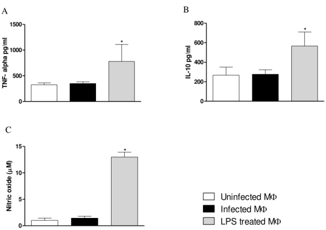
Following the evaluation of expression of these enzymes, cytokine as well as NO production by treated MФ were also measured. As shown in Fig. 5A-5C, L. amazonensis
infected MФ did not alter the production of TNF-alpha, IL-10, and NO. However, MФ treated with LPS produced significantly higher levels of these inflammatory mediators compared to control and infected groups, indicating that these cells were fully capable to respond to inflammatory stimuli but not to the parasite. Up regulation in CD73 expression was, therefore, observed independently of cytokine as well as NO production. Therefore, these data together with increase CD73 expression in infected MФ indicate that CD73 is regulated differently by L. amazonensis.
Figure 5: L amazonensis does not alter cytokine and NO production. Resident cells were collected from naïve
C57BL/6 mice and rested for 72hrs prior to infection. They were either infected with L. amazonensis or treated with
5µg/ml of LPS. Cells were then further incubated for 24hrs at 330C/5% CO2. Supernatant from all groups was
collected for the measurement of TNF-alpha, IL-10 and NO. In figures, A. TNF-alpha B IL-10 were measured by
ELISA and C. NO production in treated MФ was measured by Griess method. This result is the mean±SD of at least
3 independent experiments * p<0.05 indicates statistically significant between infected and control groups using One-way analysis of variance (ANOVA) followed by Bonferroni posttest.
. 0 500 1000 1500 * T N F - a lp h a p g /m l 0 200 400 600 800 * IL -1 0 p g /m l 0 5 10 15 * N it ri c o x id e ( M) A B C
5.6. Infected macrophages express high CD39 and CD73 in in ex-vivo studies
As an another approach, in order to demonstrate the effects of L. amazonensis on the expression of CD39 and CD73 in vivo, CFSE tagged live metacyclics were injected in the peritoneum of mice and peritoneal cell population was harvested after 24hrs of inoculation. It was found that majority of the cells from whole peritoneal cell population could be divided into CD39+CD73+ or CD39+CD73- (Fig 6B). Inside these populations, total F4/80+ cells (Fig 6C)
Isotype control
CD39+CD73+
CD39+CD73-CD39
CD73
FSC SSC
F4/80 CFSE
A
.
B
T
D C
E
. F4/80+
CD39+CD73+
CD39+CD73-0 50 100
150 *
C
F
S
E
+
M
F
I
Figure 6: L. amazonensis infected macrophages show high CD73 expression in in ex-vivo studies. 50X106
CFSE tagged live metacyclics were inoculated in the peritoneum of mice and then whole peritoneal cell population was harvested after 24hrs of inoculation. Cells were labeled with anti-mouse F4/80, anti-CD39 and anti-CD73 antibodies. Figure A. FSC X SSC shows size and granularity for whole cell population. B. Inside gated population;
cells expressing CD39 and CD73 were shown. Cells expressing CD39+CD73+ or CD39+CD73- were selected and
then examined for C. F4/80+ population and D. CFSE positivity was determined in F4/80+ population. In the bar
diagram E. MFI for CFSE in F4/80+ population is shown. Data are the mean±SD from 2 independent experiments.
*p<0.05 indicates statistically different between using Paired two tailed Student´s t- test. Two mice were used for each control group per experiment.
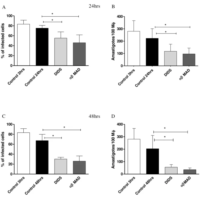
5.7. CD39 and CD73 activity determine parasite survival in infected macrophages
Figure 7: CD39 and CD73 activity determine survival of L. amazonensis. Resident MФ were obtained from
naïve mice by injecting 10ml of ice cold PBS in to the peritoneal cavity and rested for 72hrs at 370C/5% CO2 as
described in materials and methods. Cells were then infected with metacyclic forms of the parasites in a ratio of 1:3
and allowed for the parasites to interact for 3hrs at 330C/5% CO2. Extracellular parasites were washed away and the
inhibitors DIDS and αβ MAD were added against CD39 and CD73 at a concentration of 200µM respectively. In the
figure, the percentage of infection for 24hrs (A) and for 48hrs (C) and the amastigote number per 100 MФ for 24hrs
(B) and for 48hrs (D) were calculated. Control represents infected MФ treated with DMSO. Data are the mean±SD
from 3 independent experiments. * p<0.05 is statistically different between control and treated groups using repeated measures of ANOVA followed by Dunnett posttest against the control group
Cont rol 3
hrs
Cont rol 4
8hrs DIDS MAD
0 20 40 60 80 100 * * % o f in fe c te d c e ll s Cont rol 3
hrs
Cont rol 4
8hrs DIDS MAD
0 100 200 300 400 * * A m a s ti g o te s /1 0 0 M C D Cont rol 3
hrs
Cont rol 2
4hrs DIDS MAD
0 20 40 60 80 100 * * % o f in fe c te d c e ll s Cont rol 3hrs Cont rol
24hrs DIDS MA D 0 100 200 300 400 * * A m a s ti g o te s /1 0 0 M
A 24hrs B
5.8 Evaluation of cytokine and NO production from treated macrophages
In order to determine the mechanism of parasite killing in treated and untreated infected MФ, cytokine and NO production were measured. Interestingly, reduction in parasite survival in MФ was not related to the production of TNF-alpha, IL-10 or NO production as shown in Fig. 8A-8C. These results indicate that killing of the parasites under these circumstances is possibly mediated via pathways involving other than cytokine mediated NO production.
Figure 8: Inhibition of CD39 and CD73 activity does not alter cytokine and NO production by infected macrophages. Resident MФ were obtained from naïve mice as described in materials and methods. Cells were then
infected with L. amazonensis and inhibitors DIDS and αβ MAD were added after 3hrs of infection and then left
them for 48hrs. Supernatant was collected from these groups after 48hrs incubation. Control represents infected MФ
treated with DMSO. Production of A.TNF-alpha B. IL-10 C. NO in treated groups is illustrated in the figure. Data are the mean±SD from 3 independent experiments.
.
Control DIDS
MAD
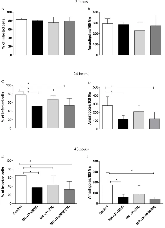
5.9 Inhibition of A2a and A2b adenosine receptors reduces L. amazonensis survival in rested macrophages
Figure 9: Survival of L. amazonensis depends on A2a and A2b receptors. MФ were harvested and rested before
infection. A2a and A2b antagonists ZM241385 and MRS1754 were added at a concentration of 5µM at the time of infection and kept throughout the infection. Percentage of infection and amastigote number were determined during
3hrs (A) and (B), 24hrs (C) and (D) and 48hrs (E) and (F) infection. Control represents infected MФ treated with
DMSO. These data indicate the mean±SD of at least 3 independent experiments. *p<0.05 represents statistical different between treated and control groups using repeated measures of ANOVA followed by Dunnett posttest against the control group
5.10 Evaluation of cytokine and NO production from treated macrophages
Similarly, as in CD39 and CD73 enzyme activity inhibition experiments, cytokine and NO production were evaluated from adenosine receptor inhibited MФ culture supernatant. As observed in Fig 8, blockade of A2a and A2b adenosine receptors did not induce cytokine and NO production as shown in Fig. 10A-C, once again suggesting a possible common downstream pathway other than NO production, involving in CD39/CD73 enzyme actions as well as adenosine receptors.
Figure 10: Inhibition of A2a and A2b receptors does not alter cytokine and NO production by infected macrophages. MФ were harvested and rested as previously described in materials and method. A2a and A2b antagonists ZM241385 and MRS1754 were added at the time of infection and kept throughout the infection. Supernatant collected from these groups after 48hrs of infection and was evaluated for A. TNF-alpha B. IL-10 by
ELISA and C. NO production by Griess method. Control represents infected MФ treated with DMSO. These data
indicate the mean±SD of 3 independent experiments.
Control M+(P+MRS) M+(P+ZM) M+(P+MRS/ZM)
5.11 Effects of HIF-1α inhibitor on growth of L. amazonensis
FM19G11 is an inhibitor to HIF-1α. Before studying its role in parasitic survival, parasite growth was determined in presence of FM19G11. It was observed that this inhibitor did not alter the growth of the parasites (Fig 11).
Figure 11: Growth Curve of L. amazonensis in presence of FM19G11 L. amazonensis was cultivated in Grace´s
medium/10% FBS. Inhibitor to HIF-1α in two different concentrations of 100nM or 200nM was added at the time of
cultivation. Cultures were incubated at 250C for 6 days and growth curve was plotted. In a control group, DMSO
was added. Results are mean±SD of 3 independent experiments.
5.12 Inhibition of HIF-1α in resident macrophages and its role inL. amazonensis infection
Infection and inflammation induced by the parasite may lead to localized hypoxic conditions in host cells. However, in vitro studies, it has been found that L. amazonensis can induce HIF-1α even when the cells are not hypoxic (Degrossoli et.al., 2007). Therefore, we evaluated possible role of HIF-1α in parasitic survival in MФ in the presence of HIF-1α inhibitor.
In order to study the possible role of HIF-1α in the survival of L. amazonensis in MФ,
72hrs rested MФ were treated with FM19G11 at a concentration of 100nM and 200nM. It was found that 3hrs of incubation with parasites in presence of the inhibitor did not affect the parasite infection and amastigote number (Fig 12A and Fig 12B). During 24hrs of infection, in presence of HIF-1α inhibitor, although no appreciable decrease in percentage of infection was observed, there was significant reduction in amastigote number per 100 MФ (Fig 12C and Fig 12D).
0 2 4 6 8
0 100 200 300
Time in days
P
ar
a
si
te
s
x
10
^5
/
m
L Control