Revista Brasileira de Hematologia e Hemoterapia
Brazilian Journal of Hematology and Hemotherapy
www.rbhh.org
Original article
Early and late oral features of chronic graft-versus-host
disease
Alessandra Oliveira Ferrari Gomes
a, Sandra Regina Torres
a, Angelo Maiolino
a,
Cesar Werneck Noce dos Santos
a, Arley Silva Junior
a,b, Maria Elvira Pizzigatti Correa
c,
Maria Cláudia Rodrigues Moreira
d, Lucio de Souza Gonçalves
aa Universidade Federal do Rio de Janeiro (UFRJ), Rio de Janeiro, RJ, Brazil b Universidade Federal Fluminense (UFF), Niterói, RJ, Brazil
c Universidade Estadual de Campinas (UNICAMP), Campinas, SP, Brazil d Instituto Nacional de Câncer (INCA), Rio de Janeiro, RJ, Brazil
A R T I C L E I N F O
Article history:
Received 16 April 2013 Accepted 20 July 2013
Keywords:
Chronic disease
Hematopoietic stem cell transplan-tation
Diagnosis, oral
Graft-versus-host disease
*Corresponding author at: Universidade Federal do Rio de Janeiro, Departamento de Patologia e Diagnóstico Oral, Avenida Brigadeiro Trompowski, s/n, Ilha do Governador 21941-590, Rio de Janeiro, RJ, Brazil.
E-mail address: [email protected] (A.O.F. Gomes).
1516-8484/$ - see front matter © 2014 Associação Brasileira de Hematologia, Hemoterapia e Terapia Celular. Published by Elsevier Editora Ltda. All rights reserved.
DOI: 10.5581/1516-8484.20140012
A B S T R A C T
Background: Chronic graft-versus-host disease is a serious complication of allogeneic hematopoietic cell transplantation, and the mouth is one of the affected sites.
Objective: The aim of this study was to evaluate the oral features of this disease after hematopoietic cell transplantation.
Methods: This was a cross-sectional multicenter study that enrolled patients submitted to transplantation. Oral evaluations used the National Institutes of Health criteria, salivary flow rates, and the range of mouth opening. Pain and xerostomia were evaluated through a visual analogue scale. Patients were divided into two groups based on the transplantation time (up to one year and more than one year).
Results: Of the 57 evaluated recipients, 44 had chronic graft-versus-host disease: ten (22.72%) in the group with less than one year after transplantation, and 34 (77.27%) in the group with more than one year after transplantation. Lichenoid/hyperkeratotic plaques, erythematous lesions, xerostomia, and hyposalivation were the most commonly reported oral features. Lichenoid/hyperkeratotic plaques were significantly more common in patients within the first year after the transplant. The labial mucosa was affected more in the first year. No significant changes occurred in the frequency of xerostomia, hyposalivation, and reduced mouth opening regarding time after transplantation.
Conclusion: Oral chronic graft-versus-host disease lesions were identified early in the course of the disease. The changes observed in salivary gland function and in the range of mouth opening were not correlated with the time after transplantation.
Introduction
Allogeneic hematopoietic cell transplantation (HCT) is associated with early and late oral complications. Most of these complications are related to graft-versus-host disease (GVHD) and may cause transitory and/or permanent sequelae. Many studies have reported oral manifestations of chronic GVHD (cGVHD),1-3 but to the authors’ knowledge, only one study has addressed the temporal outcomes of some of these features.4 Most studies focus on specific oral alterations of cGVHD, and have not used the National Institutes of Health (NIH) criteria to evaluate the oral features.
cGVHD affects from 50% to 80% of the adult patients who undergo HCT, regardless of prophylaxis and donor-patient matching techniques.2
Oral manifestations can be the first signs of cGVHD, and might be considered a disease marker.3,5 These oral manifestations are observed in the majority of cases of cGVHD,2,5-7 and are considered an important cause of morbidity and loss of quality of life in long-term survivors.5
According to the NIH Consensus Criteria for GVHD, the disease is currently diagnosed by its clinical manifestations and no longer according to time post-transplantation.8 The clinical manifestations of cGVHD are classified as diagnostic, distinctive, or common clinical features. The finding of a ‘diagnostic’ feature of cGVHD establishes the condition without further testing. In the oral cavity, these features include lichen planus-like lesions, hyperkeratotic plaques, and restriction of mouth opening due to skin sclerosis. ‘Distinctive’ features are likely to support the diagnosis of cGVHD; however, they are not enough to establish the diagnosis. The oral ‘distinctive’ features include xerostomia, mucoceles, mucosal atrophy, pseudomembranes, and ulcers. ‘Common’ features of cGVHD refer to the manifestations found in both acute GVHD and cGVHD, which include gingivitis, mucositis, erythema, and pain.8
Some clinical changes of oral cGVHD such as vasculitis-like features or a telangiectatic appearance of the mucosa, inflammation, and loss of the stippling of the attached gingiva may represent early alterations of oral cGVHD, even though they are not considered in the NIH criteria.2 Erosive lesions are observed in the most severe forms of cGVHD, and are followed by pain, which may interfere in oral hygiene and food ingestion.9 Xerostomia generally causes discomfort, but hyposalivation, that is, reduced salivary flow rates (SFR), which have more serious consequences, have not been included in the NIH criteria.10-12
Temporal features of systemic cGVHD have previously been addressed. However, although oral cGVHD has been analyzed in many publications,1-3 data regarding the temporal relationship of oral features is lacking. The present study aimed to perform a cross-sectional evaluation of the oral features of cGVHD according to the time after HCT.
Methods
This was a cross-sectional multicenter study conducted in two hospitals in Brazil: Hospital Universitário Clementino Fraga Filho (HUCFF) of the Universidade Federal do Rio de Janeiro (UFRJ) and the Center for Hematology and Hemotherapy of
the Universidade Estadual de Campinas, from January of 2008 to January of 2011. Adult patients who underwent HCT for hematological conditions were included. All patients signed an informed consent. This study was approved by the institution’s review board and conducted in accordance with the Helsinki Declaration as revised in 2008.
Demographic and clinical characteristics were assessed from medical records. After the transplantation, oral evaluations were performed by a trained dentist. The oral exam was performed using frontal light-emitting diode illumination, with the patient sitting on a chair.
Oral features were classified according to NIH criteria as diagnostic, distinctive, and common features.8 These features referred to any changes related to oral signs and symptoms (mucosal lesions, perception of changes in salivary flow rate and moisture, changes in sensitivity, and reduction of mouth opening). Oral lesions were defined as morphological changes of the oral mucosa.
The severity of oral symptoms was scored according to the NIH criteria:8 no symptoms (0); mild symptoms with disease signs not limiting the oral intake (1); moderate symptoms with disease signs, with partial limitation of oral intake (2); and severe symptoms with disease signs and major limitation of oral intake (3). Moreover, the patients graded oral sensitivity through a visual analogue scale (VAS) graduated from 0 (no sensitivity) to 10 (worst possible pain).13,14 Positive sensitivity was recorded when patients reported symptoms greater than 0 on the VAS.
Resting SFR were used to assess salivary function under standard conditions.15 Saliva samples were collected between 9:00 a.m and 11:30 a.m.16 Participants were asked not to eat until the exam was performed. Patients were instructed to spit the accumulated saliva periodically into a disposable cup for five minutes. They were instructed not to eat nor swallow during the exam.16 The liquid part of the SFR was then measured with a disposable syringe. The SFR results were determined as milliliters per minute, and reduced SFR was defined as < 0.3 mL/min.11
Oral dryness was also evaluated using a VAS graduated from 0 (no dryness) to 10 (the worst possible oral dryness). To eliminate common causes of dry mouth feeling, xerostomia was considered when the patient recorded a score > 2.13
The oral involvement of scleroderma was evaluated using a Willis compass to measure the maximum range of mouth opening (RMO). The device measured the midline distance from the border of the central upper incisors to the border of the central lower incisors. Reduced RMO was defined as < 35 mm.17
In order to analyze the temporal manifestations of cGVHD, the patients were separated in two groups: within one year post-HCT and over one year post-HCT. In general, cGVHD develops from three to 15 months after HCT,1 and the period of one year post-HCT has been used in some studies to evaluate cGVHD.18,19 The date of the transplant was an accurate datum and was available for all patients.
Results
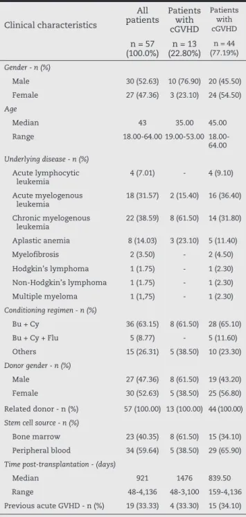
Fifty-seven patients who underwent HCT were analyzed; 27 (47.4%) women and 30 (52.6%) men. The median age was 43 years, ranging from 18 to 64 years. Among the patients submitted to HCT, 68.4% presented oral features. The clinical and demographic characteristics of the 57 HCT patients are shown in Table 1. The oral features of the HCT patients included in the study are listed in Table 2.
Forty-four (77.2%) patients developed cGVHD. Among the cGVHD patients, 88.63% presented oral features. This analysis was focused primarily on patients with cGVHD, as the majority of the HCT recipients developed this condition. Among the patients who developed cGVHD, the mouth was the most frequently affected site (88.63%), regardless of the different stem cell sources (bone marrow: 71.4%; peripheral blood: 66.7%). The most affected organs were the mouth, skin, liver, eyes, and lungs.
The most common oral features observed in cGVHD patients were oral lesions (70.45%), xerostomia (56.81%), reduced SFR (47.72%), and reduced RMO (31.81%). The most common types of oral lesions in cGVHD patients were erythema (54.54%) and lichenoid/hyperkeratotic lesions (52.27%). Lichenoid lesions are considered a diagnostic feature by the NIH criteria, and were observed in all patients that presented oral features within one year post-HCT; many patients presented more than one type of oral manifestation. The NIH classification and grading of the cGVHD patients who presented oral features are listed in Clinical characteristics
All
patients Patients with cGVHD
Patients with cGVHD
n = 57 (100.0%)
n = 13 (22.80%)
n = 44 (77.19%)
Gender - n (%)
Male 30 (52.63) 10 (76.90) 20 (45.50) Female 27 (47.36) 3 (23.10) 24 (54.50)
Age
Median 43 35.00 45.00 Range 64.00 19.00-53.00
18.00-64.00
Underlying disease - n (%)
Acute lymphocytic leukemia
4 (7.01) - 4 (9.10) Acute myelogenous
leukemia
18 (31.57) 2 (15.40) 16 (36.40) Chronic myelogenous
leukemia
22 (38.59) 8 (61.50) 14 (31.80) Aplastic anemia 8 (14.03) 3 (23.10) 5 (11.40) Myelofibrosis 2 (3.50) - 2 (4.50) Hodgkin’s lymphoma 1 (1.75) - 1 (2.30) Non-Hodgkin’s lymphoma 1 (1.75) - 1 (2.30) Multiple myeloma 1 (1,75) - 1 (2.30)
Conditioning regimen - n (%)
Bu + Cy 36 (63.15) 8 (61.50) 28 (65.10) Bu + Cy + Flu 5 (8.77) - 5 (11.60) Others 15 (26.31) 5 (38.50) 10 (23.30)
Donor gender - n (%)
Male 27 (47.36) 8 (61.50) 19 (43.20) Female 30 (52.63) 5 (38.50) 25 (56.80) Related donor - n (%) 57 (100.00) 13 (100.00) 44 (100.00)
Stem cell source - n (%)
Bone marrow 23 (40.35) 8 (61.50) 15 (34.10) Peripheral blood 34 (59.64) 5 (38.50) 29 (65.90)
Time post-transplantation - (days)
Median 921 1476 839.50
Range 48-4,136 48-3,100 159-4,136
Previous acute GVHD - n (%) 19 (33.33) 4 (33.30) 15 (34.10) GVHD: graft-versus-host disease; cGVHD: chronic graft-versus-host disease; Busulfan: Bu; Cyclophosphamide: Cy; Fludarabine: Flu.
Table 1 - Clinical and demographic characteristics of the 57 patients submitted to hematopoietic cell transplantation.
Features
Patients without cGVHD
Patients with cGVHD
p-value n = 13
(22.80%)
n = 44 (77.19%)
Oral lesions - n (%)
Lichenoid/hyperkeratotic - 23 (52.30) < 0.01 Erythema 1 (7.70) 24 (54.50) 0.03 Ulcers 1 (7.70) 11 (25.00) NS Mucoceles - 8 (18.20) NS Atrophy 4 (30.76) 17 (38.60) NS Drug-induced gingival
hyperplasia
1 (7.70) - NS Presented sensitivity - n (%) 5 (38.50) 30 (68.20) NS
Oral sensitivity (VAS) 0.02
Median 0.00 0.60 Range 0.00-4.80 0.00-8.00 Presented xerostomia - n (%) 3 (23.10) 25 (56.80) 0.05
Xerostomia (VAS) 0.01
Median 0.30 2.45 Range 0.00-7.00 0.00-10.00 Presented hyposalivation - n (%) 1 (7.70) 21 (47.70) 0.01
Resting salivary flow rates (mL/min) 0.02
Median 0.50 0.30 Range 0.08-0.92 0.00-1.00 Patients with reduced range of
mouth opening - n (%)
2 (15.40) 15 (34.10) 0.08
Range of mouth opening (mm) 0.06
Median 39.00 35.00 Range 32.00-
48.00
24.00-50.00
cGVHD: chronic graft-versus-host disease; HCT: hematopoietic cell transplantation; NS: non-significant; VAS: visual analogue scale.
Table 3. Data on the characteristics of the oral lesions, salivary function, and RMO according to the time post-transplantation are listed in Tables 4, 5, and 6, respectively.
There were no differences related to the time of transplantation regarding the features of xerostomia, SFR, or RMO. Moreover, there was no relationship between hyposalivation and gender (p-value = 0.39) or age (p-value = 0.93). Among the patients who had cGVHD, 54.5% presented concomitant xerostomia and sensitivity. Only ten (22.7%) patients had concomitant ulcers and sensitivity.
Discussion
This study was relevant due to the evaluation of all oral cGVHD features in a standardized manner. Most of the studies on oral cGVHD evaluated isolated aspects of the disease. Moreover, the present study was one of the first to evaluate these features using the NIH criteria. Besides the subjective way that the NIH defines these criteria, objective evaluations were performed for the signs and symptoms of oral cGVHD. To the best of the Oral lesions
Less than one year of HCT
n = 9 (23.07%)
More than one year
of HCT n = 30 (76.92%)
p-value n (%) n (%)
Classification of oral features
Diagnostic 9 (100.00) 15 (50.00) < 0.01 Distinctive 7 (77.80) 24 (80.00) NS Common 6 (66.70) 20 (66.70) NS
Severity of oral features
No symptoms 1 (11.10) 9 (30.00) NS Mild 3 (33.30) 13 (43.30) NS Moderate 5 (55.60) 8 (26.70) NS Some patients presented more than one kind of oral feature and in different oral sites.
HCT: hematopoietic cell transplantation; NS: non-significant.
Table 3 - Classification and grading of oral features of chronic graft-versus-host disease according to the National Institutes of Health criteria (n = 39).
Lesions
Less than one year
of HCT
More than one year
of HCT
p-value
n = 9 (23.07%)
n = 30 (76.92%)
Type of oral lesiona - n (%)
Lichenoid/hyperkeratotic 9 (100.00) 14 (46.70) < 0.01 Erythema 5 (55.60) 19 (63.30) NS Ulcers 3 (33.30) 8 (26.70) NS Mucoceles 4 (44.40) 4 (13.30) 0.06 Oral sensitivity - n (%) 7 (77.80) 23 (76.70) NS
Oral sensitivity (VAS) NS
Median 0.70 0.15 Range 0.00-1.70 0.00-6.00
Anatomical site of oral lesionsa - n (%)
Vermillion border 3 (33.30) 8 (26.70) NS Labial mucosa 5 (55.60) 5 (16.70) 0.03 Tongue 8 (88.90) 16 (53.30) NS Buccal mucosa 8 (88.90) 18 (60.00) NS Palate 5 (55.60) 10 (33.30) NS Gingiva 5 (55.60) 21 (70.00) NS HCT: hematopoietic cell transplant; NS: non-significant; VAS: visual analogue scale.
a Some patients present more than one kind of oral feature and in
different oral sites.
Table 4 - Early and late characteristics of oral lesions in chronic graft-versus-host disease (n = 39).
Salivary flow rate and perception
Less than one year
of HCT
More than one year
of HCT
p-value n = 9
(23.07%)
n = 30 (76.92%)
Xerostomia - n (%) 7 (77.80) 18 (60.00) NS
Xerostomia (VAS)
Median 2.10 3.00 NS Range 0.00-8.80 0.00-10.00 Hyposalivation - n (%) 6 (66.70) 15 (60.00) NS
Resting salivary flow rate (mL/min)
Median 0.25 0.27 NS Range 0.00-0.36 0.00-0.80
HCT: hematopoietic cell transplant; VAS: visual analogue scale; NS: non-significant.
Table 5 - Early and late characteristics of salivary function in chronic graft-versus-host (n = 39).
Range of mouth opening
Less than one year of HCT
More than one year
of HCT
p-value n = 9
(23.07%) (76.92%)n = 30
Patients with reduced range of mouth opening
1 (11.10) 13 (43.30) NS
Range of mouth opening (mm) NS
Median 39.50 32.50 Range
33.00-44.00
24.00-48.00 HCT: hematopoietic cell transplant; NS: non-significant.
authors’ knowledge, there is no study correlating the frequency and the severity of oral cGVHD lesions with the time post-HCT. Only one study evaluated the temporal correlation of salivary flow and taste changes with time post-HCT, but the authors also included patients without GVHD in the analysis.4
In the present study, the frequency of cGVHD was consistent with data reported in the literature, even when the majority received GVHD prophylaxis to prevent the disease.9 Among cGVHD patients, the mouth was the most affected site, regardless of the time post-HCT. When tracking oral aspects of cGVHD patients, it is observed that the morphological changes of the existing alterations vary along the course of the disease. Considering the temporal differences in oral manifestations of cGVHD, the patients in this study were divided into two groups; more than and less than one year after HCT.
Diagnostic features were significantly more frequent in cGVHD patients in the less than one year group; the labial mucosa was more affected in the first year after transplantation. In the literature, lichenoid lesions, considered a diagnostic feature, have been observed in over 80% of cGVHD patients.1 In the present study, nearly half of the cGVHD patients presented lichenoid oral lesions/hyperkeratotic plaques. However, when the temporal factor was considered, cGVHD patients with less than one year post-HCT presented significantly more lichenoid lesions/hyperkeratotic plaques. This finding suggests the presence of time-related changes regarding this type of oral lesion. Healthcare professionals should be aware of these lesions as the first oral sign of cGVHD.
Oral pain has been reported as a frequent symptom in cGVHD,20 and is generally associated with ulcerations.14 In fact, the majority of the cGVHD patients in the present study presented mild to moderate symptoms. However, only a few patients presented sensitivity related to the oral ulcers. Moreover, there were no significant differences in the oral symptomatology of cGVHD patients in relation to the time post-HCT. No significant temporal differences were observed for the other oral signs or symptoms.
Reduced SFR is related to several systemic conditions, such as Sjögren’s syndrome, diabetes mellitus, hepatitis C, and some immunological diseases, as well as some medications.10 After transplantation, late alterations in salivary function are related to cGVHD.12,21,22 Severe involvement of the salivary glands causes permanent destruction of the salivary parenchyma, and oral dryness may be observed.2,21,22 In cGVHD, there is a progressive reduction in the production of saliva due to parenchymal atrophy of the minor salivary glands.12,21,22 Apparently, this progressive salivary dysfunction was not observed in the cGVHD population of the present study, as there were no significant temporal differences observed in SFR. The understanding of this finding should be a focus of future studies.
Although older patients are more likely to have hyposalivation, the most likely causes are medications and disease.23 In the studied population, there were no significant differences between the reduced SFR and age.
The feeling of dry mouth does not necessarily correlate to signs of hyposalivation.12 In the present study, 9.1% of cGVHD patients with xerostomia presented normal SFR. This may be
explained by the changes in the sialochemistry or by sensory alterations.24
There was no correlation between oral mucosa changes and salivary dysfunction in the patients with oral cGVHD of the present study. This is in agreement with a study that reported that salivary gland and oral mucosa involvement are separate features of cGVHD.12
Scleroderma is a rare and late manifestation of cGVHD.25 Sclerodermatous plaques on the lips and in the oral tissues may result in the impairment of joint mobility, leading to a permanent restriction of mouth opening.26,27 In the present study, there were no significant differences in the RMO of the cGVHD patients in relation to the time post-HCT.
The frequency of oral alterations in patients who received HCTs was high, even among those who did not develop cGVHD (61.5%). This figure is higher than the prevalence of oral alterations in the Brazilian population (22.0%).28 The oral alterations observed in HCT recipients without cGVHD may be associated with the treatment for the hematologic condition, such as cyclosporine-related gingival hyperplasia.29 The oral erythema and ulceration observed in these patients may result from injuries that frequently occur in the oral mucosa. The four cases of atrophy in patients with no history of cGVHD may represent early cases of the disease. Among these four patients, only one had less than one year post-transplantation; in this case, acute GVHD was considered in the differential diagnosis. Oral sensitivity was significantly higher in patients with cGVHD, which may be explained by the fact that cGVHD patients have more oral features. Xerostomia and hyposalivation were also significantly higher in patients who developed cGVHD than in other HCT recipients; the mean SFR of HCT recipients who did not develop cGVHD was considered to be within the normal limits.30 Among the patients who did not develop GVHD, there was only one who presented hyposalivation and three who presented xerostomia. This may be due to the fact that early salivary gland dysfunction in post-HCT may be related to toxicity of the conditioning regimen.11,13,15
One of the limitations of this study was its sample size. Although the study was performed in two hematological centers, there were still difficulties in recruiting patients due to the fact that, currently, few patients receive HCT. The cross-sectional analysis may also present some limitation, especially because the majority of patients had more than one year of post-HCT. Future studies should prospectively evaluate the oral features of cGVHD, correlating them to features in other organs.
Conclusion
cGVHD patients with less than one year of transplantation when compared to those with more than one year post-HCT.
Conflicts of interest
The authors declare no conflicts of interest.
Acknowledgments
This research was supported by the Conselho Nacional de Desenvolvimento Científico (CNPq: #472817/2007-8) and the Fundação de Amparo à Pesquisa do Estado do Rio de Janeiro (FAPERJ: #E-26/170.794/2007 grant to S. R. Torres)
R E F E R E N C E S
1. Nicolatou-Galitis O, Kitra V, Van Vliet-Constantinidou C, Peristeri J, Goussetis E, Petropoulos D, et al. The oral manifestations of chronic graft-versus-host disease (cGVHD) in paediatric allogeneic bone marrow transplant recipients. J Oral Pathol Med. 2001;30(3):148-53.
2. Schubert MM, Correa ME. Oral graft-versus-host disease. Dent Clin North Am. 2008;52(1):79-109.
3. Noce CW, Gomes A, Copello A, Barbosa RD, Sant’anna S, Moreira MC, et al. Oral involvement of chronic graft-versus-host disease in hematopoietic stem cell transplant recipients. Gen Dent. 2011;59(6):458-62.
4. Boer CC, Correa ME, Miranda EC, de Souza CA. Taste disorders and oral evaluation in patients undergoing allogeneic hematopoietic SCT. Bone Marrow Transplant. 2010;45(4):705-11.
5. Imanguli MM, Pavletic SZ, Guadagnini JP, Brahim JS, Atkinson JC. Chronic graft-versus-host disease of oral mucosa: review of available therapies. Oral Surg Oral Med Oral Pathol Oral Radiol Endod. 2006;101(2):175-83.
6. Flowers ME, Parker PM, Johnston LJ, Matos AV, Storer B, Bensinger WI, et al. Comparison of chronic graft-versus-host disease after transplantation of peripheral blood stem cells versus bone marrow in allogeneic recipients: long-term follow-up of a randomized trial. Blood. 2002;100(2):415-9. 7. Lee SJ, Flowers ME. Recognizing and managing chronic
graft-versus-host disease. Hematology Am Soc Hematol Educ Program. 2008:134-41.
8. Filipovich AH, Weisdorf D, Pavletic S, Socie G, Wingard JR, Lee SJ, et al. National Institutes of Health consensus development project on criteria for clinical trials in chronic graft-versus-host disease: I. Diagnosis and staging working group report. Biol Blood Marrow Transplant. 2005;11(12):945-56.
9. Woo SB, Lee SJ, Schubert MM. Graft-vs.-host disease. Crit Rev Oral Biol Med. 1997;8(2):201-16.
10. Torres SR, Peixoto CB, Caldas DM, Silva EB, Akiti T, Nucci M, et al. Relationship between salivary flow rates and Candida counts in subjects with xerostomia. Oral Surg Oral Med Oral Pathol Oral Radiol Endod. 2002;93(2):149-54.
11. Jensen SB, Pedersen AM, Reibel J, Nauntofte B. Xerostomia and hypofunction of the salivary glands in cancer therapy. Support Care Cancer. 2003;11(4):207-25.
12. Imanguli MM, Atkinson JC, Mitchell SA, Avila DN, Bishop RJ, Cowen EW, et al. Salivary gland involvement in chronic graft-versus-host disease: prevalence, clinical significance, and recommendations for evaluation. Biol Blood Marrow Transplant. 2010;16(10):1362-9.
13. Pai S, Ghezzi EM, Ship JA. Development of a Visual Analogue Scale questionnaire for subjective assessment of salivary dysfunction. Oral Surg Oral Med Oral Pathol Oral Radiol Endod. 2001;91(3):311-6.
14. Fall-Dickson JM, Mitchell SA, Marden S, Ramsay ES, Guadagnini JP, Wu T , St John L, Pavletic SZ; National Institutes of Health Chronic Graft-versus-Host Disease Study Group. Oral symptom intensity, health-related quality of life, and correlative salivary cytokines in adult survivors of hematopoietic stem cell transplantation with oral chronic graft-versus-host disease. Biol Blood Marrow Transplant. 2010;16(7):948-56.
15. Márton K, Madléna M, Bánóczy J, Varga G, Fejérdy P, Sreebny LM, et al. Unstimulated whole saliva flow rate in relation to sicca symptoms in Hungary. Oral Dis. 2008;14(5):472-7. 16. Nagler R, Barness-Hadar L, Lieba M, Nagler A. Salivary
antioxidant capacity in graft-versus-host disease. Cancer Invest. 2006;24(3):269-77.
17. Dijkstra PU, Huisman PM, Roodenburg JL. Criteria for trismus in head and neck oncology. Int J Oral Maxillofac Surg. 2006;35(4):337-42.
18. Allen JL, Fore MS, Wooten J, Roehrs PA, Bhuiya NS, Hoffert T, et al. B cells from patients with chronic GVHD are activated and primed for survival via BAFF-mediated pathways. Blood. 2012;120(12):2529-36.
19. Arora M, Klein JP, Weisdorf DJ, Hassebroek A, Flowers ME, Cutler CS, et al. Chronic GVHD risk score: a Center for International Blood and Marrow Transplant Research analysis. Blood. 2011;117(24):6714-20. Comment in: Blood. 2011;117(24):6408-9.
20. Treister NS, Cook EF Jr, Antin J, Lee SJ, Soiffer R, Woo SB. Clinical evaluation of oral chronic graft-versus-host disease. Biol Blood Marrow Transplant. 2008;14(1):110-5.
21. Nagler R, Marmary Y, Krausz Y, Chisin R, Markitziu A, Nagler A. Major salivary gland dysfunction in human acute and chronic graft-versus-host disease (GVHD). Bone Marrow Transplant. 1996;17(2):219-24.
22. Nagler RM, Nagler A. Salivary gland involvement in graft-versus-host disease: the underlying mechanism and implicated treatment. Isr Med Assoc J. 2004;6(3):167-72. 23. Ship JA, Pillemer SR, Baum BJ. Xerostomia and the geriatric
patient. J Am Geriatr Soc. 2002;50(3):535-43.
24. Torres SR, Lotti RS, Peixoto CB, Graça PA, Lima ME, Pina CC, et al. Efficacy of a questionnaire about xerostomia for the detection of hyposalivation. Rev Assoc Paul Cirurg Dent. 2002;56(3):227-31.
incidence, predictors and outcome. Haematologica. 2006;91(2):258-61.
26. Peñas PF, Jones-Caballero M, Aragüés M, Fernández-Herrera J, Fraga J, García-Díez A. Sclerodermatous graft-vs-host disease: clinical and pathological study of 17 patients. Arch Dermatol. 2002;138(7):924-34.
27. Tamagawa R, Takenaka H, Katoh N, Shimazaki C, Bamba H, Kishimoto S. Sclerodermatous chronic graft-versus-host disease presenting with dysphagia. Eur J Dermatol. 2006;16(1):92-3. Comment on: Eur J Dermatol. 2003;13(2):171-6.
28. Pedrazas CH, Azevedo MN, Torres SR. Oral events related to low-dose methotrexate in rheumatoid arthritis patients. Braz Oral Res. 2010;24(3):368-73.
29. Jean SM, Sharma P, Taylor D, Mook D. Cyclosporine-induced gingival overgrowth in New Zealand White rabbits (Oryctolagus cuniculus). Comp Med. 2009;59(4):357-62. 30. Sreebny L. Saliva - salivary gland hypofunction (SGH). FDI