B
RADIOISOTOPIC EVALUATION OF
BONE REPAIR AFTER EXPERIMENTAL
SURGICAL TRAUMA
AVALIAÇÃO RADIOFARMACOLÓGICA DO REPARO ÓSSEO APÓS TRAUMA
CIRÚRGICO PADRONIZADO
Ana Cristina BREITHAUPT-FALOPPA, PhD
Department of Maxillofacial Surgery, Prosthesis and Traumatology, University of Sao Paulo School of Dentistry, Sao Paulo (SP), Brazil.
Pedro Fernandes LARA, PhD
Department of Pharmacology. Institute of Biomedical Sciences, University of Sao Paulo, Sao Paulo (SP), Brazil. Division of Radioisotope Diagnostic Imaging, Beneficencia Portuguesa Hospital, Sao Paulo (SP), Brazil.
Marinilce Fagundes dos SANTOS, PhD
Department of Histology and Embryology, Institute of Biomedical Sciences, University of Sao Paulo, Sao Paulo (SP), Brazil.
Ricardo Martins OLIVEIRA-FILHO, PhD
Department of Pharmacology. Institute of Biomedical Sciences, University of Sao Paulo, Sao Paulo (SP), Brazil.
Oswaldo CRIVELLO JUNIOR, PhD
Department of Maxillofacial Surgery, Prosthesis and Traumatology, University of Sao Paulo School of Dentistry, Sao Paulo (SP), Brazil.
ACKGROUND: Scientific approach of the bone reaction after surgical procedures provides valuable information on methods and techniques. The purpose of this study was to follow this process using a radioisotope marker of bone remodelling. MATERIAL AND METHODS: Two bone cavities were created (one for every tibia) in adult Wistar male rats using a 0.5 mm spherical burr; left tibial cavities were filled with bovine freeze-dried bone; the right ones were left unfilled for control. Scintigrams were done with sodium methylene diphosphonate (MDP) labelled with radioactive pertechnetate (99mTcO
4
–) to evaluate the inflammatory response and the local osteoblastic activity.
The evolution of bone repair was additionally evaluated by light microscopy. RESULTS: Our results have shown that the highest bone activity was recorded between the 7th and the 14th day after surgery. The morphological analysis confirmed the results obtained with radioisotope analysis and did not reveal significant differences regarding the evolution of bone repair between the filled and the unfilled defects. CONCLUSION: We confirmed that 99mTc
-MDP is a valuable tool to study bone repair, as it was able to show subtle alterations of bone activity even in lesions as small as those created herein (0.5 mm wide, 0.5 mm deep).
UNITERMS: Bone repair; Scintigraphy; Light microscopy; Freeze-dried bone; Surgical trauma; Rat.
INTRODUCTION
There is a vast literature about the process of bone repair, and much of it deals with biomaterials which have been developed in an attempt to accelerate that process and to avoid donor site morbidity. The first studies on bone repair and bone substitutes are dated back to the beginning of the 20th century
8,27.
Despite the enormous evolution in the scientific approach of biological questions, information given by conventional clinical methods of assessing healing response is rather fragmentary. Radiographic exams, for instance, do not show
the changes occurring at the very beginning of the process; the clinical manifestations not only take time to be observed, but also are highly influenced by subjective factors like mood and others.
By contrast, bone scintigraphy with 99mTc–labeled
detect bone alterations is by far higher than conventional or panoramic X-ray exams 13,10. Bone uptake of the 99mTc complexes
varied from 40-60% at 30 min postinjection and the uptake in soft tissue is minimum 4. The uptake of radioactive material
depends on the regional blood supply and on the degree of metabolic activity of the newly forming bone, and behaves as a probe of osteogenic activity 2,20,21. Radionuclide bone imaging
may accurately monitor the revascularization and bone regeneration after the bone graft implantation 11. The uptake of
this radiopharmaceutical provides an opportunity to evaluate bone activity and can be a potential research tool, especially in animal models.
Many biomaterials exist which have been useful to stimulate the bone repair of surgically-created defects and occasionally alleviate discomfort and pain related to the first post-surgical days 24. Freeze-dried bone is one of those materials, and it has
been indicated in a number of clinical situations as for the filling of large cystic/tumoral cavities or of those alveolar sockets resulting from the removal of included/impacted teeth
7. The effectiveness of freeze-dried bone to reduce the time
required for bone repair as compared to autogenous bone grafts was reported in the early 50s 16, 22. Although it has been stated 9 that freeze-dried bone has the potential to function physically
as a nidus for the growth of appositional new bone in alveolar sockets following tooth removal, clinical reports demonstrated that the filling of surgical defects with this material does not alter the evolution of the local inflammatory reaction 7,14.
In the present investigation we studied the efficacies of the filling of surgically created defects in rat tibiae with freeze-dried bovine bone. This was done by following the regional uptake of 99mTc-methylene diphosphonate (99mTc-MDP). Radionuclide
countings were coupled with histological analysis in order to evaluate the effectiveness of 99mTc-MDP uptake as a reliable
marker of bone remodelling.
MATERIALS AND METHODS
Animals and treatment
72 adult, male Wistar rats (180-220 g) were obtained from animal facilities of the Institute of Biomedical Sciences, University of Sao Paulo. The NIH guidelines for the care and use of laboratory animals have been observed; the experimental design was approved by the Ethics Committee, Faculty of Medicine, University of São Paulo6. Chloral hydrate was used
as the general anesthetic agent (400 mg/kg, ip) and standard aseptic techniques were applied. Round unicortical defects were created in the metaphysis of both tibiae according to Virolainen, et al.30 (1997), except that a 0.5 mm cylindrical burr
was used, avoiding invasion of the subjacent bone marrow (Figure 1A). The defects on the left tibiae were filled with freeze-dried bone granules (Osteon®, Cirumedica, Cotia, SP), moisted
with sterile phosphate-buffered saline (PBS) immediately prior to use. The defects of the right tibiae were left unfilled, for control. The soft tissues were sutured in two layers with silk thread.
Radiopharmaceutical injection and sample collection
The animals were sacrificed at 1, 3, 7, 14, 21 or 28 days after surgery. Two hours before death under deep ether anesthesia, 500 µCi (18.5 MBq) of 99mTc-MDP (Group 1, n = 36) or of
Na99mTcO
4 (Group 2, n = 36) were injected intravenously in 0.2
ml saline. The tibiae were dissected free from any adherent tissue and two segments were cut out, using a steel disc: one of them contained the defect and the other one was a contiguous, proximally adjacent segment, without defect. These segments were then labeled as follows: Lof = left, operated, filled; Lcc = left, contiguous, control; Rou = right, operated, unfilled; Rcc = right, contiguous, control. One segment of every
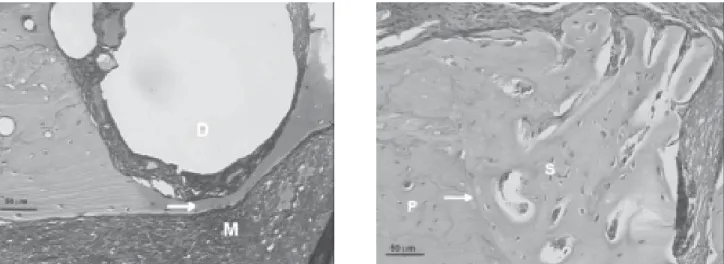
FIGURE 1- Photomicrographs of the surgically-created (unfilled) defects in rat tibia. In A, 1 day after surgery. The arrow
indicates a thin layer of cortical bone between the bottom of the defect (D) and the bone marrow (M). In B, 21 days after
surgery. The arrow points a divisory line between the newly formed bone (primary bone, P) and the old, normal bone
femur was also cut out, at a middle position, and studied as a naïve control.
Radioactivity countings and histological analysis
All samples were weighed and immediately immersed in 2.0 ml of 10 % formaldehyde solution (operated segments) or distilled water (intact segments). Radioactivity was determined in a Packard Cobra-II® gamma counter and expressed as counts/
min (cpm) per mg of fresh weight. The samples from group 1 animals (injected with 99mTc-MDP) were fixed in 10% formalin,
decalcified in 60% formic acid, dehydrated and embedded in Paraplast®. Serial
7-µm thick sections were stained with
haematoxylin-eosin for light microscopy examination. Pictures were taken with a CCD® camera (MTI) using a NIH Image
software and processed in a Power Macintosh computer using Adobe Photoshop® software.
Statistical analysis
Results were analysed by one-way analysis of variance and the Tukey-Kramer multiple comparisons test. A 2.01 version Graph Pad In statTM software was used for this purpose. When
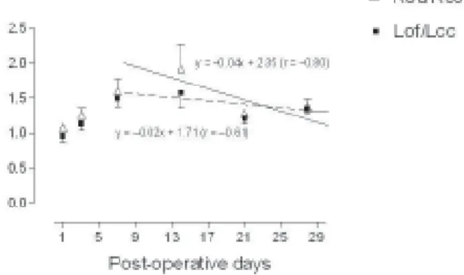
appropriate, the Student’s t test was also used. In curves describing the ratios of 99mTc-MDP uptake (see Figure 6), the
points in the segments comprising 14–28 post-operative days were fitted by linear regression, and the angular coefficients were then compared by analysis of variance.
RESULTS
Since Na99mTcO
4 cannot bind directly to any ligand 23, the
results obtained with its i.v. injection give information about the regional blood flow. Accordingly, Figure 2 shows that blood flow within bone was essentially similar in all segments in all post-operative (P.O.) days. A slight, yet significantly enhanced flow (about +67%) was detected in the 1st P.O. day, an effect seen not only in the operated segments (Lof, Rou) but also in the contiguous, defect-free controls (Lcc, Rcc).
The affinity of MDP towards bone can be clearly seen in Figure 3, which shows that, in femur, countings of 99mTc-MDP
are ca. 1,500% higher (P<0.001) than those of Na99mTcO 4 at the
3rd P.O. day. However, we observed a significant, progressive fall of that difference, and at the 28th day it was reduced to ca. 730% (P<0.001).
The uptake of 99mTc-MDP in bearing and in
defect-free tibial segments is compared in Figure 4. The operated fragments bound the MDP tracer significantly more than did the contiguous fragments. At the 7th P.O. day the difference reached ca. 57%, and this progressively felt down to 34% at the 28th day. Essentially undistinguishable results were observed in fragments containing the defects filled with freeze-dried bone and in their contiguous, control counterparts (Figure 5). At the 7th P.O. day the difference of radiotracer uptake between operated and non-operated areas was about 51% and at the 28th day it was about 42%. On the other hand, no remarkable differences were detected when the ratios of MDP uptake in filled defects were compared to those in unfilled cavities, being both situations corrected as a function of the local blood flow (Figure 6).
Notwithstanding, it is interesting to notice that not only there is an obvious exacerbation of 99mTc-MDP uptake around
the 14th P.O. day, but also the slope of the ‘recovery’ part of the curve (from the 14th up to the 28th day) is slightly faster for the unfilled cavities (Rou/Rcc, dashed line) than for the filled ones
FIGURE 2- Uptake of Na99mTcO
4 in tibiae and femurs of rats. The tracer (ca. 500 µCi) was injected i.v. and the animals
were sacrificed 2 h later. Radioactivity (in cpm) was counted
in fragments of tibiae which included the surgical defect
and in contiguous, defect-free segments. A distant, naïve
bone fragment (femur) was also counted, for control. Left
tibial defects were filled with freeze-dried bone; the right
ones were left unfilled, and groups of animals (n = 6 in
every group) were sacrificed at the indicated times after
surgery. Abbreviations: Lof = left, operated, filled; Lcc = left,
contiguous, control; Rou = right, operated, unfilled; Rcc =
right, contiguous, control; F = femur. Values are mean ±
SEM (cpm/mg wet weight)
FIGURE 3- Uptake of 99mTc-MDP and Na99mTcO
4 in femurs of rats. The tracers (ca. 500 µCi) were injected i.v. in parallel
groups of animals which were sacrificed 2 h later. Values
are mean ± SEM (cpm/mg wet weight) of data from 6
animals for every group. Asterisks indicate significant
differences regarding the corresponding points of
Na99mTcO
(Lof/Lcc, solid line).
Light microscopy examination of our material did not reveal significant differences of the bone repair process regarding the presence or absence of foreign material in the osseous cavities. Figure 1B shows, at the 21st P.O. day, the almost completely recovered defect in an unfilled cavity. The pictures seen with filled cavities (not shown) were essentially identical.
DISCUSSION
Surgically-created defects in rat tibia constitute a good model for studies on bone repair for such reasons as easy surgical access, convenient bone cortical thickness, great volume of bone marrow, and others 1,26,28,30.
In the experimental model used herein, the defect dimensions were small enough to allow proper bone regeneration. Being so, the cavity could not be classified as a ‘critical bone defect’, i.e. an intra-osseous wound with poor healing evolution 3. In
fact, the 0.5 mm depth of the cavity avoided invading the
underlying bone marrow (Figure 1A) and thus, in the filled defects (‘Lof’, see legend to Figure 2), it was assured that the biomaterial stayed inside cavity, without being expelled out as it would be the case if the blood flowed from bone marrow
3,26,28,30.
The observed increases in bone blood flow in the reference region (femur, F) might not be due to some direct, local reaction following the surgical act, but would be rather consequent to a regional vascular effect, triggered by the surgical procedure. In fact, there are several peculiarities about blood supply in mineralized tissues which are presently far from being completely understood 12,29.
It is well established that diphosphonates avidly bind to hydroxylapatite and can thus cause various cellular effects in bone cells25. Although the exact explanation for this
phenomenon is at present unresolved, it is presumable that the fading of the aforementioned ‘vascular effect’ triggered at bone level (even in distant bones) by the surgery may play a role.
The uptake in the operated segments shows that the tracer was actively uptaken by surgically-stimulated bone around
FIGURE 4- Uptake of 99mTc-MDP and Na99mTcO
4 in right tibiae of rats. The tracers (ca. 500 µCi) were injected i.v. in parallel
groups of animals which were sacrificed 2 h later.
Radioactivity (in cpm) was counted in defect-free segments
(control, panel A) and in segments containing unfilled
surgical defects (panel B). Values are mean ± SEM (cpm/
mg wet weight) of data from 6 animals for every group.
Asterisks indicate significant differences regarding the
corresponding points of Na99mTcO
4 uptake (*P<0.05; **P<0.01)
FIGURE 5- Uptake of 99mTc-MDP and Na99mTcO
4 in left tibiae of rats. The tracers (ca. 500 µCi) were injected i.v. in parallel
groups of animals which were sacrificed 2 h later.
Radioactivity (in cpm) was counted in defect-free segments
(control, panel A) and in segments containing the surgical
defects filled with freeze-dried bone (panel B). Values are
mean ± SEM (cpm/mg wet weight) of data from 6 animals
for every group. Asterisks indicate significant differences
regarding the corresponding points of Na99mTcO
the 7th day after surgery, and this occurred at a time point when cellular activity and organic matrix production conceivably were at their maximum levels17,18,19-30. In addition, it is known
that increased osteoblastic activity results in an increased deposition of the radiopharmaceutical into the affected area in comparison to the nearby or contralateral normal osseous tissues20,21.
As far as MDP uptake is concerned, the filling of osseous defects with freeze-dried bone did not interfere on the time course of the bone healing evolution. In other words, it seems likely that even if the filling of a surgically-created bone cavity with a freeze-dried bone preparation may conveniently elicit osteoconduction, the elapsed time for tissue healing may not be shortened or the clinical symptoms alleviated. Our findings confirm that bovine osseous grafts do not interfere with bone repair nor do they elicit ortotopical bone formation when the dimensions of the defect are not critical. Concerning the light microscopy, similar results have also been reported in other experimental approaches in the literature5.
CONCLUSIONS
In conclusion, our data support the view that the measurement of the uptake of 99mTc-MDP can provide valuable
information on the evolution of bone reaction after graft procedures and be used as a research tool in animal models.
RESUMO
Este trabalho objetivou estudar a evolução temporal do processo de reparo ósseo em tíbia de rato, após trauma cirúrgico padronizado. A incorporação do radiofármaco 99mTc-MDP na
região afetada foi tomada como medida indireta da intensidade
de reação tecidual; foi feito também acompanhamento histológico do processo de reparo. Foram realizadas cirurgias nas duas tíbias de 72 animais divididos em 2 grupos, sendo sacrificados em diferentes dias pós-operatórios (1, 3, 7, 14, 21 e 28 dias p.o.). As cavidades criadas nas tíbias esquerdas foram preenchidas com osso liofilizado bovino, e as direitas serviram como controle (não preenchidas). Grupos paralelos de animais foram injetados com 99mTc para avaliar a influência do fluxo
sangüíneo regional nos resultados. Duas horas após a injeção dos radiofármacos os animais foram sacrificados, a radiatividade foi contada tanto nos fragmentos das tíbias contendo os defeitos cirúrgicos como em fragmentos intactos de fêmur e de tíbias, como controle. Os resultados indicam que a maior atividade do tecido ósseo ocorreu entre 7 e 14 dias p.o. O emprego do radiofármaco mostrou ser de valor na avaliação do reparo dada sua sensibilidade. Não houve efeito significativo da presença de osso liofilizado sobre a evolução do reparo ósseo.
UNITERMOS: Reparação óssea; Radiofármaco; Microscopia óptica; Osso liofilizado; Trauma cirúrgico; Ratos.
ACKNOWLEDGEMENT
This investigation was supported by a grant to ACBF (FAPESP Fellowship no. 98/13353-7).
REFERENCES
1- Alberius P, Gordh M, Lindberg L, Johnell O. Effect of cortical perforations of both graft and host bed on onlay incorporation to the rat skull. Eur J Oral Sci 1996; 104:554–61.
2- Alexander JM. Radionuclide bone scanning in the diagnosis of lesions of the maxillofacial region. J Oral Surg 1976; 34:249–56.
3- Bach DE, Burguess LPA, Zislis T, Quigley N, Hollinger JO. Cranial, iliac and demineralized bone freeze-dried bone grafts of the mandible in dogs. Arch Otolaryngol Head Neck Surg 1991; 117:390–5.
4- Banerjee S, Samuel G, Kothari K, Unni PR, Sarma HD, Pillai MR. Tc-99m and Re-186 complexes of tetraphosphonate ligands and their biodistribution pattern in animal models. Nucl Med Biol. 2001 Feb; 28(2): 205-13.
5- Batista PS, Sant’Ana Filho M. Microscopic evaluation of healing in osseous cavities submitted to implants of lyophilized bovine bone (Bio-Oss) in femur of female rats. Rev Pos-Graduacao Fac Odontol Univ S Paulo 2001; 8:62–9.
6- Bayne K. Developing guidelines on the care and use of animals. Ann N Y Acad Sci 1998; 30:105–10.
7- Becker N, Urist M, Becker BE, Jackson W, Parry DA, Bartold M, Vicenzzi G, De Georges D, Niederwanger M. Clinical and histological observations of sites implanted with intraoral autogenous bone grafts or allografts: 15 human case reports. J Periodontol 1996; 67:1021–33.
FIGURE 6- Ratios of 99mTc-MDP uptake in tibiae of rats. The
straight lines represent the linear regression calculations
of the descending part of the curves. Dashed line refers to
right tibial (with unfilled defects) values; solid line refers to
the left tibial (with defects filled with freeze-dried bone)
values. The tracer (ca. 500 mCi) was injected i.v. and the
animals were sacrificed 2 h later. Abbreviations: see legend
8- Brown WL, Brown CP. Preliminary report on experimental bone and periosteal transplantation. Surg Gynecol Obstet 1913; 27:681–9.
9- Brugnami F, Then PR, Moroi H, Leone CW. Histologic evaluation of human extraction sockets treated with demineralized freeze-dried bone allograft (DFDBA) and cell occlusive membrane. J Periodontol 1996; 67:821–5.
10- Bush FM, Harrington WG, Harkins SW. Interexaminer comparison of bone scintigraphy and panoramic radiography of temporomandibular joints: correlation with signs and symptoms. J Prosthet Dent 1992; 67:246–51.
11- Chen B, Pei GX, Wang K, Fan YX, Wang H, Jin D, Wei KH. Repair of tibial defect with tissue-engineered bone graft and radionuclide bone imaging in goats. Di Yi Jun Yi Da Xue Xue Bao 2002 ;22(11):966-9.
12- Clemens TL. Vasoactive agents and bone metabolism. In: Bilezekian JP, Raisz LG, Rodan GA (editors). Principles of Bone Biology. San Diego: Academic Press, 1996. p.597–605.
13- Craemer TD, Ficara AJ. The value of the nuclear medical scans in the diagnosis of temporomandibular joint disease. J Oral Surg 1986; 58:382–5.
14- Crivello Jr O, Lara PF, Oliveira-Filho RM, Menecheli-José AP, Galantier, C. Avaliação cintigráfica da reparação alveolar pós-cirúrgica com osso liofilizado. In: Proceedings of the 14a. Reunião da Sociedade
Brasileira de Pesquisas Odontológicas 1997; p. 162.
15- Cronhjort M, Sääf M, Sjönberg HE, Schnell PO, Jacobsson H. Influence of the phosphate balance on the activity distribution of
99mTc-hydroxy-methylene diphosphonate. Experimental studies in
the mouse. Acta Radiol 1998; 39:427–33.
16- Dubau R, Meunier JP, Demarty R, Henaff F. Une nouvelle méthode de conservation des greffons osseux par dessiccation sous vide à partir de l’élat congelé (lyophilisation). Presse Méd 1952; 60:1402.
17- Garcia DA, Jansons D, Kapur KK. Bone-imaging and semiconductor probe measurements of technetium-99m-polyphosphate in the detection of periapical pathology in dog. Arch Oral Biol 1976; 21:167–74.
18- Genant HK, Bautovich GJ, Singh M, Lathrop KA, Harper PV. Bone-seeking radionuclides: an in vivo study of factors affecting skeletal uptake. Radiology 1974; 113:373–82.
19- Hutchinson IL, Cullum ID, Langford JA, Jarritt PH, Ell PJ, Harris M. The investigation of osteoradionecrosis of the mandible by 99mTc-methylene diphosphonate radionuclide bone scans. Br J Oral Maxillofac Surg 1990; 28:143–9.
20- Katzberg RW, O’Mara RE, Tallents RH, Weber DA. Radionuclide skeletal imaging and single photon emission computed tomography in suspected internal derangement of the temporomandibular joint. J Oral Maxillofac Surg 1984; 42:782–7.
21- Kelly JF, Cagle JD, Stevenson JS, Adler GJ. Technetium-99m radionuclide bone imaging for evaluating mandibular osseous allografts. J Oral Surg 1975; 33:11–7.
22- Kreuz FP, Hyatt GW, Turner TC, Bassett AL. The preservation and clinical use of freeze-dried bone. J Bone Joint Surg 1951; 33A:863– 72.
23- Lever SZ. Technetium and rhenium compounds. In: Wagner Jr HN (editor). Principles of Nuclear Medicine. Philadelphia: Saunders 2nd ed 1995. p.213–20.
24- Lewandrowski KU, Gresser JD, Wise DL, Trantolo DJ. Bioresorbable bone graft substitutes of different osteoconductivities: a histologic evaluation of osteointegration of poly(propylene glycol-co-fumaric acid)-based cement implants in rats. Biomaterials 2000, 21(8):757-64.
25- Miller SC, Jee WSS. The effect of dichloromethylene diphosphonate, a pyrophosphate analog, on bone and bone cell structure in the growing rat. Anat Rec 1979; 193:439–62.
26- Okamoto T, Garcia Jr IR, Magro-Filho O, Storti SC. Implante de osso anorgânico em cavidade óssea: estudo histológico em ratos. Rev Odontol UNESP 1994; 23:213–4.
27- Phemister DB. The fate of transplanted bone and regenerative power of its various constituents. Surg Gynecol Obstet 1914; 19:303– 33.
28- Rodriguez y Baena R, Zaffe D, Brusotti C, Marchetti C, Botticelli A, Rizzo S. Materiali osteoconduttori: sperimentazione animali e analisi strumentali II. Minerva Stomatol 1997; 46:635–47.
29- Solheim E, Pinholt EM, Talsnes O, Larsen TB, Kirkeby OJ. The relationship between revascularisation and osteogenesis in fresh or demineralised bone grafts. Eur Surg Res 2001; 33:42–6.
30- Virolainen P, Elima K, Metsäranta M, Aro HT, Vuorio E. Incorporation of cortical bone allografts and autografts in rats. Acta Orthop Scand 1998; 69:537–44.
Recebido para publicação em: 12/08/2003 Encaminhado para reformulações em: 19/09/2003 Pronto para publicação em: 12/12/2003
Correspondence should be addressed to: Prof. Dr. Oswaldo Crivello Junior
Department of Maxillofacial Surgery, Prosthesis and Traumatology
University of São Paulo School of Dentistry Av. Prof. Lineu Prestes 2227
05508-900 São Paulo (SP), Brazil Email: [email protected]