T
INFLUENCE OF THE EDTA, ND:YAG LASER
AND ASSOCIATION OF BOTH ON THE FILLING
OF ARTIFICIAL LATERAL ROOT CANALS
INFLUÊNCIA DO EDTA, DO LASER DE ND: YAG E DA ASSOCIAÇÃO DE
AMBOS NA OBTURAÇÃO DE CANAIS LATERAIS ARTIFICIAIS
Fernanda Gomes de MORAES, DDS, MSc
Post Graduate Student – Bauru Dental School – University of Sao Paulo.
Clovis Monteiro BRAMANTE, DDS. MSc, PhD
Associate Professor of Endodontics – University of Sao Paulo, Bauru Dental School.
Ivaldo Gomes de MORAES, DDS, MSc, PhD
Assistant Professor of Endodontics – University of Sao Paulo, Bauru Dental School.
Everdan CARNEIRO, DDS, MSc
Post Graduate Student – Bauru Dental School – University of Sao Paulo.
Renato MENEZES, DDS, MSc
Post Graduate Student – Master degree – Bauru Dental School – University of Sao Paulo.
his study aimed at evaluating the influence of EDTA, Nd:YAG laser and the combination of both for filling of artificial lateral root canals. Forty-five human mandibular premolars were employed, on which three artificial lateral root canals were prepared by means of a reamer with a similar diameter to a K file #15. The teeth were instrumented through the stepback technique employing Gates Glidden burs at the middle and cervical thirds and manual files at the apical portion, and irrigation with 1% sodium hypochloride. The teeth were divided in three groups: Group 1 –EDTA for 5 minutes; Group 2 –application of Nd:YAG laser at 15 Hz, 100 mJ and 1.5 Watts; and Group 3 – association of both. Roots were filled through the Tagger’s hybrid technique, radiographed and the radiographs were digitized. Scores were assigned to the filling of the lateral root canals. Statistical analysis revealed no significant differences between the entire groups and also on the analysis of each third.
UNITERMS: Lasers; Root canal obturation; Edetic acid.
INTRODUCTION
Filling of the root canals is the final stage of the clinical procedures of cleaning, reaming and disinfection of the root canal and should seal the entire system as hermetically as possible.
According to Cohen2, De Deus3 and Leonardo; Leal11, proper filling should be compact and complete and accomplished with inert or antiseptic materials that are able to assure a hermetic sealing. A perfect obturation should fulfill the following requirements: closure, filling of space and antisepsis. Filling may be accomplished just with cement or cement associated to other materials, especially gutta-percha, depending on the obturation technique employed. Anatomically, the root canal system comprises small
canals additionally to the main canal, which do not allow direct access during the biomechanical preparation because of their positioning and also their diameters. Such canals may be the explanation for unsuccessful endodontic therapies because they house necrotic remnants, microorganisms and/or exudate. According to Weine22, the contents of the inflamed and/or infected lateral canals may originate pain during the endodontic treatment and may also imitate periodontal diseases. Moreover, the number of lateral canals filled during the endodontic treatment is smaller than their frequency. Lack of obturation of the small canals that allow communication between the main canal and the periodontium may give rise to perpetuation of a lesion, therefore leading to failure of the endodontic treatment25.
procedures performed during the biomechanical preparation is fundamental to allow their filling with the obturation materials.
Besides obturation of these ramifications, the obturation material should also be able to seal the dentinal tubuli, therefore imprisoning bacteria that might have remained viable inside the dentinal mass, after the biomechanical preparation.
Thus, removal of the smear layer from the walls of the root canals is fundamental to allow intermeshing between the obturation material and the dentinal mass. EDTA has been the most largely employed substance for that purpose, which is able to eliminate the smear layer both from the openings of the ramifications and from the dentinal tubuli. Currently, technological advancements have led to the wide employment of laser on both Medicine and Dentistry, which may also be applied for preparation of the root canal walls before obturation.
After its creation by Maiman21 in 1960, the laser has been increasingly studied. There are many types of laser, e.g. the Nd:YAG laser. Soon in 1972, Weichman and Johnson21 made use of the Nd:YAG laser for irradiation of the apical foramen through the root canal and observed a change on the dentinal structure. Many studies demonstrated the employment and action of the Nd:YAG laser on dental structures. Depending on the adjustment of its parameters, interesting and useful outcomes may be achieved for dental treatment, such as: treatment of dentinal hypersensitivity9, 14, pulpotomy9, sterilization of the root canal9, cleaning9, 16, root canal obturation9, apicectomy8 9, 17, 18, treatment of root fractures9, sterilization of instruments9
and modification of the dentinal wall, where permeability is reduced due to the dentinal melting9, 12, 13, 19. This melting might obliterate the openings of both dentinal tubuli and ramifications.
Despite of the important role played by such imprisonment of the bacteria present on the dentinal mass remaining from the anterior stages of endodontic treatment before obturation, on the other hand the partial or total obliteration of the openings of the ramifications may impair or prevent its complete filling, which is an unfavorable aspect.
Therefore, it was interesting to conduct an evaluation of the employment of the Nd:YAG laser inside the root canals as to its capacity to melt the smear layer or the dentin itself, therefore obliterating the openings of the lateral canals, impairing or preventing their obturation. This study aimed at comparing the occurrence of obturation of artificial lateral root canals prepared on teeth treated with EDTA, Nd:YAG laser or EDTA associated to the Nd:YAG laser after canal instrumentation.
MATERIAL AND METHODS
A total of 45 human extracted mandibular premolars stores at 10% formaldehyde solution with a single canal were employed. The crowns were removed and three lateral canals
were prepared on one of the proximal walls, being one at each root third, by means of a rotary reamer #15 perpendicular to the canal axis.
After preparation of the lateral root canals, instrumentation of the root canals was initiated. Odontometry was visually accomplished with a K file #10 with a stop.
The canals were initially instrumented with Gates Glidden burs #2 to #5 on the middle and cervical thirds. Instrumentation of the apical third was manually accomplished with K files by means of the stepback technique, employing the file #40 as the working file. After accomplishment of the apical stop, the programmed stepback on cervical direction was initiated, on which each increase in file size corresponded to a 1-mm decrease in length, up to finalization with a file #60.
At every change of file, irrigation was performed with a 5cc Luer Lock syringe with a 30X4 cannula, employing a 1% sodium hypochlorite solution (Milton solution). After instrumentation was completed, the canals were irrigated with 5ml of saline solution.
After instrumentation and final irrigation with saline, the roots were randomly divided in 3 groups, with 15 roots each, according to the treatments that would be performed on the root canal walls.
Group I – Application of EDTA (17%) filling the root canals, followed by agitation with the working file and maintenance for 5 minutes. After this time, the root canals were irrigated with saline solution.
Group II – Application of the Nd:YAG laser (Pulse Master 600 IQ. American Dental Technologies, provided by the UNESP Dental School at Sao Jose dos Campos), after drying of the root canals, with the following parameters: 15 Hz, 100mJ and 1,5 Watts for 7 seconds with helicoidal movements at a apical to cervical direction with four repetitions, adding up to 28 seconds, transmitted by a 300-mm optic fiber.
Group III – Application of EDTA on the root canals for 5 minutes and irrigation with saline solution as accomplished for Group I. After this period of time, the canals were dried with absorbent paper points and the Nd:YAG laser was applied in the same manner as for Group II.
After the proper treatment, the canals were filled by means of the Tagger’s hybrid technique, with AH Plus cement and gutta-percha cones, and the coronal opening was sealed with Cimpat.
After obturation, radiographs were taken from the roots on the buccolingual direction for analysis of the obturation of the artificial lateral root canals.
Evaluation of the filling of the lateral canals was completed through observation of the radiographic images, which were digitized and analyzed by means of the software Digora. Analysis of the filling of the lateral canals was performed according to the following scores:
0 = complete filling
2 = extrusion of the obturation material with lack of filling of the lateral canal
3 = empty lateral canal with no evidence of penetration of material
Data were submitted to the Kruskal Wallis test, because it is a non-parametric test that compares two or more groups and the analysis was subjective.
RESULTS
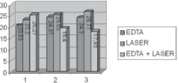
Graph I demonstrates the values related to the mean positions representing the filling of the lateral canals according to the experimental groups, regardless of the third analyzed.
Graph II demonstrates the values related to the mean positions representing the filling of the lateral canals according to the groups, separated by thirds:
The results demonstrated that there was no statistically significant difference between groups or between thirds.
DISCUSSION
The technological evolution throughout the last decades has been remarkable. Laser is one of these evolutions, which has been developing and catching the curiosity and the interest of clinicians and researchers. Specifically on this study, the main interest was to observe its effect on the openings of the lateral canals and on the dentinal walls, with or without a smear layer.
The temperature reached by the dentinal structure is also related to the power and other parameters of the laser, such as: frequency, timing of radiation, amount of energy. The higher these parameters, the larger will be the increase in temperature5, 10, 23. Lan10 evaluated the increase in temperature of the external root surface on the apical third during application of the Nd:YAG laser with different parameters and concluded that with 10mJ of energy and 20pps, 80mJ and 25pps, and 60mJ and 30pps, the temperature did not exceed 10oC, which is considered the maximum limit to avoid damage to the periodontal ligaments. Bahcall et al.1 evaluated the biological effect on the periodontal ligament
of teeth submitted to application of the Nd:YAG laser with a power of just 3W, 25pps for periods of time below 30 seconds and observed damages such as cell necrosis on the periodontal ligament soon on the first day, followed by an increase in he number and volume of osteocytes and osteoclasts, and ankylosis and lysis of the cement at 30 days.
These reports allow the selection of the following parameters on the present study: 1.5W of power, 100mJ of energy and 15Hz of frequency for a period of 28 seconds, as suggested by Gutknecht, et al.6, 7.
The observed results demonstrated that the treatments applied to the root canal walls did not significantly interfere with the obturation of the artificial lateral root canals in general (Figure 1) or on the individual analysis of each third independently (Figure 2), in disagreement with the findings of Levy12 and Miserendino; Levy, Rizoiu13, who observed partial or complete obliteration of the openings of lateral canals besides the dentinal tubuli. However, it should be highlighted that the power employed by these investigators was also much larger than that of the present study. Levy12 made use of a power of 35W and frequency of 20Hz, whereas Miserendino, Levy, Rizoiu13 applied a power of 5W with a frequency of 50Hz.
Evaluation of the results, comparing the specific treatments accomplished on the root canal walls, might lead to the assumption that the smear layer present on the root canal walls in Group II, which did not employ the EDTA, might be a sufficient resource of material to yield obliteration of the openings of the lateral walls by its presence itself or due to a larger amount of material to be melted by the laser when compared to Group III. This was not observed, allowing the conclusion that the smear layer may have lost its effect or may have been eliminated by the application of laser.
The change in dentinal structure is directly related to an increase in the laser power21. Through observation of these factors, it may be concluded that, on the mentioned studies, the dentin may have been sufficiently melted to obstruct the openings of the lateral canals. This did not occur on the present study, certainly because of the smaller level of power employed.
FIGURE 1- Graph demonstrating the mean positions
achieved in relation to the scores assigned to the obturation of the lateral canals, regardless of the third evalueted
FIGURE 2- Graph demonstrating the mean positions
achieved in relation to the scores assigned to the obturation of the lateral canals, according to the thirds:
The lack of obstruction of the openings of the artificial lateral root canals allowed their obturation in most cases. Some canals were partially obturated, probably due to the inclusion of air bubbles during obturation, as observed by Villegas, et al.20, and also because of the presence of some portion of smear layer that might partially obliterate the canal lumen.
The obliteration occurring at least partially in some cases was not enough to completely prevent penetration of the obturation material. The compression exerted by the condenser during obturation by means of the Tagger’s hybrid technique pushes the material against the root canal walls and consequently towards the lateral canals, therefore filling them. Moreover, the AH Plus cement is highly flowable and this property allows its penetration in the small gaps that might exist due to the partial obliteration yielded by the presence of the smear layer.
Since the results did not demonstrate superiority of any treatment applied to the dentinal walls in relation to the others, this might suggest that, considering the cost/benefit relationship, utilization of the EDTA would be the most appropriate. However, the action of the laser is not restricted to the structural portion of the dentin. It also has some bactericide property4, 6, 8, 15. However, the reduction in the number of bacteria also depends on the energy of radiation, as well as on the bacterial species present4.
Some disadvantages should also be remembered, such as an increase in temperature that might lead the cells on the periodontal ligament to necrosis, occurrence of ankylosis1, and also alterations on the layer of odontoblastic cells24, a change in the chemical composition of the collagen on the dentinal matrix, and consequently an increase in the mineral content (calcium phosphate).
CONCLUSION
According to the methodology employed on the present study, it was concluded that no significant differences could be observed as to the presence or absence of obturation of the artificial lateral root canals after treatment of the root canal walls with EDTA, Nd:YAG laser or the association of both.
ACKNOWLEDGEMENT
To the UNESP campus at Sao Jose dos Campos, especially to Professor PhD Marcia Valera, for allowance of the laser device. To CNPq, for funding this study.
RESUMO
Este estudo teve como objetivo avaliar a ação do EDTA, do laser de Nd:YAG e a associação de ambos na obturação dos canais laterais artificiais. Foram utilizados 45 dentes pré-molares inferiores humanos, nos quais foram
confeccionados três canais laterais com o auxílio de um alargador com diâmetro compatível de uma lima do tipo K número 15. Os dentes foram instrumentados pela técnica regressiva utilizando brocas de Gates Glidden nos terços cervical e médio e limas manuais no terço apical e irrigação com hipoclorito de sódio a 1 %. Os dentes foram divididos em três grupos: Grupo I EDTA por 5 minutos, Grupo II aplicação de laser de Nd:YAG com 15 Hz, 100mJ e 1,5 Watts e Grupo III a associação de ambos. As raízes foram obturadas pela técnica Híbrida de Tagger, radiografadas e as radiografias escaneadas. Para o preenchimento dos canais laterais foram dados escores. A análise estatística mostrou não haver diferença significante entre os grupos analisados globalmente e nem quando analisados por terços.
UNITERMOS: Laser; Obturação do canal radicular; EDTA.
REFERENCES
1- Bahcall J, Howard P, Miserendino L, Walia H. Preliminary investigation of the histologycal effects of laser endodontic treatment on the periradicular tissues in dogs. J Endod 1992 Feb; 18(2):47-51.
2- Cohen S, Burns R C. Caminhos da polpa. Rio de Janeiro: Guanabara Koogan; 1998.
3- De Deus Q D. Endodontia. Rio de Janeiro: MEDS; 1992.
4- Folwaczny M, Mehl A, Jordan C, Hickel R. Antibacterial effects of pulsed Nd:YAG laser irradiation at different energy setting in root canals. J Endod 2002, Jan; 28(1):24-9.
5- Fraunhofer J A V, Allen D J. Thermal effects associated with the Nd:YAG dental laser. Angle Orthod 1993 Apr; 63(4):299-303.
6- Gutknecht N, Moritz A, Conrads G, Sievert T, Lampert F. Bactericidal effect of the Nd:YAG laser in vitro root canals. J Clin Laser Med Surg 1996 Apr; 14(2):77-80.
7- Gutknecht N, Kaiser F, Hassan A, Lampert F. Long – term clinical evaluation of endodontically treated teeth by Nd:YAG lasers. J Clin Laser Med Surg 1996 Jan; 14(1):77-80.
8- Hardee M W, Miserendino L J, Kos W, Walia H. Evaluation of the antibacterial effects of intracanal Nd:YAG laser irradiation. J Endod 1994 Aug; 20(8):377-80.
9- Kimura Y, Wilder– Smith P, Matsumoto, K. Lasers in endodontics: a review. Int Endod J 2000 May; 33(3):173-85.
10- Lan W H. Temperature elevation on the root surface during Nd:YAG laser irridiation in the root canal. J Endod 1999 Mar; 25(3):155-6.
11- Leonardo M R, Leal J M. Endodontia: tratamento de canais radiculares. São Paulo: Panamericana; 1998.
13- Miserendino L J, Levy G, Rizoiu I M. Efects of Nd:YAG laser on the permeability of root canal wall dentin. J Endod 1995 Feb; 21(2):83-7.
14- Morlock B J, Pippin D J, Cobb C M, Killoy W J, Rapley, J W. The effect of Nd:YAG laser exposure on root surfaces when used as an adjunct to root planning: an in vitro study. J Periodontol 1992 July; 63(7):637-41.
15- Moshonov J, Orstavik D, Yamauchi S, Pettiette M, Trope M. Nd:YAG laser irradiation in root canal disinfection. Endod dent Traumat 1995 Oct.; 11(5):220-4.
16- Saunders W P, Whitters C J, Strang R, Moseley H, Payne A P, McGadey J. The effect of an Nd:YAG pulsed laser on the cleaning of the root canal and the formation of a fused apical plug. Int Endod J 1995 July; 28(4):213-20.
17- Stabholz A, Khayat A, Ravanshad S H, McCarthy D W, Neev J, Torabinejad M. Effects of Nd:YAG laser on apical seal of teeth after apicoectomy and retrofill. J Endod 1992 Aug; 18(8):371-5.
18- Stabholz A, Khayat A, Weeks D A, Neev J, Torabinejad M. Scanning electron microscopic study of the apical dentine surfaces lased with Nd:YAG following apicectomy and retrofill. Int Endod J 1992 Nov; 25(6):288-91.
19- Türkmen C, Günday M, Karaçorlu M, Basaran B. Effect of CO2, Nd:YAG and ArF Excimer lasers on dentin morphology and pulp chamber temperature: an in vitro study. J Endod 2000 Nov; 26(11):644-8.
20- Villegas J C, Yoshioka T, Kobayashi C, Suda H. Obturation of accessory canals after four different final irrtigation regimes. J Endod 2002 July; 28(7):534-6.
21- Weichman J A, Johnson F M, Nitta L K. Laser use in endodontics part II. Oral Surg oral Med Oral Patho1972 Nov; 31(5):828-30.
22- Weine F S. The enigma of the lateral canal. Dent Clin North Amer 1984 Oct; 28(4):833-52.
23- White J M, Fargan M C, Goddis H E. Intrapulpal temperatures during pulsed Nd:YAG treatment of dentin in vitro. J Periodontol 1994 Mar; 65(3):255-9.
24- Wigdor H, Abt E, Ashrafi S, Walsh JR J T. The effect of lasers on dental hard tissues. J Am Dent Assoc 1993 Feb; 124(2):65-70.
25- Xu G, Zhang Z. Filling of the lateral canal. Oral Surg 1984 Aug; 58(2):221-4.
Recebido para publicação em: 09/06/2003 Enviado para reformulações em: 06/08/2003 Pronto para publicação em: 26/08/2003
Corresponding author: Fernanda Gomes de Moraes