*e-mail: [email protected]
Trabalho apresentado no 1º Congresso da Sociedade Brasileira em Materiais, Rio de Janeiro, Julho de 2002.
Preparation and Biocompatibility of Poly
(Methyl Methacrylate) Reinforced with Bioactive Particles
Marivalda de Magalhães Pereiraa*, Rodrigo Lambert Oréficea, Herman Sander Mansura, Miriam Teresa Paz Lopesb, Regina Maria De Marco Turchetti-Maiab,
Anilton Cesar Vasconcelosb
aDepartamento de Engenharia Metalúrgica e de Materiais
bInstituto de Ciências Biológicas, Universidade Federal de Minas Gerais,
Rua Espírito Santo 35/206, 30030-160 Belo Horizonte - MG, Brazil
Received:October 3, 2002; Revised: April 6, 2003
Calcium phosphates and bioactive glasses have been used in many biomedical applications for more than 30 years due basically to their bioactive behavior. However, ceramics are too brittle for applications that require high levels of toughness and easy processability. In this work, a biphasic calcium phosphate (BCP) and a bioactive glass composition (BG) were combined with polymers to produce composites with tailorable properties and processability. The BCP particles were syn-thesized by a precipitation technique. The BG particles were produced by sol-gel processing. The BCP particles were treated with a silane agent to improve the compatibility between particles and the polymer matrix. Dense samples were produced by hot pressing (200 °C) a mixture of 30 wt.% of particles in poly (methyl methacrylate). The samples produced were characterized by X-ray diffraction, infrared spectroscopy and scanning electron microscopy. Mechanical proper-ties were evaluated by a three point bending test. Samples were also submitted to in vitro bioactivity test and in vivo toxicity test. Results showed that the production of the composites was success-fully achieved, yielding materials with particles well dispersed within the matrices. Evaluation of the in vivo inflammatory response showed low activity levels for all composites although compos-ites with silane treated BCP particles led to milder inflammatory responses than composcompos-ites with non-treated particles.
Keywords: biomaterials, composites, poly(methyl methacrylate), bioactive glass, calcium phosphate
1. Introduction
Several materials have been developed with the objec-tive of regeneration or substitution of bone structures in-jured or lost by diseases or accidents. However, a gap re-mains to be filled since no synthetic material used until now presents characteristics close to the natural tissue, attend-ing both biological aspects as well as mechanical require-ments. Among these materials bioactive ceramics have been
largely studied and used1,2, due to their osteoconduction
properties and the ability of promoting the formation of a continuous bone-ceramic interface, therefore allowing an implant fixation mechanism. Bioceramics usually have
mechanical properties quite different from those of natural tissues, particularly a high elastic modulus and a low tough-ness. This fact restricts the use of these materials in a wide range of applications. One alternative that is being
consid-ered and studied is the production of composites3-5.
Poly-mer matrices reinforced with a bioactive phase can poten-tially combine the typical bioactive behavior of some bioceramics with mechanical properties closer to human tissues. In this work the polymer used to produce the com-posites was poly (methyl methacrylate) which has been
largely used as a biomedical material6. In combination with
proper-ties closer to bone tissue. The composites were prepared using different bioactive ceramic particles, a bioactive glass (BG) and a biphasic calcium phosphate (BCP). In one type of composite produced the BCP particles were treated with a silane agent to improve the compatibility between parti-cles and the polymer matrix. The goal of the study was to evaluate the structural and mechanical characteristics of these composites. Also the toxicity and the inflammatory response capability of these new composites were
investi-gated. An in vivo test was performed by surgical
implanta-tion of the biomaterial in the subcutaneous tissue of mice, followed by histological studies in pre-established intervals of four, seven and fourteen days after the implantation, to evaluate the induced inflammatory response.
2. Materials and Methods
Commercial PMMA granules (from Metacril) was used as the polymer matrix. The bioceramic particles were pro-duced as follows. All reagents used were analysis grade.
A bioactive glass with molar composition 60% SiO2,
36% CaO and 4% P2O5 was synthesized by the sol-gel
method. Tetraethylorthosilicate (TEOS), triethylphosphate (TEP) and tetra-hydrated calcium nitrate were used as the silica, phosphorous and calcium source respectively. The
hydrolysis was performed in the presence of 2N HNO3
so-lution and H2O/TEOS molar ratio equal to 8. The reagents
were mixed for 1 h and the sol formed was cast in polyethylene containers. The samples were aged at 60 °C, dried up to 170 °C, and heat treated at 700 °C. For the prepa-ration of the biphasic calcium phosphate particles calcium hydroxide and monobasic calcium phosphate solutions were mixed at ambient temperature for 30 min, filtered and dried at 100 °C for 24 h. The filter cake was heat treated at 1100 °C for 3 h and then ground.
The ground materials were separated in the 37 to 150 µm range. Part of the BCP particles was immersed in a silane solution, filtered and dried before being mixed with the polymer material. The mixture of 30% weight of bioactive particles and PMMA was compression molded at 15 MPa for 15 min at 170 °C.
The bioactive particles and the obtained composite sam-ples were analyzed by Scanning Electron Microscopy and X-ray diffraction analysis (XRD). The mechanical behavior of the composites was evaluated by the three point bending test using samples 40 × 5 × 3 mm and 0.75 mm/min cross head speed. Three samples of each condition were tested. To evaluate the bioactivity of the composites samples were
immersed in a simulated body fluid (SBF)7 at 37 °C for
different time periods. The surface area/volume ratio was
fixed in 0.7cm-1. The samples were analyzed by Fourier
Transform Infrared Spectroscopy using the diffuse reflect-ance method (DRIFT).
The in vivo test to evaluate the toxicity and inflamma-tory response was performed in five weeks old mice, ac-cording to ASTM Standards F 763-87 and F 981-93. The animals, divided in three groups of five animals each, were
previously dewormed with Porocin and bathed with
Triatox for ectoparasite control, 48 h before the
experi-ment. The animals were sedated with Diazepan (8 mg/kg,
ie 240 µg/30 g) via intra peritoneal and anesthetized with
Thionembutal (30 mg/kg, ie 1 mg/30 g) using the same
route, with five minutes interval between them. The mate-rial was surgically implanted in the subcutaneous tissue of the interescapular region at the animal’s back. Control inci-sions were also made in the caudal position. The animals were euthanized 4, 7 and 14 days after implantation. Histo-logical sections of the materials-tissue interface were rou-tinely processed and analyzed by optical microscopy.
3. Results
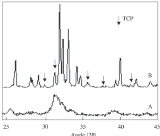
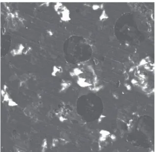
Figure 1 shows SEM micrographs of the obtained com-posites revealing that their production was successfully achieved, yielding materials with particles well dispersed within the matrices. A higher magnification shows that the BCP particles have several pores or voids, probably origi-nated during sample preparation, since small particles were observed in the solution during ultrasonic cleaning of the polished samples. This suggests that the sintering proce-dure was not efficient and that a higher sintering tempera-ture may be necessary. The XRD spectra of the BCP parti-cles presented in Fig. 2 indicates the presence of the two crystalline phases, hydroxyapatite (HA) and b-tricalcium phosphate (TCP). The proportion of the two phases present, determined based on the area under the peaks, is 80% HA and 20% TCP.
The mechanical properties of the composites determined by the three point bending test are presented in Table 1. The stress × strain curve shows that the composites have a brit-tle behavior, similar to the polymer used. The introduction of the glass particles increased the elastic modulus com-pared to the polymer, as expected. The same behavior was not observed for the composites reinforced with BCP parti-cles. All composites presented a decrease in strength rela-tive to the polymer PMMA. This decrease in strength may be related to the presence of particles-matrix interfacial de-fects. No significant difference was observed in the mechani-cal properties of the composites produced with BCP parti-cles treated with silane compared with composites contain-ing no treated particles.
Some important applications of biomaterials are related to the replacement or repair of hard tissues that are not load carrier (such as in bones located at the face). For this type of application, biomaterials need to have stiffness similar to bone, so they can resemble bone for aesthetic purposes. On the other hand, they do not require high strength, since they will not be responsible for carrying load. In these cases, increasing the elastic modulus of polymers can have a posi-tive impact in producing bioacposi-tive materials that combine bioactive behavior with elastic modulus comparable to bone. It is important to emphasize that other bioactive materials
(such as hydroxyapatite and bioglasses) have elastic modu-lus too high, when compared to bone. Therefore, although a decrease in strenght compared to the pure polymer was observed, the increase in elastic modulus presented by the PMMA/BG composites leaded to materials closer in stiff-ness to bone and potential materials for non load bearing applications.
The FTIR spectra of samples before and after immer-sion in SBF are presented in Figs. 3 and 4. In the unreacted PMMA/BG composite samples the band present at
1700 cm-1 corresponds to the C=O vibration related to the
polymer matrix, the bands at 1100 and 482 cm-1 corresponds
Figure 2. X-ray diffration spectra of BCP particles after drying at 100 °C (A) and after heat treatment at 1100 °C for 3 h (B). Peaks indicated by arrows correspond to tricalcium phosphate (TCP); other peaks correspond to hidroxyapatite (HA).
Table 1. Mechanical properties of the composites, determined by tree point bending test.
Material Strength (MPa) Elastic modulus
(GPa)
PMMA 91 1.7
PMMA/BG 64 6.0
PMMA/HA 38 1.4
to the Si-O-Si stretching and bending vibrations respectively, characteristic of the glass particles. After immersion in SBF solution the major modifications observed are: a pronounced decrease in intensity of the bands related both to the poly-mer matrix and to the glass particles and the appearance of
two bands at 598 and 566 cm-1, assigned as P-O bending
vibrations, relative to the formation of a low crystallinity calcium phosphate layer. This layer is well established after 20 h immersion in SBF and was also observed by SEM analysis (Fig. 5). The formation of this layer in the early stages has been considered indicative of a high reactivity of the material and an indication of its bioactivity1,8.
In the case of the PMMA/BCP composites the triplet in
the region of 602-550 cm-1, assigned as P-O bending
vibra-tion, is characteristic of a crystalline hydroxyapatite, the major phase present in the PMMA/BCP composite. No sig-nificant modification in the FTIR spectra was observed af-ter immersion in SBF, even afaf-ter 8 weeks.
Histological sections showed a typical mild inflamma-tory response, with predominant fibroblasts and macrophages scattered involving the implant, with low ac-tivity levels and a quick resolution for all composites. Illus-trative sections are shown in Fig. 6 for the PMMA/BG com-posites. At four days after the implantation a subacute in-flammatory response was observed, but with mild cellular response. At seven days post surgery, the implanted mate-rial was surrounded by a fibrous capsule, with dense fibro-plasia and active macrophages sometimes fused in giant cells containing fragments of the implanted material. At 14 days after the implantation the inflammatory response was prac-tically well resolved with significant reduction in the number of macrophages.
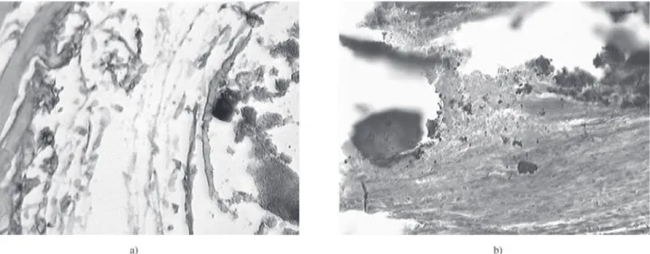
Animals implanted with silane treated particles PMMA/ BCP composites showed milder inflammatory responses than composites with non-treated particles (Fig. 7). There-fore it was proposed that the silane treatment on particles Figure 4. FTIR spectra of composites PMMA/BCP before and
after immersion in SBF for 0 (A), 1 (B) and 8 weeks (C).
Figure 6. Histological sections of tissue-implant interface formed after surgical insertion of PMMA/BG composite samples for 4 (a), 7 (b) and 14 (c) days. HE stain, magnifications: 40, 4 and 10× respectively.
improved their adhesion toward the polymer matrix, reduc-ing particle pull out durreduc-ing in vivo test, resultreduc-ing in improved biocompatibility.
The mild inflammatory response observed in the
sub-cutaneous in vivo analysis show the non toxic nature of the
materials produced. Further in vivo analyses of the PMMA/
BG composite interaction with bone tissue leading to bone bonding should be performed to confirm the bioactive na-ture of these composites.
4. Conclusions
The production of the composites was successfully achieved yielding materials with particles well dispersed within the matrices. The elastic modulus increased in the composites reinforced with BG particles but not in the com-posites reinforced with BCP particles. Although a decrease in strength compared to the pure polymer was observed, the increase in elastic modulus presented by the PMMA/BG composites leaded to materials closer to bone in terms of stiffness and potential materials for non load bearing applications.
The formation of an HCA layer in vitro was observed in the PMMA/BG composites indicating the high reactivity of
the material and its bioactivity. Further in vivo analyses of
this composite interaction with bone tissue leading to bone
Figura 7. Histological sections of tissue-implant interface after surgical insertion of PMMA/BCP (a) and PMMA/BCP/Sil (b) composite samples 7 days post surgery. HE stain, magnification 40×.
bonding should be performed to confirm the bioactive
na-ture of the composites. Evaluation of the in vivo
inflamma-tory response showed low activity levels for all composites, and their non toxic behavior. Composites with silane treated BCP particles led to milder inflammatory responses than composites with non-treated particles.
References
1. Hench, L J Amer Cer Soc, v. 8, p. 1705-1728, 1998.
2. Ducheyne, P.; Qiu, Q. Biomaterials, v. 20, p. 2287-2303,
1999.
3. Wang, M.; Hench, L.L.; Bonfield, W. Journal of
Bio-medical Materials Research, v. 42, p. 577-586, 1998.
4. He, D.; Jiang, B. J. Appl. Polym. Sci., v. 49, p. 617-621,
1993.
5. Bonfield, W. Proceedings of the 11th International
Sym-posium on Ceramics in Medicine, Bioceramics , v. 11, p. 37-40, 1998.
6. Mousa, W.F.; Kobayashi, S.S.; Kamimura, M. et al.,
Biomaterials, v. 21, p. 2137-2146, 2000.
7. Kokubo, T.; Kushitani, H.; Sakka S.; Kitsugi,T.;
Yamamuro, T. J Biomed Mater Res, v. 24, p. 721-734,
1990.
8. Salinas, A.J.; Roman, J.; Vallet-Regi, M. et al.,