Sulfated-polysaccharide fraction extracted from red algae
Gracilaria birdiae
ameliorates trinitrobenzenesulfonic
acid-induced colitis in rats
Tarcisio V. Britoa, José P. R. P. Netoa, Rafael S. Prudêncioa, Jalles A. Batistaa, José S. C. Júniora, Renan O. Silvaa, Álvaro X. Francob, Karoline S. Aragãob, Pedro M. G. Soaresb,
Marcellus H. L. P. Souzab, Luciano S. Chavesc, Ana L. P. Freitasc, Jand-V. R. Medeirosaand André L. R. Barbosaa
aLAFFEX – Laboratory of Experimental Physiopharmacology, Biotechnology and Biodiversity Center Research (BIOTEC), Federal University of Piauí,
Parnaíba, PiauíbLAFICA – Laboratory of Pharmacology of Inflammation and Cancer, Department of Physiology and Pharmacology andcLaboratory
of Proteins and Carbohydrates of Marine Algae, Department of Biochemistry and Molecular Biology, Federal University of Ceará, Fortaleza, Ceara, Brazil
Keywords
antioxidant; colitis;Gracilaria birdiae; sulfated polysaccharide; TNBS
Correspondence
André L. Reis Barbosa, Biotechnology and Biodiversity Center Research, Av. São Sebastião, n° 2819, CEP, Parnaíba, 64202020 Piauí, Brazil.
E-mail: andreluiz@ufpi.edu.br
Received December 4, 2013 Accepted January 18, 2014
doi: 10.1111/jphp.12231
Abstract
Objectives The aim of this study was to evaluate the protective effect of the sulfated-polysaccharide (PLS) fraction extracted from the seaweed Gracilaria
birdiaein rats with trinitrobenzenesulfonic acid (TNBS)-induced colitis.
Methods In the experiments involving TNBS-induced colitis, rats were pretreated with polysaccharide extracted fromG. birdiae(PLS: 30, 60 and 90 mg/kg, 500μL p.o.) or dexamethasone (control group: 1 mg/kg) once daily for 3 days starting before TNBS instillation (day 1). The rats were killed on the third day, the portion of distal colon was excised and washed with 0.9% saline and pinned onto a wax block for the evaluation of macroscopic scores. Samples of the intestinal tissue were used for histological evaluation and assays for glutathione (GSH) levels, malonyldialdehyde (MDA) concentration, myeloperoxidase (MPO) activity, nitrate and nitrite (NO3/NO2) concentration and cytokines levels.
Key findings PLS treatment reduced the macroscopic and microscopic TNBS-induced intestinal damage. Additionally, it avoided the consumption of GSH, decreased pro-inflammatory cytokine levels, MDA and NO3/NO2concentrations
and diminished the MPO activity.
Conclusions Our results suggest that the PLS fraction has a protective effect against intestinal damage through mechanisms that involve the inhibition of inflammatory cell infiltration, cytokine releasing and lipid peroxidation.
Introduction
The study for natural products with pharmacological prop-erties has significantly contributed to the discovery of com-pounds with important applications.[1,2] Recently, marine
algae have attention as a source of bioactive substances for the development of new drugs.[3]In particular, algae are a
very important and commercially valuable resource for the food industry; they also serve as soil conditioners and are used in traditional folk medicine due to their known health benefits.[4,5]
Red seaweeds produce an innumerous variety of sulfated galactans and are rich sources of sulfated polysaccharides (PLS). PLS from theGracilariagenus are composed mainly
of the alternating 3-linked-β-D-galactopyranose unit (Gal) and the 4-linked-3,6-anhydro-α-L-galactopyranose unit (AnGal). The Gal unit can be substituted for either methyl or sulfate ester radicals.[6]
Recent studies have shown that PLSs extracted from marine red algae demonstrated antioxidant and anti-inflammatory effects,[7,8] decreasing the production and
releasing of free-radical scavengers and preventing oxidative damage in the living organism.[9]However, few studies have
correlated the protective effect of PLSs from seaweeds to the intestinal damages associated with the colitis induced by trinitrobenzenesulfonic acid (TNBS).
And Pharmacology
The inflammatory bowel diseases (IBD) refer essentially to two different diseases: Crohn’s disease and ulcerative colitis (UC). Although the aetiology of IBD remains unclear, there is evidence that it involves immune, genetic and environmental factors, which are related to the initia-tion and development of colitis.[10,11]In the IBD, there is an
increasing number of inflammatory cells that are found in areas of the intestine with chronic inflammation, resulting in overproduction of a variety of pro-inflammatory media-tors including eicosanoids, platelet-activating factor, releas-ing of pro-inflammatory cytokines and specimens reactive of oxygen and nitrogen metabolites.[10–13]
Currently, there is no effective therapy to cure the disease but the mainstream treatment depends on reduction of the abnormal inflammation in the colon lining, and thereby relieves the symptoms of disease. The treatment depends on the severity of the disease; therefore treatment is adjusted for each individual.[14]Most people with mild or moderate
IBD are treated with corticosteroids to reduce inflammation and relieve symptoms.[15]Nearly 25% of patients with UC
requiring steroids therapy become steroid dependent after 1 year, and virtually all develop steroid-related adverse events.[16]
Considering the data above, it is plausible that the red algae and their derivative, the PLS, demonstrated marked antioxidant and anti-inflammatory effect, decreasing the production of free-radical scavengers and preventing oxida-tive damage. Thus, this study aimed at evaluating the pro-tective effect of PLS fraction extracted from red algae
G. birdiae in the trinitrobenzenesulfonic acid
(TNBS)-induced colitis in rats.
Methods
Extraction of the PLS fraction
The extraction of the polysaccharide of G. birdiae was accomplished at the Laboratory of Biochemistry of Sea Algae at the Department of Biochemistry and Molecular Biology of the Federal University of Ceará. The marine red algae Gracilaria was collected at Flecheiras beach, Trairí, Ceará, Brazil, in September 2006, geographical localization: 03°13′25′S and 39°16′65″W. A voucher specimen (No. 40781) was deposited in the Herbarium Prisco Bezerra in the Department of Biological Sciences, Federal University of Ceará, Brazil. The samples were cleaned of epiphytes, washed with distilled water and stored at−20°C. The extrac-tion procedure of polysaccharide was performed according with method previously described.[17]
Chemical structure of PLS fraction
The chemical structure was previously described.[18,19]Total
sugar content of each fraction was determined according to
the method of Dubois.[20] Protein fractions were obtained
by Bradford’s method.[21]Sulfate content was determined by
the barium chloride gelatine method,[22]and the
monosac-charide contents of red seaweed galactans was obtained by reductive hydrolysis.[23]
Animals
Male Wistar rats (160–180 g) deriving from Federal Univer-sity of Piauí (UFPI). The animals were housed at 25±2°C under a 12/12 h light/dark cycle and were deprived of food for 12–16 h before the experiments, with free access to water. Experiments were conducted in accordance with current established principles for the care and use of research animals (National Institutes of Health guidelines) and were approved by Ethics Committee of the Federal Uni-versity of Piauí (protocol n° 036/12).
Induction of colitis
Colitis was induced (n=6 rats/group) by intracolonic single instillation of a solution of 20 mg of TNBS in 50% ethanol (EtOH), and the animals were previously anesthe-tized with ketamine (80 mg/kg; intramuscularly, i.m.) and xylazine (10 mg/kg, i.m.). Control groups received an equivalent volume of saline. A rubber catheter was inserted into the rectum, 8 cm distal to the anus, and the TNBS solu-tion was introduced. Animals were placed head down in a vertical position for 30 s and then returned to their cages. Three days after the induction of colitis, the rats were killed and the experimental protocols executed as described below.
Treatment protocols
In the experiments involving TNBS-induced colitis, rats were treated with PLS (30, 60 and 90 mg/kg; orally, p.o.) or dexamethasone (1 mg/kg; subcutaneously, s.c.) once daily for 3 days before and after TNBS instillation (only on first day). On the third day after induction of TNBS-colitis, the rats were killed, and the abdomens were then opened, and after the identification of the intestine, the portion of distal colon was excised and washed with 0.9% saline. Then this segment, measuring 5 cm, were weighed to determinate the colon oedema. The results were expressed in increase in colon weight (g)/5 cm ratios, compared with a normal control group, without colitis. The evaluation of macro-scopic scores of lesion was performed by modifying the criteria previously described (Table 1).[24] Additionally,
samples of intestinal tissue were then removed for the measurement of glutathione (GSH) concentration,[25]
malonyldialdehyde (MDA) level,[26] myeloperoxidase
(MPO) activity,[27]nitrate and nitrite (NO
concentra-tion[28] and cytokine levels[8] in inflamed colon tissue.
Samples were fixed in 10% formalin for histopathological analysis.
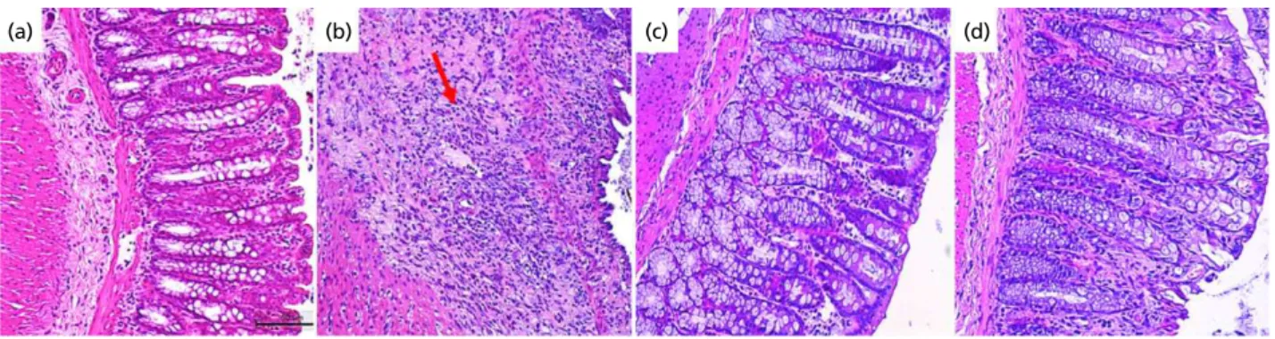
Histological evaluation
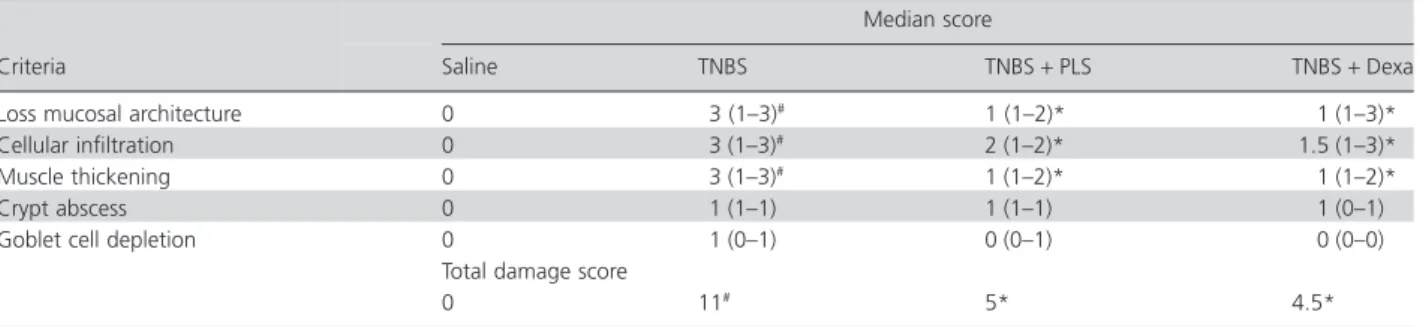
For histological evaluation, the intestinal tissue was fixed in formalin for 24 h. After that, the samples were stored in a solution of 70% alcohol. The samples were embedded in paraffin and sectioned, and the sections were depar-affinized, stained with haematoxylin and eosin, and then examined under a microscope (blind analysis). The laminas were analysed according to the criteria described previ-ously,[29] in which scores are assigned to the following
parameters: loss of mucosal architecture (score of 0–3), cel-lular infiltration (score of 0–3), muscle thickening (score of 0–3), crypt abscess (score of 0–1) and goblet cell depletion (score of 0–1).
Myeloperoxidase assay
This assay was performed to evaluate the neutrophil infil-tration into the intestinal mucosa during TNBS-induced colon damage. Tissue samples were collected and homog-enized (50–100 mg) in 50 mm 50 mm K2HPO4 buffer
(pH 6.0) containing 13.72 mm
hexadecyltrimethylam-monium bromide. Then, homogenate was centrifuged (40 000×g for 7 min at 4°C), and the supernatant assayed by spectrophotometry for MPO activity determination at 450 nm. The results were expressed as the MPO units per milligram of colon tissue.[27]
GSH levels
The GSH levels in the fragments of intestinal tissue were determined according to the method described previously with modifications.[25]
MDA concentration
The MDA concentration was measured using the method described previously with modifications.[26]
Measurement of nitric oxide
(NO3/NO2) concentration
Homogenate of intestinal tissue of the animals was incu-bated in a microplate with nitrate reductase for 12 h to convert nitrate (NO3) to nitrite (NO2). Nitric oxide
produc-tion was determined by measuring nitrite concentraproduc-tions in an ELISA plate reader at 540 nm using the Griess method.[28]Results were expressed as micromoles of nitrite
using the internal standard curve.
Cytokine measurements
Samples of intestinal tissue were collected homogenized in sterile saline. After that, the interleukin IL-1βand tumour necrosis factor (TNF)-αlevels were measured using ELISA kits according to the manufacturer’s recommendations.[8]
The homogenates were centrifuged at 0.8 g at 4°C for 10 min, and supernatants were stored at−80°C until further analysis. The results were expressed as picograms per milli-litre of homogenate (pg/ml).
Statistical analysis
The results were expressed as means±SEM. Statistical sig-nificance of differences between the groups was determined by one-way analysis of variance (ANOVA) followed by the Student–Newman–Keuls test. For categorical variables, the Kruskal−Wallis test followed by Dunn’s test was performed. P<0.05 was defined as statistically significant.
Results
PLS structure
The PLS fraction isolated from the red algaeG. birdiaewas previously identified.[15,16] This galactan is an agar-type
polysaccharide composed mainly by β-D-galactopyranose linked to 3,6-anhydro-α-L-galactose with low methyl sub-stituted groups. The structure is formed by →4–3,6 anhydro-α-L-galp (1→3)β-D-galp 1→segments, with the possibility of anα-L-galp unit substituted at the 6-position for a sulfate ester.[16]In addition, the molar mass
distribu-tion was found to be within 2.6×10⁶and 3.75×10⁵g/mol,
while the soluble carbohydrate, protein and sulfate contents were 85.5%, 2.5% and 8.4%, respectively.[15,16]
PLS reduced TNBS-induced macroscopic lesion scores
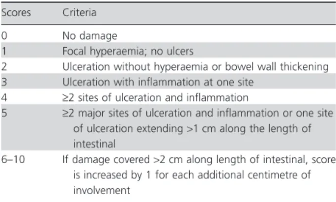
In Figure 1, we can observe that the administration intracolon of TNBS induced a significant (P<0.05) increase of colon macroscopic intestinal lesions (18.00±2.2 scores of lesion) when compared with saline group (0.60±0.04 scores of lesion). The treatment with PLS (30, Table 1 Criteria for macroscopic scoring intestinal lesion
Scores Criteria
0 No damage
1 Focal hyperaemia; no ulcers
2 Ulceration without hyperaemia or bowel wall thickening 3 Ulceration with inflammation at one site
4 ≥2 sites of ulceration and inflammation
5 ≥2 major sites of ulceration and inflammation or one site of ulceration extending>1 cm along the length of intestinal
60 and 90 mg/kg, p.o.) reduced (30 mg/kg: 4.40±1.03 scores of lesion; 60 mg/kg: 3.20±1.02 scores of lesion; 90 mg/kg: 2.80±0.42 scores of lesion) the damage scores of macroscopic lesions in the colon tissue, with the maximal effect observed at a dose of 90 mg/kg. Similar effects were produced by dexamethasone (5.50±1.65 scores of lesion) administration, a drug of choice for treating IBD.
Histopathological evaluation
TNBS-induced colitis promotes change in histological find-ings characterized by severe intestinal damage, a massive
inflammatory cell infiltration, ulceration and muscle thick-ening. The histological examination of colon sections from those rats that received only TNBS into the colon revealed the presence of crypts showing extensive epithelial destruc-tion (Figure 2 and Table 2). The histological evaluadestruc-tion of colons from rats treated with PLS 90 mg/kg revealed a pro-nounced reduction in the inflammatory response with moderate loss of epithelial cells and minimal inflammatory infiltration into the colonic tissue, resulting in a decreased microscopic damage score and showing a reduction of 45% in the total damage score, as compared with TNBS group.
PLS reduced TNBS-induced increase in wet weight of the colon
In Figure 3, we observed that TNBS induced a significant (P<0.05) increase in the colon weight (1.03±0.03 g; 75%) of the injured area in 5 cm of the bowel tissue, as compared with saline group (0.26±0.01 g) or Dexa Group (0.68± 0.05). However, pretreatment with PLS 90 mg/kg signifi-cantly reversed (0.55±0.05 g) the colon injury after TNBS colonic instillation.
PLS reduced TNBS-induced MPO activity
Figure 4 shows that the TNBS into the colon determined MPO activity in the concentration of 11.78±2.393 units of MPO/mg of colon tissue, while the group treated with PLS 90 mg/kg decreased an activity of this enzyme at 2.578±1.415 UMPO/mg of colon tissue, which was equiva-lent to a reduction of 78%.
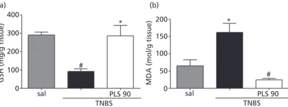
PLS treatment interferes with TNBS-induced GSH and MDA levels in rat intestine
As showed in Figure 5A, the colitis produced a significant (P<0.05) decrease in colonic GSH content (91.56± 14.96 mg/g of tissue) as compared with the saline group
25
#
* *
* * 20
15
10
5
0
sal 30 60
TNBS
90 Dexa
Macroscopic scores of lesion
Figure 1 The polysaccharide fraction extracted fromGracilaria birdiae reduces TNBS-induced macroscopic intestinal damage. Rats were treated with saline (sal: control), PLS (30, 60 and 90 mg/kg, p.o.) or dexamethasone (1 mg/kg, s.c.) once daily for 3 days. On the third day, the rats were killed, and the abdomens were opened, and there was the identification of the intestine for the evaluation of macroscopic scores of lesion. The results are expressed as the mean±SEM of the macroscopic scores of 5–7 animals per group. *P<0.05 versus TNBS group; #P<0.05 versus saline group (ANOVA followed by the
Newman–Keuls post hoc test).
(a) (b) (c) (d)
(290.20±16.74 mg/g of tissue). The treatment for 3 days with PLS in dose of 90 mg/kg prevent the consumption of GSH (286.30±57.07 mg/g of tissue), which remained at a much higher concentration as compared with TNBS control group. On the another hand, Figure 5B shows that the oxi-dative stress in colonic mucosa induced by TNBS was evi-denced by the significant increased of MDA (162.5± 25.63 nmol/g of tissue) concentration when compared with saline group (65.07±17.93 nmol/g of tissue). PLS 90 mg/kg
was able to inhibit the increase of MDA (23.95±
4.72 nmol/g of tissue) levels in inflamed mucosa tissue.
Nitric oxide (NO3/NO2) concentration
As shown in Figure 6, the TNBS group showed an increased level of NO3/NO2in colon tissue (0.23±0.03μm) when this
group was compared with saline group (0.08±0.01μm).
The treatment of PLS reduces the levels of nitrate and nitrite to (0.08±0.00μm) in the intestinal damage caused
by TNBS instillation into the colon.
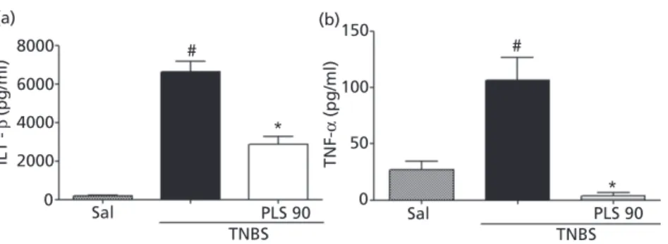
PLS decreased TNBS-induced IL-1βand
TNF-α production
Figure 7 shows that the intracolonic administration of TNBS induced an increased of IL-1β (panel A; 6630± 561.5 pg/ml) and TNF-α (panel B; 106.8±19.97 pg/ml) levels, comparing this group with saline group (IL-1β: 187.4±31.65 pg/ml; TNF-α: 26.99±7.771 pg/ml). More-over, the treatment with PLS 90 mg/kg significantly reduced the concentrations of these cytokines (IL-1β: 2872.00±267.20 pg/ml; TNF-α: 26.99±2.67 pg/ml). Table 2 Histological assessment of bowel damage
Criteria
Median score
Saline TNBS TNBS+PLS TNBS+Dexa
Loss mucosal architecture 0 3 (1–3)# 1 (1–2)* 1 (1–3)*
Cellular infiltration 0 3 (1–3)# 2 (1–2)* 1.5 (1–3)*
Muscle thickening 0 3 (1–3)# 1 (1–2)* 1 (1–2)*
Crypt abscess 0 1 (1–1) 1 (1–1) 1 (0–1)
Goblet cell depletion 0 1 (0–1) 0 (0–1) 0 (0–0)
Total damage score
0 11# 5* 4.5*
PLS, polysaccharide; TNBS, trinitrobenzenesulfonic acid. Score for histological damage expressed as mean±SEM (n=5). #P<0.05 versus saline
group; *P<0.05 versus TNBS group (Kruskal–Wallis non-parametric test and Dunn’s test were used for multiple comparisons of histological analyses).
1.5
1.0
#
*
*
0.5
W
et weight
(g/cm of colon)
0.0
sal PLS 90
TNBS
Dexa
Figure 3 The PLS fraction extracted from Gracilaria birdiaereduces the wet weight of the colons of the rats treated with TNBS. Rats were treated with saline (sal: control), PLS (90 mg/kg, p.o.) or dexametha-sone (1 mg/kg, s.c.) once daily for 3 days. On the third day, the rats were killed, and the abdomens were then opened. After identification of the intestine, samples of tissue (5 cm) were collected for the evalu-ation of wet weight of the colons. The results are expressed as the mean±SEM of 5–7 animals per group. *P<0.05 versus TNBS group;
#P<0.05 versus saline group (ANOVA followed by the Newman–Keuls
post-hoc test).
15
10
MPO (U/mg tissue)
5
0
sal
*
#
PLS 90 TNBS
Figure 4 Effect of the polysaccharide fraction extracted from Gracilaria birdiaeon the activity of intestinal tissue MPO in a rat model of TNBS-induced colitis. Animals were treated with saline (sal: control), PLS (90 mg/kg,p.o.) once daily for 3 days. On the third day, the rats were killed, the samples of colon were collected and MPO activity was evaluated. The results are expressed as the mean±SEM of 5–7 animals per group. *P<0.05 versus saline group;#P<0.05 versus TNBS group
Discussion
The inflammatory bowel diseases (IBD) refer essentially to two different, yet closely related conditions: Crohn’s disease and UC.[30] The development of inflammatory bowel
disease (IBD) is characterized by frequent episodes of diar-rhoea, abdominal pain, blood in the stool and weight loss over a period of months to years.[31]This leads to a
discom-fort and low quality of life for the person who bears this disease.
IBD is related to an abnormally exacerbated immune system to otherwise innocuous stimuli which are not properly abrogated by the feedback system normally downregulating the mucosal response towards luminal factors.[12]Early events of IBD occur long before symptoms
appear. Thus, study in humans often needs to be
retrospec-tive, what can cause many errors. That has led to studies of animal models of IBD to determine early events in the disease process and to test new treatments.[32,33]
In this context, TNBS is a substance used as a valuable model to investigate the pathophysiological mechanism of IBD. Histological modifications occur in TNBS-induced colitis resemble that of IBD in terms of ulceration, inflam-mation and leucocyte infiltration. Although the acid prop-erty of TNBS is partly responsible for the mucosal damage, TNBS has been shown to elicit intestinal protein antigenic-ity by functioning as a hapten,[34] thereby developing an
immune and inflammatory responses. The early acute phase of TNBS-induced inflammation is presumably essential to establish the continued progression of the colitis that is mediated by the T-cell dependent immunity.[35]
Many marine algae and their isolated constituents have shown beneficial therapeutic properties including analgesic, anti-inflammatory and gastroprotective actions.[30,36,37]
Knowing that marine algae are an important therapeutic tool against gastrointestinal injury, this study aims to verify the effect of PLS fraction extracted from G. birdiae in TNBS-induced colitis in rats.
The results of this study demonstrated, for the first time, the effectiveness of the PLS action extracted fromG. birdiae against TNBS-induced large intestine damage on the macroscopic and histological examinations in rats. These parameters were used as an indicator of disease-associated intestinal wall thickening and intensity of inflammation.[38]
According to our results, we can infer that PLS fraction produce anti-inflammatory effect in TNBS-induced colon damage in rats.
Another parameter studied was the wet weight of the colon in animals with or without colitis. Our results dem-onstrated that the PLS 90 mg/kg decreased the wet weight of the colon. TNBS-induced colitis resembles that of IBD in terms of ulceration, inflammation and leucocyte infiltra-tion,[39]causing an intense injury leading to an increase in
400
(a) (b)
300
200
200
150
100
50
0 100
GSH (mg/g tissue) MDA (mol/g tissue)
0 sal
#
#
* *
PLS 90 TNBS
sal PLS 90
TNBS
Figure 5 Effect of the PLS fraction extracted fromGracilaria birdiaeon glutathione levels (a) and MDA concentration (b) in TNBS-induced colitis. Animals were treated with saline (sal: control), PLS (90 mg/kg p.o.) once daily for 3 days. On the third day, the rats were killed, the samples of colon were harvest, and glutathione level was evaluated. The results are expressed as the mean±SEM of 5–7 animals per group. *P<0.05 versus TNBS group;#P<0.05 versus saline group (ANOVA followed by the Newman–Keuls post-hoc test).
0.3
0.2
0.1
NO
3
(
µ
M
)
0.0
sal
#
*
PLS 90 TNBS
Figure 6 Effect of the polysaccharide fraction extracted from Gracilaria birdiaeon intestinal tissue concentration of nitric oxide (NO3/
NO2). In the assay, animals were treated with saline (sal: control), PLS
(90 mg/kg, p.o.) once daily for 3 days. On the third day, the rats were killed, the samples of colon were collected, and NO3/NO2concentration
was measured. The results are expressed as the mean±SEM of 5–7 animals per group. *P<0.05 versus TNBS group; #P<0.05 versus
volume and weight of damaged intestine. Corroborating to the previous results, our substance decreased the inflamma-tory process in the colonic mucosa.
We can observe in our results the improvement of micro-scopic scores of lesion in the treatment with PLS (90 mg/ kg). This substance also decreases the inflammatory response resulting and minimal inflammatory infiltration into the colonic tissue. Neutrophils were attributed as the cells responsible for disrupting epithelial integrity and causing colon injury in IBD.[40] Thus, the reduction of
neutrophil infiltration in the animals treated with PLS con-tributes to the reduction in neutrophil-mediated colon tissue injury during colitis.
Corroborating with our results above, PLS fraction also decreased MPO activity. Neutrophil infiltration into the lamina propria is a common feature of colitis and probably accounts for significant non-specific injury from this disease.[40,41]During the migration, the neutrophils released
MPO to penetrate into the injury tissue. MPO is an enzyme mainly found in azurophilic granules of neutrophils that produce the microbicidal molecule hypochlorite, a strong oxidant, upon the reaction with H2O2and Cl−. Moreover, it
can serve as a good marker of inflammation, tissue injury and neutrophil infiltration in gastrointestinal tissues.[42,43]
Thus, we can infer that the PLS decrease the mucosal damage by decreasing free radicals production derived of neutrophil infiltration during the inflammation in the colon.
Oxidative stress has been proposed to play an important role in the pathogenesis of inflammatory bowel disease and is related to neutrophil infiltration within the inflamed colon mucosa. The recruitment and activation of neutro-phils during acute inflammation contribute to the over-production of reactive oxygen and nitrogen species that overwhelm the tissue antioxidant protective mechanisms, resulting in oxidative stress, which perpetuates inflamma-tion of the colon.[44]Therefore, this study investigated three
oxidative stress markers: GSH and MDA and NO3/NO2
concentration.
Antioxidant compounds play an important role in various pathological conditions, including inflammation, neurodegenerative diseases and cancer.[45,46]It has been
sys-tematically reported in the literature that PLSs showing antioxidant activity, such as those extracted from marine algae, protect against cell death due to their ability to degrade excessive reactive oxygen species.[47] Nevertheless,
few studies have correlated antioxidant potential with TNBS-associated injury of the intestinal tissue.
Our results demonstrated that the PLS fraction increased the levels of GSH in animals with TNBS-induced colitis. GSH, an endogenous antioxidant, protects the cells against oxidative damage, keeping the sulfhydryl groups (-SH) of proteins reduced and preventing them from reacting with free radicals.[48]Concentrations of endogenous antioxidants,
such as GSH, are decreased significantly in patients with inflammatory bowel disease and in experimental models of colitis.[49,50] According this result, we can infer that PLS
decreased the mucosa damage acting in the production and action of endogenous antioxidants.
Oxidative damage in colon tissue is a potential aetiologi-cal or triggering factor for IBD, because the detrimental effects of reactive oxygen molecules have been well estab-lished in the inflammation process.[51]MDA is a product of
lipoperoxidative processes that take place as a consequence of the colonic oxidative insult.[52]Our results demonstrated
for the first time that the PLS reduced the MDA concentra-tion in the colonic inflamed mucosa. Thus, we can infer that the action of PLS to reduce the inflammatory response in the colon is seemingly related to the decrease of oxidative stress.
The damage in intestinal mucosa induced by TNBS was accompanied with high levels of free radicals derivate of the degradation of NO (NO3/NO2 radicals).[53] Our
result demonstrated that the PLS treatment decreased the
8000
(a) (b)
150
100
50
TNF-α
(pg/ml)
0
# #
*
* 6000
4000
2000
IL1
-β
(pg/ml)
0
Sal PLS 90 Sal PLS 90
TNBS TNBS
concentration of NO3/NO2in the intestinal mucosa of the
rats treated with TNBS. NO is a free radical with moderate reactivity. Its production in large quantities via the upregulation of inducible nitric oxide synthase (iNOS) can inhibit key enzymes in the mitochondrial electron trans-port chain.[53,54] In addition to this, high levels of nitric
oxide from activated iNOS are toxic and can damage the tissue directly by the peroxynitrite formation after reaction with superoxide.[55] Thus, we infer the PLS has protective
action against intestinal injury by decreasing the formation of peroxynitrite.
The inflammatory cells infiltration in the colitis is
dependent of the overproduction of several
pro-inflammatory mediators, for example cytokines.[13] Our
results demonstrated that PLS 90 mg/kg decreased the con-centration of these cytokines in inflamed intestinal mucosa. Cytokines, such as IL-1β and TNF-α, are increased in inflamed tissue, including the mucosa of IBD lesions.[56,57]
These mediators are responsible for many of the features of inflammation; an important feature of UC is the recruit-ment of neutrophils and mononuclear cells. Leucocyte adherence and recruitment are increased in the micro-vessels in chronic disease,[58] mediated in part by the
upregulation of adhesion molecules towards vascular endothelial cells by TNF-αand IL-1β. Moreover, increased
levels of tissue-specific and inflammatory chemokines enhance leucocyte migration.[56]Thus, these results revealed
that the PLS has an action to reduce the inflammatory response through inhibition of pro-inflammatory cytokines in the intestine injury resulted from TNBS-induced colitis in rats. A possible mechanism may involve the downregulation of the inflammatory response by inhibiting the synthesis and release of pro-inflammatory mediators.
Conclusions
In conclusion, our results suggest that PLS has a protective effect in the TNBS-induced colitis in rats, through mecha-nisms that involve the inhibition of inflammatory cell infiltration, reduction in the oxidative stress and of pro-inflammatory cytokines concentration. Thus, we suggest that PLSs may have potential applications in the develop-ment of novel therapeutic targets against the inflammatory bowel disease in humans.
Acknowledgements
The authors are grateful to the Brazilian Agency for Scien-tific and Technological Development-CNPq (Brazil) and the technical assistance of Maria Silvandira Freire França.
References
1. Sousa FCFet al. Medicinal plants and
their bioactive constituents: a scien-tific review of bioactivity and poten-tial benefits in the anxiety disorders in
animal models. Rev Bras Farmacogn
2008; 18: 642–654.
2. Correa MFP et al. Natural products
from plant origin potentially useful
in the asthma therapy. Rev Bras
Farmacogn2008; 18: 785–797.
3. Qi H et al. Antioxidant activity of
different molecular weight sulfated
polysaccharides from Ulva pertusa
Kjellm (Chlorophyta). J Appl Phycol
2005; 17: 527–534.
4. Yang YF et al. Growth of Gracilaria
lemaneiformisunder different cultiva-tion condicultiva-tions and its effects on nutrient removal in Chinese coastal
waters. Aquaculture 2006; 254: 248–
255.
5. Jiao Get al. Chemical structures and
bioactivities of sulfated
polysac-charides from marine algae. Mar
Drugs2011; 9: 196–223.
6. Rees DA et al. Biogenesis of
3,6-anhydro-L-galactose. Biochem J1961;
81: 347–352.
7. Silva RO et al. A
sulfated-polysaccharide fraction from seaweed Gracilaria birdiae prevents naproxen-induced gastrointestinal damage in
rats.Mar Drugs2012; 10: 2618–2633.
8. Brito TV et al. Anti-inflammatory
effect of a sulphated polysaccharide fraction extracted from the red algae Hypnea musciformis via the
suppres-sion of neutrophil migration by
the nitric oxide signalling pathway. J Pharm Pharmacol 2013; 65: 724– 733.
9. Souza MCR et al. Antioxidant
activ-ities of sulfated polysaccharides from
brown and red seaweeds.J Appl Phyco
2007; 19: 153–160.
10. Podolsky DK et al. Pride and
preju-dice: inflammatory bowel disease
models and drug development. Curr
Opin Gastroenterol2000; 16: 295–296.
11. Elson CO et al. Experimental models
of inflammatory bowel disease.
Gas-troenterology1995; 109: 1344–1367.
12. Fiocchi C. Inflammatory bowel
disease: etiology and pathogenesis. Gastroenterology1998; 115: 182–205.
13. Katz JAet al. Pathogenesis of
inflam-matory bowel disease. Curr Opin
Gastroenterol1999; 15: 291–297.
14. Botoman VA et al. Management of
inflammatory bowel disease.Am. Fam.
Phys1998; 57: 57–68.
15. Hanauer SBet al. Postoperative
main-tenance of Crohn’s disease remission with 6-mercaptopurine, Natural prod-ucts in treatment of ulcerative colitis and peptic ulcer 119 mesalamine, or
placebo: a 2-year trial.
Gastroenterol-ogy2004; 127: 723–729.
16. Faubion WAet al. The natural history
of corticosteroid therapy for inflam-matory bowel disease: a
population-based study. Gastroenterology 2001;
121: 255–260.
17. Farias WRet al. Structure and
sulfated galactans from invertebrates. J Biol Chem2000; 275: 29299–29307.
18. Souza BWSet al. Chemical
characteri-zation and antioxidant activity of sulfated polysaccharide from the red
seaweed Gracilaria birdiae. Food
Hydrocoll2012; 27: 287–292.
19. Maciel JS et al. Structural
characteri-zation of cold extracted fraction of soluble sulfated polysaccharide from
red seaweed Gracilaria birdiae.
Car-bohydr Polym2008; 71: 559–565.
20. DuBois Met al. Colorimetric method
for determination of sugars and
related substances. Anal Chem 1956;
28: 350–356.
21. Bradford MM. A rapid and sensi-tive method for the quantitation of microgram quantities of protein uti-lizing the principle of protein-dye
binding.Anal Biochem1976; 72: 248–
254.
22. Lloyd AGet al. Infrared studies on
phate esters. I. Polysaccharide
sul-phates.Biochim Biophys Acta1961; 46:
108–115.
23. Stevenson TT, Furneaux RH. Chemi-cal methods for the analysis of sul-phated galactans from red algae. Carbohydr Res1991; 210: 277–298.
24. Morris GP et al. Hapten-induced
model of chronic inflammation and
ulceration in the rat colon.
Gastroen-terology1989; 96: 795–803.
25. Sedlak J, Lindsay RH. Estimation of total, protein-bound, and nonpro-tein sulfhydryl groups in tissue with
Ellman’s reagent.Anal Biochem1968;
24: 1992–2005.
26. Mihara M, Uchiyama M. Determina-tion of malonaldehyde precursor in
tissues by thiobarbituric acid test.Ana
Biochem1978; 86: 271–278.
27. Bradley PP et al. Measurement of
cutaneous inflammation: estimation of neutrophil content with an enzyme
marker. J Invest Dermatol 1982; 78:
206–209.
28. Green LC et al. Analysis of nitrate,
nitrite, and [15N]nitrate in biological
fluids. Anal Biochem1982; 126: 131–
138.
29. Appleyard CB, Wallace JL. Reactiva-tion of hapten-induced colitis and its prevention by anti-inflammatory
drugs.Am J Physiol1995; 269: 119–
125.
30. Podolsky DK. Pride and prejudice: inflammatory bowel disease models
and drug development. Curr Opin
Gastroenterol2000; 16: 295–296. 31. Farmer RG. Clinical features,
labora-tory findings and course of Crohn’s
disease. In: Kirsner JB, ed.
Inflamma-tory Bowel Disease. Philadelphia, PA: WB Saunders, 1988: 175–184. 32. Strober W. Animal models of
inflam-matory bowel disease: an overview. Dig Dis Sci1985; 30: 3–10.
33. Sartor RB et al. Granulomatous
enterocolitis induced by purified
bacterial cell wall fragments.
Gastro-entero1ogy1985; 89: 587–595.
34. Neurath Met al. TNBS-colitis.Int Rev
Immunol2000; 19: 51–62.
35. Zhang ZMD et al. Critical role of
IL-17 receptor signaling in acute
TNBS-induced colitis.Inflamm Bowel
Dis2006; 12: 382–388.
36. Chaves LS et al. Anti-inflammatory
and antinociceptive effects in mice of a sulfated polysaccharide fraction extracted from the marine red algae Gracilaria caudata.Immunopharmacol Immunotoxicol2013; 35: 93–100.
37. Damasceno SRB et al. Role of the
NO/K ATP pathway in the protective effect of a sulfated-polysaccharide
fraction from the algae Hypnea
musciformis against ethanol-induced
gastric damage in mice. Rev Bras
Farmacogn2013; 23: 320–328.
38. Lee JY et al. Inhibitory effects of
Geijigajakyak-Tang on trinitrobenzene
sulfonic acid-induced colitis.J
Ethno-pharmacol2009; 126: 244–251. 39. Grisham MB, Yamada T. Neutrophils,
nitrogen oxides, and inflammatory
bowel disease.Ann N Y Acad Sci1992;
664: 103–115.
40. Nosál’ová V et al. Effect of
N-acetylcysteine on colitis induced by
acetic acid in rats. Gen Pharmacol
2000; 35: 77–81.
41. Casagrande R et al. Protective effect
of topical formulations containing quercetin against UVB-induced
oxida-tive stress in hairless mice. J
Photochem Photobiol B 2006; 84: 21–27.
42. Bamias G et al. Cytokines in the
pathogenesis of ulcerative colitis.
Discov Med2011; 60: 459–467.
43. Martín AR et al. Resveratrol, a
polyphenol found in grapes, sup-presses oxidative damage and stimu-lates apoptosis during early colonic
inflammation in rats.Biochem
Phar-macol2004; 67: 1399–1410.
44. Cuzzocrea S et al. Antioxidant
ther-apy: a new pharmacological approach in shock, inflammation and ischemia/
reperfusion injury. Pharmacol Rev
2001; 53: 135–159.
45. Zhao B. Natural antioxidants protect neurons in Alzheimer’s disease and
Parkinson’s disease. Neurochem Res
2009; 34: 630–638.
46. Costa LSet al. Biological activities of
sulfated polysaccharides from tropical
seaweeds.Biomed Pharmacother2010;
64: 21–28.
47. Urso ML, Clarkson PM. Oxidative stress, exercise, and antioxidant
sup-plementation. Toxicology 2003; 189:
41–54.
48. Amirshahrokhi K et al. The effect of
methylsulfonylmethane on the
experi-mental colitis in the rat. T Appl
Pharmacol2011; 253: 197–202.
49. Tahan G et al. Melatonin expresses
powerful anti-inflammatory and
antioxidant activities resulting in
complete improvement of
acetic-acid-induced colitis in rats. Dig Dis
Sci2011; 56: 715–720.
50. Spitz DR et al. Metabolic oxidation/
reduction reactions and cellular
responses to ionizing radiation: a unifying concept in stress response
biology. Cancer Metastasis Rev 2004;
23: 311–322.
51. Pavlick KP et al. Role of reactive
metabolites of oxygen and nitrogen
in inflammatory bowel disease. Free
Radic Biol Med2002; 33: 311–322.
52. Pacher P et al. Nitric oxide and
peroxynitrite in health and disease. Physiol Rev2007; 87: 315–424.
53. Kolios Get al. Nitric oxide in
inflam-matory bowel disease: a universal messenger in an unsolved puzzle. Immunology2004; 113: 427–437.
54. Martínez-Flórez S et al. Quercetin
activation and nitric oxide produc-tion in interleukin-1beta-activated rat
hepatocytes.J Nutr 2005; 135: 1359–
1365.
55. Kaulersch Wet al. Polyclonal nature of
the intestinal lymphocyte populations in inflammatory bowel disease: a molecular genetic evaluation of the immunoglobulin and T cell antigen
receptors. Gastroenrerology 1988; 95:
364–370.
56. Fiocchi C. Lymphokines and the
intes-tinal immune response. Role in
inflammatory bowel disease.Immunol
Invest1989; 18: 91–102.
57. Hatoum OA et al. The intestinal
microvasculature as a therapeutic
target in inflammatory bowel disease. Ann N Y Acad Sci2006; 1072: 78–97. 58. Mora JR, von Andrian UH. T-cell
homing specificity and plasticity: new
concepts and future challenges.Trends