Trends Psychiatry Psychother. 2013;35(4) – 238-251
Review Article
rends
in Psychiatry and PsychotherapyT
© APRS
Electroencephalographic indings in panic disorder
Achados eletroencefalográicos no transtorno do pânico
Marcele Regine de Carvalho,1 Bruna Brandão Velasques,2 Maurício Cagy,3 Juliana Bittencourt Marques,4 Silmar Teixeira,5
Antonio Egidio Nardi,6 Roberto Piedade,7 Pedro Ribeiro8
Abstract
Some studies have reported the importance of electroencepha -lography (EEG) as a method for investigating abnormal parame -ters in psychiatric disorders. Different indings in time and fre -quency domain analysis with regard to central nervous system arousal during acute panic states have already been obtained. This study aimed to systematically review the EEG indings in panic disorder (PD), discuss them having a currently accepted neuroanatomical hypothesis for this pathology as a basis, and identify limitations in the selected studies. Literature search was conducted in the databases PubMed and ISI Web of Kno-wledge, using the keywords electroencephalography and panic disorder; 16 articles were selected. Despite the inconsistency of EEG indings in PD, the major conclusions about the absolute power of alpha and beta bands point to a decreased alpha po -wer, while beta power tends to increase. Different asymmetry patterns were found between studies. Coherence studies poin -ted to a lower degree of inter-hemispheric functional connecti -vity at the frontal region and intra-hemispheric at the bilateral temporal region. Studies on possible related events showed changes in memory processing in PD patients when exposed to aversive stimuli. It was noticed that most indings relect the current neurobiological hypothesis of PD, where inhibitory deicits of the prefrontal cortex related to the modulation of amygdala activity, and the subsequent activation of subcortical regions, may be responsible to trigger anxiety responses. We approached some important issues that need to be considered in further researches, especially the use of different methods for analyzing EEG signals.
Keywords: Electroencephalography, panic disorder, neurobiology, brain mapping.
Resumo
Alguns estudos relataram a importância da eletroencefalograia (EEG) como método de investigação de parâmetros anormais em transtornos psiquiátricos. Achados diferentes na análise do domí -nio do tempo e da frequência em relação à excitabilidade do sis -tema nervoso central durante estados agudos de pânico já foram obtidos. O objetivo deste estudo foi revisar sistematicamente os achados de EEG no transtorno do pânico (TP), discuti-los com base em uma hipótese neuroanatômica atualmente aceita para essa pa -tologia e identiicar limitações nos estudos selecionados. A pesquisa bibliográica foi realizada nas bases de dados PubMed e ISI Web of Knowledge, utilizando as palavras-chave eletroencefalograia e transtorno do pânico; 16 artigos foram selecionados. Apesar da in -consistência dos achados de EEG no TP, as principais conclusões sobre a potência absoluta das bandas alfa e beta apontam para uma diminuição da potência em alfa, enquanto em beta a potência tende a aumentar. Diferentes padrões de assimetria foram encon -trados entre estudos. Os estudos de coerência apontaram para um menor grau de conectividade funcional inter-hemisférica na região frontal e intra-hemisférica na região temporal bilateral. Estudos de potenciais eventos relacionados demonstraram mudanças no pro -cessamento da memória em pacientes com TP quando expostos a estímulos aversivos. Notou-se que a maioria dos resultados relete a atual hipótese neurobiológica do TP, nos quais déicits inibitó -rios do córtex pré-frontal em relação à modulação da atividade da amígdala, e a subsequente ativação de regiões subcorticais, podem ser responsáveis por desencadear respostas de ansiedade. Foram abordadas algumas questões importantes que precisam ser consi -deradas em futuras pesquisas, especialmente o uso de diferentes métodos de análise de sinais de EEG.
Descritores: Eletroencefalograia, transtorno do pânico, neurobio -logia, mapeamento encefálico.
1 Laboratory of Panic and Respiration, Institute of Psychiatry (IP), Universidade Federal do Rio de Janeiro (UFRJ), Rio de Janeiro, RJ, Brazil. National Science and Technology Institute for
Translational Medicine (INCT-TM), Rio de Janeiro, RJ, Brazil. Brain Mapping and Sensory Motor Integration (BMSMI), IP, UFRJ, Rio de Janeiro, RJ, Brazil. 2 Brain Mapping and Sensory Motor
Integration, IP, UFRJ, Rio de Janeiro, RJ, Brazil. Instituto de Neurociências Aplicadas (INA), Rio de Janeiro, RJ, Brazil. Neuromuscular Research Laboratory, Instituto Nacional de Traumatologia
e Ortopedia (INTO), Rio de Janeiro, RJ, Brazil. 3 BMSMI, IP, UFRJ, Rio de Janeiro, RJ, Brazil. Department of Epidemiology and Biostatistics, Institute of Community Health, Universidade Federal
Fluminense (UFF), Rio de Janeiro, RJ, Brazil. 4 BMSMI, IP, UFRJ, Rio de Janeiro, RJ, Brazil. INA, Rio de Janeiro, RJ, Brazil. 5 BMSMI, IP, UFRJ, Rio de Janeiro, RJ, Brazil. Department of Physiotherapy,
Policlínica Piquet Carneiro, Universidade do Estado do Rio de Janeiro (UERJ), Rio de Janeiro, Brazil. Laboratory of Physical Therapy, Universidade Veiga de Almeida (UVA), Rio de Janeiro, Brazil.
6 Laboratory of Panic and Respiration, IP, UFRJ, Rio de Janeiro, RJ, Brazil. INCT-TM, Rio de Janeiro, RJ, Brazil. 7 BMSMI, IP, UFRJ, Rio de Janeiro, RJ, Brazil. 8 BMSMI, UFRJ, Rio de Janeiro, RJ,
Brazil. INA, Rio de Janeiro, RJ, Brazil. Department of Bioscience, School of Physical Education, UFRJ, Rio de Janeiro, RJ, Brazil.
Financial support: none. Submitted Mar 20 2013, accepted for publication Jul 05 2013. No conlicts of interest declared concerning the publication of this article.
Trends Psychiatry Psychother. 2013;35(4) – 239 EEG indings in PD - de Carvalho et al.
Introduction
Panic disorder (PD) is a chronic illness characterized by spontaneous severe anxiety attacks associated with somatic and vegetative symptoms and it usually occurs in comorbidity with other psychiatric disorders. The presence of agoraphobia in PD points out a more severe illness and a worse prognosis, requiring special therapeutic approaches.1
The cognitive functioning of PD patients is characterized by a catastrophic misinterpretation of bodily sensations. This abnormal cognitive processing is crucial for developing
or maintaining the disorder.2,3 The cognitive model
proposes that PD patients misinterpret somatic symptoms as dangerous. This induces anxiety and an increase in physiological activity, thus making somatic symptoms more likely to happen. Then, this vicious circle presumably induces panic attacks (PAs). Cognitive distortions characterized by a negative or catastrophic anticipation of the future have also proved to be present.4
Several etiological hypotheses have been proposed for PD, but its pathophysiology has not been consistently deined. Dysfunctions of different brain areas, such as the amygdala, locus ceruleous, hippocampal area, and prefrontal cortex,5 have been suggested to play a role in the pathophysiology of PD. The neuroanatomical hypothesis of PD by Gorman et al.6 assumes that the fear mechanisms of the sensitive central nervous system originate at the central nucleus of the amygdala. Deicits in the relay and coordination of upstream and downstream sensory information could result in increased amygdala activation, with consequent behavioral and autonomic responses.
Hypotheses on the neurocircuitry involved in fear and anxiety have been inluenced by evidence of neuroimaging techniques, especially functional magnetic
resonance imaging (fMRI).6 In neuroscience, it is
very useful to obtain data through different functional techniques; fMRI, recognized for its spatial resolution, can help identifying small deep subcortical structures involved in the neurocircuitry of PD and they are of great importance with regard to PD symptoms. Conversely, electroencephalography (EEG), recognized by its high temporal resolution, can collect neuroelectric data from cortical structures and it is also able to relect the activation of subcortical structures on the cerebral cortex
in real time.7 These possibilities allow a better analysis
of affective states and cognitive processing in anxiety disorders such as PD.
Electroencephalographic studies have reported different indings with regard to central nervous system arousal during acute panic attacks.8 EEG is a technique that measures electrocortical activity over the scalp. Speciically, EEG registers the electrical activity of billions of nerve cells and the connections between them
in the brain cortex.7 Some studies have underlined the
importance of EEG as a method for investigating some abnormal parameters in psychiatric disorders, such as bipolar disorder, depression, and autism. There are two main methods of EEG signal analysis: frequency domain analysis (Figure 1), represented by the description of
energy distribution in the signal as a frequency function9;
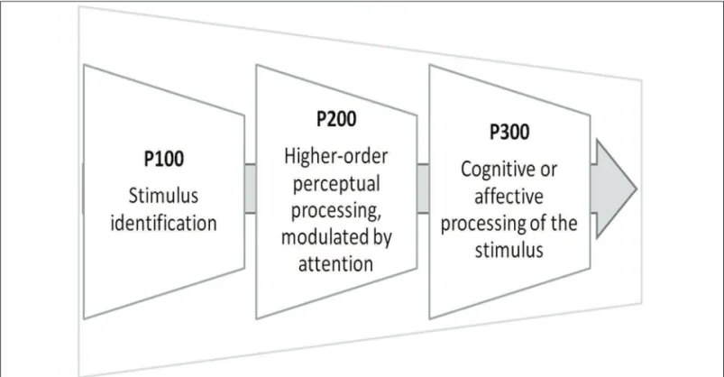
and time domain analysis (Figure 2), represented by the event-related brain potential (ERP).10
240 – Trends Psychiatry Psychother. 2013;35(4) EEG indings in PD - de Carvalho et al.
Method
Literature search was conducted in the databases PubMed and ISI Web of Knowledge search engines using the keywords electroencephalography and panic disorder; it was conducted in February 2013. Two independent reviewers (MRC and BV) performed study selection procedures and data extraction. The search did not took into account just the publication date, only articles published in English were included. The search in PubMed identiied 177 abstracts and 17 abstracts were found in ISI Web of Knowledge; 34 full-text articles were reviewed after abstract selection. Articles containing information on sleep states, atypical PAs, and seizures were not included, in order to exclude confusing conditions that could bias the indings about PD subjects. Sixteen original research articles on EEG analysis of frequency domain and time domain were selected. The main themes of PD articles were: spectral power, coherence, and asymmetry of EEG bands – concerning frequency domain analysis; and ERP, auditory evoked potential (AEP) – concerning time domain analysis. The lack of a search strategy in the databases PsychINFO and EMBASE was a limitation of this study.
Results
This section presents the main results that connected PD to EEG; it is organized according to the analytical method of the EEG signal: frequency domain analysis The frequency domain analysis is divided into the
EEG bands absolute power, relative power, coherence, and asymmetry, which relect the readiness of different oscillating circuits. This analysis predicts subsequent cognitive and perceptual ability in a frequency-speciic
domain.11 Absolute power is deined as the total energy
in an electrode located at a cortical area in a speciic
frequency.12 Relative power represents the percentage
of power in any band compared with the total power in EEG. Coherence is a linear synchronization measure between two signals recorded at different brain locations. It may help understanding the functional
connectivity between different regions of the brain.13,14
Finally, asymmetry is deined as the difference between homologous electrodes in relation to the EEG absolute power. It detects the energy balance between the two hemispheres and the cortical areas.15 The other kind of analysis, the ERPs, consists of brain responses that are linked in time to a given event, as a strategy to examine information processing. Early components of the ERP, such as P100 (a positive wave that occurs at around 100 ms), are associated with stimulus identiication, whereas late components of the ERP, such as P300 (a positive wave that occurs at around 300 ms), may relect cognitive or affective processing of the stimulus.16,17
As there is a lack of information on EEG correlates of PD in the literature, this study aimed to systematically review EEG indings in PD, discuss them in the light of a currently accepted neuroanatomical hypothesis for this disorder, and pinpoint limitations of the selected studies.
Trends Psychiatry Psychother. 2013;35(4) – 241 EEG indings in PD - de Carvalho et al.
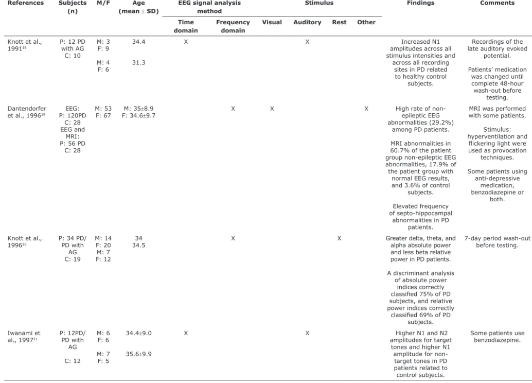
(Figure 3) and time domain analysis. Findings are also summarized in chronological order (Table 1).
Studies on spectral power, coherence, and asymmetry
Wise et al.11 conducted a quantitative
electroen-cephalographic (qEEG) and autonomic assessment of 52 PD patients, with or without agoraphobia. EEG data were acquired under open-eye and closed-eye rest condi -tions, and spectral power was assessed within 4 frequen -cy bands: theta, alpha-1, alpha-2, and beta. The results showed attenuated spectral power for alpha-1, alpha-2, and beta related to control subjects, especially under closed-eye condition. Alpha-1 attenuation was more ap -parent and suggested alertness and engaged attention. The authors’ explanation on this data is that in the ab -sence of experimentally-based information processing participants may have experienced spontaneous cognition and, thus, they could be more vigilant to body sensations, making anxiety likely to happen. This study also showed that regions often related to fear and anxiety, bilateral temporal and right frontal cortices, reduced beta spectral power in PD patients.6 Frontal alpha-1 asymmetry was also observed in PD under the closed-eye condition and it was related to a negative emotional state that is associ-ated with preponderance of right-sided cortical activity.
Knott et al.20 used qEEG to compare 34 PD patients
(with or without agoraphobia) to healthy control subjects, measuring signals from multiple scalp sites. PD patients exhibited greater overall delta, theta, and alpha absolute power and lesser beta relative power. According to the authors, the higher theta amplitude observed may be a relection of greater cortical activation, since PD patients report high levels of anxiety. Increased alpha is not a result commonly found in anxiety states, but, along with elevated delta and theta power, this may suggest that a primary role of alpha is regulating/inhibiting
the excessive subcortical arousal that is relected by an increase in slow-wave activity associated with the temporal cortex and the limbic system. Non-localizing EEG band observations related to PD patients may lead to the conclusion that they originate from deeper subcortical regions, possibly within the brainstem. Regarding the classiications of PD patients and healthy participants, a discriminant analysis of absolute power indices correctly classiied 75% of subjects, and relative power indices correctly classiied 69% of subjects.
Arikan et al.26 assessed EEG spectral power and eye
blink electrical startle responses of 39 patients with different psychiatric disorders, among which 10 were PD patients. They used EEG percent power of each frequency band to calculate the arithmetical difference between open and closed eyes as representative of EEG reactivity to eye opening. The authors observed an elevated beta reactivity at the left central area for PD, and this may suggest an increased startle relex since high beta relative power is related to increased anxiety.
Knott et al.22 assessed qEEG in 37 PD patients with
or without agoraphobia and healthy control subjects while listening to neutral and relaxation audiotapes. They observed a theta and alpha increase in the control group during relaxation, with no change in the activity of frequencies among the patient group. This result may suggest that the relaxation process was effective, since increase in these frequencies is associated with decreased electrocortical arousal states. According to the authors, these indings could provide some support to the abnormal electrocortical response related to PD symptomatology. Patients exhibited less beta and more theta activity in relation to control subjects, regardless of the condition. The authors also draw attention to the increased resting theta that may be associated with PA, highlighting a tendency for spontaneous attacks in patients during an electrocortical proile of decreased central nervous system
2
4
2
–
T
re
n
ds
Psy
ch
ia
tr
y
Psy
ch
oth
e
r.
2
0
1
3
;3
5
(4
)
EE
G
in
di
ngs
in
PD
-
de
C
ar
va
lh
o
et
al.
References Subjects
(n)
M/F Age
(mean ± SD)
EEG signal analysis method
Stimulus Findings Comments
Time domain
Frequency domain
Visual Auditory Rest Other
Knott et al.,
199118
P: 12 PD with AG
C: 10
M: 3 F: 9
M: 4 F: 6
34.4
31.3
X X Increased N1
amplitudes across all
stimulus intensities and across all recording
sites in PD related
to healthy control
subjects.
Recordings of the late auditory evoked
potential.
Patients’ medication was changed until
complete 48-hour
wash-out before testing.
Dantendorfer
et al., 199619 P: 120PDEEG:
C: 28 EEG and
MRI: P: 56 PD
C: 28
M: 53
F: 67 F: 34.6M: 35±±8.99.7 X X X High rate of non-epileptic EEG
abnormalities (29.2%) among PD patients.
MRI abnormalities in
60.7% of the patient group non-epileptic EEG abnormalities, 17.9% of the patient group with
normal EEG results, and 3.6% of control
subjects.
Elevated frequency
of septo-hippocampal abnormalities in PD
patients.
MRI was performed with some patients.
Stimulus: hyperventilation and
lickering light were used as provocation
techniques.
Some patients using anti-depressive
medication,
benzodiazepine or
both.
Knott et al.,
199620
P: 34 PD/ PD with
AG C: 19
M: 14 F: 20 M: 7 F: 12
34 34.5
X X Greater delta, theta, and
alpha absolute power
and less beta relative
power in PD patients.
A discriminant analysis
of absolute power
indices correctly
classiied 75% of PD subjects, and relative power indices correctly
classiied 69% of PD subjects.
7-day period wash-out
before testing.
Iwanami et
al., 199721
P: 12PD/ PD with
AG
C: 12
M: 6 F: 6
M: 7 F: 5
34.4±9.0
35.6±9.9
X X Higher N1 and N2
amplitudes for target
tones and higher N1
amplitude for non-target tones in PD patients related to control subjects.
Some patients use benzodiazepine.
Table 1 - EEG studies on PD
T
re
n
ds
Psy
ch
ia
tr
y
Psy
ch
oth
e
r.
2
0
1
3
;3
5
(4
)
–
2
4
3
EE
G
in
di
ngs
in
PD
-
de
C
ar
va
lh
o
et
al.
Knott et al.,
199722
P: 37 PD/ PD with
AG
C: 20
M: 17 F: 20
M: 7 F: 13
34.7±9.1
35.8±3.7
X X Theta and alpha
increased in the
control group during
relaxation.
Less beta and more
theta in PD in relation to control subjects regardless of tape
condition.
3 to 7-day period
wash-out before testing.
Pauli et al.,
199716
P: 15 PD
C: 15
M: 4 F: 11
M: 4 F: 11
35.5±9.6
35.3±7.6
X X Larger P300 amplitudes
to the presentation of
body-related words in
PD patients.
Body-related words led to a relatively small
positive peak in healthy control subjects.
Positive slow wave more marked at
central and parietal
sites and at the right
hemisphere than at the left hemisphere in PD patients related to
control subjects.
2-week period
wash-out before testing,
except for 3 patients.
Dengler et al.,
199923
P: 15 PD/ PD with
AG
M: 4
F: 11 35.9±9.4 X X Body-related word stimuli elicited an
enhanced positive
cortical slow wave
when compared to
non-somatic word
stimuli in PD patients.
Comorbidities: D, SP, GAD, history of AAD.
Hanaoka et
al., 200524
P: 18 PD/ PD with
AG
C: 18
M: 9 F: 9
M: 9 F: 9
31.5±9.4
31.2±10.5
X X Signiicantly lower
coherence in F3-F4, C3-C4, P3-P4, F7-T5, and F8-T6 electrode pairs in PD related to control
subjects.
Positive correlations were observed between coherence
measures and the
severity of panic
attacks for the higher
alpha band.
Never medicated
patients.
All right-handed
subjects.
(cont.)
2
4
4
–
T
re
n
ds
Psy
ch
ia
tr
y
Psy
ch
oth
e
r.
2
0
1
3
;3
5
(4
)
EE
G
in
di
ngs
in
PD
-
de
C
ar
va
lh
o
et
al.
Pauli et al., 200517
P: 16PD/ PD with
AG
C: 16
M: 5 F: 11
M: 5 F: 11
39.1±9.2
38.4±8.7
X X In the explicit memory
test, PD patients compared to healthy participants exhibited
enhanced discrimination scores and faster reaction times for
panic words, although recognition of panic
words was generally
worse when compared
to neutral words.
All patients were
medication-free at the time of examination.
Pauli et al., 200525
P: 25PD/ PD with
AG
C: 25
M: 9 F: 16
M: 9 F: 9
39.7
40.3
X X PD patients exhibited
enhanced ERP
potentials triggered by panic-related words as compared to neutral
threshold words at frontal electrodes in
100-200 ms after
stimulus presentation and enhanced positive
potentials in the time
window 200-400 ms
after word presentation.
Healthy subjects identiied more
panic-related than neutral words and exhibited enhanced late ERP
potentials triggered by panic-related words
related to neutral words
when compared to PD patients.
Comorbidities: Ph, SP, GAD.
Arikan et al., 200626
P: 10 PD *M/F
ratio = 0.4
29.1 X X Latency for the
right-sided contralateral
secondary component of the eye blink relex
was inversely correlated
with eye opening
reactivity in the beta range measured at the
left central region in PD patients.
7-day period wash-out
before testing.
Ghisoli et al.,
200627
P: 28PD
C: 28
M: 10 F: 18
M: 10 F: 18
43.3±10.1
39.7±7.8
X X Higher P50 ratios in
PD patients when compared to healthy
subjects.
Some patients in use of anti-depressive
medication or both
benzodiazepine and anti-depressive
medication.
(cont.)
T
re
n
ds
Psy
ch
ia
tr
y
Psy
ch
oth
e
r.
2
0
1
3
;3
5
(4
)
–
2
4
5
EE
G
in
di
ngs
in
PD
-
de
C
ar
va
lh
o
et
al.
Gordeev,
200828
P: 77 PD/ PD with
AG
C: 28
M: 24 F: 53
M: 9 F: 19
31.6±3.1
30.7±2.5
X X X Decreases in the power
density of the alpha
rhythm and increases
in the power density of
the beta 1 rhythm in
the right hemisphere in PD patients with AG.
Increase in the power
density of the theta
rhythm in the temporal
areas of the right
hemisphere in patients without AG.
Lower P300 wave
amplitudes and impairments of P300
habituation in both
hemispheres in PD with and without AG. Gordon et al.,
201029 P: 48 PD
C: 1908 M: 13
F: 35
M: 971 F: 937
_
_
X X PD patients did not
show any signiicant deviance in alpha
asymmetry from the normative control
group for either the open-eye or closed-eye
conditions.
The patient groups
were evaluated in
comparison to a
normative database.
Lopes et al.,
201030
P: 15 PD/ PD with AG
M: 3
F: 2 30.7±8.5 X X Increased alpha relative power on right and
central frontal sites
after the 35% CO2 induced panic attack.
Reduced right-sided frontal-orbital asymmetry in the beta
band in the presence of a panic attack.
Increased beta relative
power values on right temporal side during the induced panic
attack.
Decrease in the occipital-frontal intra-hemispheric coherence
in the delta band.
Delta inter-hemispheric
asymmetry in the left
occipital site
Stimulus: induced panic attack with a
35% CO2 challenge
test.
No use of psychotropic
medication (2 to 5 weeks wash-out
for anti-depressive
medication; 1 week for anxiolytic medication).
All right-handed
subjects.
(cont.)
246 – Trends Psychiatry Psychother. 2013;35(4) EEG indings in PD - de Carvalho et al.
W ise e t a l. , 2 0 1 1 1 1 P: 5 2 PD / PD w ith A G M : 15 F: 3 7 35.34 ± 13.37 X X Atte nu ate d spe ctr al pow er f or a lph a-1, al ph a-2 an d be ta re la te d to con tr ol su bj ects, e spe ci al ly in th e cl ose d-e y e con di ti on . B ila te ra l te m por al a nd ri gh t fr on ta l cor ti ce s re du ce d be ta spe ctr al pow er in PD pa tie nts. Fr on ta l a lph a-1 asy m m etr y in PD pa tie nts du ri ng th e cl ose d-e y e con di ti on . Com orbi di ti es : P h, S P, O CD , P T S D , G A D , M D D , D , A A B, BN . 20 P w it h a nt i-de pre s-si ve m edi ca ti on. A A D = a lcoh ol a bu se /de pe nde nce ; A G = a gor aph obi a; B N = bu lim ia n er vosa ; C = con tr ol su bj ects; D = dy sth ym ia ; EE G = e le ctr oe nce ph al ogr am ; F = fe m al e; G A D = ge ne ra liz ed an xi et y di sor de r; M = m al e; M D D = m aj or de pr essi ve di sor de r; M R I = m agn eti c re son an ce im agi ng; O C D = obse ssi ve -com pu lsi ve di sor de r; P = pa tie nts; PD = pa ni c di sor de r; Ph = si m pl e ph obi a; S D = sta nda rd de vi ati on ; S P = soci al ph obi a; PT S D = post -tr au m ati c str ess di sor de r.
arousal. During these attacks, patients generally exhibit less fast wave and more slow wave activity.
In a study using magnetic resonance imaging (MRI) to measure the frequency and quality of brain abnormalities in PD, EEG was also applied to detect PD patients with a high probability of morphologic brain abnormalities, using three provoking techniques: irst, in a resting state; subsequently, using hyperventilation; and inally with lickering lights. PD patients were screened with routine EEG examinations and they were divided into two matched subgroups, according to their EEG indings: 28 patients with non-epileptic EEG abnormalities, 28 patients with normal EEG results; and 28 healthy control subjects. PD patients showed a high rate of non-epileptic EEG abnormalities (29.2%). MRI abnormalities were also found in 60.7% of the non-epileptic EEG abnormalities patients, in 17.9% of the patients with normal EEG results, and in 3.6% of the control subjects. An elevated frequency of septo-hippocampal abnormalities were found in PD patients. EEG screening was effective to identify patients with a high probability of morphologic brain damages, and it also characterized all patients who
showed MRI changes in the limbic system.19
Another study measured qEEG power, coherence, and asymmetry in 15 PD patients during an induced PA with a 35% CO2 challenge test and under a rest condition. Under a double-blind study design, both 35% CO2 mixture and atmospheric compressed air were randomly assigned to PD patients. Five main indings were highlighted. The irst was an increased alpha relative power at the right and central frontal sites after the 35% CO2 induced PA, something that could mean low frontal activity during that anxiogenic state. The authors revealed a decreased right orbitofrontal beta band activation in the presence of a PA. Beta is the dominant rhythm in alert or anxious subjects. These indings may be a signal of small recruitment at the
anterior frontal area under a PA situation.30
Another data of this study pointed out an increased beta relative power on right temporal side during the induced PA, explained by the high arousing state of this experience. Decreased delta coherence at the right and left occipital-frontal intra-hemispheric areas was found and it may demonstrate an imbalance in the frontal and occipital site communication, leading or contributing to a weak regulation at inferior regions by the frontal area. The last inding was related to delta asymmetry in the occipital cortex. The left occipital site presented more delta rhythm than the right occipital cortex. This inding is justiied by the possibility that subjects have opened their eyes at the moment when PA was performed, thus, they may have focused their attention on the somatic experience.30
A study investigating EEG coherence compared 18 PD patients to healthy control subjects from the same
(con
t.
Trends Psychiatry Psychother. 2013;35(4) – 247 EEG indings in PD - de Carvalho et al.
improve over the follow-up period and they seemed to beneit more from the therapeutic programs than patients without this characteristic.23
Another study with 15 PD patients and 15 healthy control subjects, which measured ERPs using the above mentioned paradigm, revealed that PD patients recognized more body-related words than non-somatic patients. The body-related words also elicited larger P300 amplitudes (600 to 800 ms after stimulus) in PD patients. The P300 amplitudes of the healthy control subjects seemed to be unaffected by the two word types. The body-related words led to a relatively small positive peak in healthy control subjects. The positive slow wave of the PD patients, when compared to that of the healthy control subjects, was more marked at central and parietal sites and at the right hemisphere than at the left hemisphere. The authors concluded that PD patients have a lower threshold for body-related stimuli and that the results were consistent with cognitive models of PD, and certain bodily sensations are perceived and processed in an affective or catastrophic manner that differentiates PD patients from healthy control subjects.16
Pauli et al.25 also investigated 25 PD patients and
25 matched healthy subjects. The task consisted of the presentation of panic-related and neutral words under two conditions: above the perception threshold (words were presented supraliminaly: 1,000 ms) and at threshold condition (words were presented at individually determined perception thresholds). The authors observed an early processing abnormality of PD patients when words were presented at perception threshold. The patients exhibited enhanced ERPs triggered by panic-related words as compared to neutral words at frontal electrodes in 100-200 ms after stimulus presentation, and they exhibited overall enhanced positive potentials at the time window of 200 to 400 ms after word presentation. The main inding was that healthy subjects identiied more panic-related than neutral words and they exhibited a late enhanced ERP triggered by panic-related words panic-related to neutral words when compared to PD patients. The authors explained that the threshold and above threshold conditions may have inluenced the responses of healthy participants and they may have led them to respond to panic-related words when compared to PD patients, except for early ERP components.25
Another study using a lexical decision test examined implicit and explicit memory in PD patients through two different tasks: lexical decision and recognition tasks; 16 PD patients and 16 healthy control subjects matched for sex, age, and education participated in the experiment. ERPs registered during the lexical decision task were analyzed as a sensitive electrophysiological measure of information recall. In the recognition task (i.e., age group at rest and it also analyzed the relationship
of coherence measures both to the duration of disorder and the severity of PA. The results showed that PD patients, when compared to healthy control subjects, had signiicantly lower coherence in the F3-F4, C3-C4, P3-P4, F7-T5, and F8-T6 electrode pairs. With these results, they found a lower degree of inter-hemispheric connectivity at the frontal region and a lower degree of intra-hemispheric connectivity at the bilateral temporal region. There was no interaction between electrode pairs and frequency bands. Positive correlations were observed between coherence measures and the severity of PA for the higher alpha band, something that could mean that chronic condition or frequent PA may be related to pathophysiological central nervous system changes.24
Gordon et al.29 studied brain laterality with EEG at
rest across six clinical disorders, including PD. EEG alpha asymmetry was assessed at the frontal-central region for closed and open eye conditions. The clinical groups were evaluated in comparison to a normative database. PD patients did not show any signiicant deviance, in alpha asymmetry, from the normative control group for both conditions, but a non-signiicant level laterality for the anxiety groups suggested a propensity towards a particular lateralization, although still remaining within the normative range. This inding is in line with a rather left lateralized pattern generally observed in anxiety disorders, which means a decreased left hemisphere activity.
Despite the inconsistency of the indings in studies, results show that absolute alpha and beta power point to a smaller intensity of the alpha rhythm, while high-frequency beta rhythms are greater. Different asymmetry patterns were found among the studies. The coherence studies pointed to a lower degree of inter-hemispheric functional connectivity at the frontal region and intra-hemispheric functional connectivity at the bilateral temporal region.
Studies on event-related brain potential
248 – Trends Psychiatry Psychother. 2013;35(4) EEG indings in PD - de Carvalho et al.
processing executed by a brainstem nuclei, which receives auditory input prior to processing. Second, the enhanced N1 amplitude may relect a frontally-mediated attention cognitive dysfunction, already observed in PD patients. The larger N1amplitudes may relect a hippoactivity in the prefrontal cortex (PFC) (the frontal cortex may exert inhibitory inluences on LAEPs), with decreased descending inhibitory action as a response to acoustic stimuli. The last hypothesis of the authors is that N1 elevated amplitudes may relect a non-speciic processing arousal and a state-dependent ‘anxiety’ response of the patient group at the time of testing.18
Ghisoli et al.27 conducted a study with 28 PD patients, 28 healthy subjects, and 28 schizophrenic subjects. All the subjects executed an auditory task using a double-click paradigm, where two stimuli are presented with a gap of 500 ms. The authors investigated P50, which is a positive wave with small amplitude, occurring about 50 ms after an auditory stimulus that has been used as an index of sensory gating. The amplitude of the second peak, when compared to the irst one, is usually attenuated in healthy subjects. The impaired suppression of P50 suggests a diminished capacity to ilter out irrelevant stimuli. PD subjects showed higher P50 ratios when compared to healthy subjects and higher amplitude of the second peak, pointing to a diminished sensory gating in PD patients.
Iwanami et al.21 also investigated auditory ERP
recorded from Fz, Cz, Pz, C3, and C4 electrodes. Twelve PD patients and 12 age-matched control subjects participated in a standard two-tone discrimination task: rare target sound and a frequent non-target sound were presented and the subjects were required to press a button in response to the rare target sounds. Variance analysis revealed that the N1and N2 amplitudes for target sounds and the N1 amplitude for non-target sounds were signiicantly larger in the PD patients than those in the control subjects. The larger N1 and N2 amplitudes in the PD patients are suggestive of early information processing change in PD patients. The P300 component was elicited in response to target stimuli in both groups. These groups did not signiicantly differ in P300 amplitude or latency, something that may relect the preservation of the controlled steps involved in information processing.
The results demonstrated that the studies on ERP showed different objectives, but some of them could demonstrate changes in memory processing in PD patients when they were exposed to aversive stimuli.
Discussion
This study mainly aimed to systematically review the distinct electroencephalographic indings in PD, explicit memory test), PD patients exhibited enhanced
discrimination scores and faster reaction times for panic words when compared to healthy participants, although recognition of panic words was generally less accurate when compared to neutral words. The results also showed that PD and healthy participants did not differ in terms of response time or ERP measures of implicit memory. The authors concluded that the memory bias of PD patients may have been too weak to be relected in
the ERPs registered during lexical decisions.17
Auditory evoked potentials
AEPs are a subclass of ERPs. They may be used to trace the signal generated by a sound through the ascending auditory pathway. The N1 component relects selective attention and the N2 relects automatic attention and/or stimulus deviation.18
Gordeev28 investigated the P300 wave in 77 patients with PA with and without agoraphobia and 28 healthy subjects. The P300 was generated by auditory stimulation. P300 amplitudes were signiicantly lower in patients with and without agoraphobia. The authors also identiied an impairment of P300 habituation in both hemispheres for the PD patients. As P300 changes in amplitudes are related to changes in attention levels, the low P300 wave amplitude in patients may be evidence of directed attention and short-term memory impairments.
Another study evaluated sensory reactivity in PD patients by means of scalp recordings of the late auditory evoked potential (LAEP). LAEPs were separately averaged in response to four acoustic intensities. The analysis focused on group and electrode-site differences in the negative and positive component amplitudes of the LAEPs. PD patients showed increased N1 amplitudes across all stimuli intensities and across all recording sites when compared to healthy control subjects.18 The temporopolar cortex is a region directly involved in the integration of evaluative response to novel or unpleasant environmental stimulation. The authors propose that it may be involved in PD because of its sensitivity to incoming cues that are, then, characterized as potentially
threatening.18 LAEP components are generated from the
primary auditory cortex of the temporal lobes, which is connected to the para-hippocampal gyrus, responsible for managing inputs from sensory association areas. These results provide indirect evidence of a temporal lobe origin for the N1 peak. The increased N1 indings may relect a ‘defensive’ response anticipated by projections coming from hyper-excitable noradrenergic brainstem nuclei and/or septoamygdalar centers.
Trends Psychiatry Psychother. 2013;35(4) – 249 EEG indings in PD - de Carvalho et al.
of enhanced arousal.40 Beta activity is hypothesized to be
expressed more strongly if the maintenance of the status quo is intended or predicted. Abnormal enhancement of beta activity is likely to lead to an abnormal persistence of the status quo and a deterioration of lexible behavioral
and cognitive control.40
The study carried out by Gordeev28 argues that marked depression in the alpha rhythm activity, combined to a signiicantly increased beta activity in PD patients with agoraphobia may evidence: 1) a decrease in the thalamo-cortical system synchronization; 2) an increase in extrathalamic inluences from the mesencephalic reticular formation; 3) a relation to the high levels of the reactive and endogenous anxiety and depression of patients. The hypothesis is consistent with the idea that inhibitory deicits of the PFC in modulating subcortical regions are responsible for triggering autonomic, neuroendocrine, and behavioral anxiety symptoms. This dificulty may be related to the alpha band decreased activity, which could affect top-down control. Activation of the limbic circuitry through the mesencephalic reticular formation modulation contributes to the imbalance of the whole system. This may be linked to the increased activity of beta, also related to inlexible cognitive catastrophic predictions, which increase anxiety precisely because of the cited decline in inhibitory control.
Still according to results of frequency domain analysis, the studies under investigation showed different asymmetry patterns. However, an increased right frontal hemisphere activation (like a reduced frontal alpha amplitude) may be expected in PD patients and it may relect an acute emotional reaction, which seems to be associated with the prevalence of negative emotions and
to an activation of the avoidance-withdrawal system.41
The coherence studies pointed to a lower degree of inter-hemispheric functional connectivity at the frontal region and intra-hemispheric functional connectivity at the bilateral temporal region.24,30 As coherence helps understanding functional connectivity between different regions of the brain, the results show a neural function state that is coherent with the pathophysiology of PD. Phillips et al.42 identiied two circuits for emotion regulation: a ventral and a dorsal network. The ventral network (including the amygdala, insula, ventral striatum, ventral anterior cingulate gyrus, and PFC) is supposed to be involved in the identiication of the emotional signiicance of a stimulus, the production of affective states, and the automatic regulation of emotional responses. The dorsal network (including the hippocampus, dorsal anterior cingulated gyrus, and PFC) is supposed to allow the effortful regulation of affective
states and subsequent behavior. Beauregard43 also
underlines the importance of the left PFC in metacognitive according to frequency and time domain analysis,
and discuss them based on a currently accepted neuroanatomical hypothesis of this disorder. Most of the revised studies relect the neurobiological6 and psychological2-4 hypothesis of the PD.
The most common EEG changes in PD patients are found in the frontal and in the temporal cortexes.24,30-32 It is worth emphasizing that the EEG indings indicate the possibility of brain changes, but this method is not adequate to determine exactly the brain structures
involved.30,33 Observations of EEG bands in PD could
not be restricted to speciic regions, they must be
well distributed.20 This characteristic of neuroelectric
signals leads to the hypothesis that they do not originate from cortical and limbic structures, but from subcortical regions, possibly in the brainstem. This hypothesis is supported by neurobiological theories of PD, which integrate independent or interactive areas in the pathophysiology of PD. One of the most accepted theories about the fear circuitry involved in PD is the neurobiological hypothesis proposed by Gorman et al.6
According to Gorman et al.,6 a disability in the coordination of stimuli from the cortex and brainstem could lead to an abnormal activation of the amygdala, with a behavioral, autonomic, and neuroendocrine stimulation. The studies on fMRI concerning PD seem to show changes in activation of some regions that are included in this hypothesis, especially the amygdala and the PFC. Amygdala activation may be a consequence of misinterpretation of sensory information and it allows the cascade of neural events that is experienced as a PA. Decreased activity in the PFC may relect a disability in the top-down control of fear response (i.e. impaired control over the amygdala iring).34
The major indings on alpha and beta rhythms in anxiety point to a lower intensity of the alpha rhythm
while high-frequency beta rhythms are greater.11,20,28,35-38
Although these are not the only indings, they are the most reliable among the different studies and they are also consistent with the hypothesis of the fear neurocircuitry. Alpha event-related synchronization is assumed to relect top-down and inhibitory control processes. In addition to this, a decreased alpha power is closely related to active cognitive processing, relecting excitatory brain processes. Thus, diminished alpha power in PD may relect a dysfunction in thalamic-cortical circuits that is associated with an inability to inhibit irrelevant information, a role played especially by the PFC.39
250 – Trends Psychiatry Psychother. 2013;35(4) EEG indings in PD - de Carvalho et al.
and executive top-down processes, which refer to the ability to monitor and control the information processing needed to produce voluntary action, as the selection and control of behavioral strategies, especially in the inherent response tendency inhibition.43 These results may be interpreted as a frontal cortex dysfunctional modulation of subcortical and brainstem activity in PD.
Studies on ERP demonstrated changes in memory processing in PD patients when they were exposed to aversive stimuli. Similarly, previous studies using fMRI as the main technique identiied memory changes in PD patients. They observed an activation of the posterior cingulate cortex and dorsolateral PFC when PD patients were exposed to threat-related stimuli. These indings are consistent with the hypothesis that PD patients engage in enhanced memory processing of threat-related stimuli. Gordeev28 identiied signiicantly lower P300 wave amplitudes and P300 habituation impairment in both hemispheres in patients with and without agoraphobia. The low P300 wave amplitude in PD patients may be evidence of directed attention and short-term memory impairments.28 Another study examined ERPs during a lexical decision test and a recognition task in order to examine implicit and explicit memory, respectively. The authors did not ind a difference between control and PD groups for implicit memory, but they found enhanced discrimination scores and faster reaction times (explicit memory processing) for panic words in the PD group.25
There is not a consensus about most indings of EEG studies in PD, but, as it can be noticed, some of them support the current neurobiological hypothesis of the fear neurocircuitry of PD, indicating an impairment in frontal areas to attain cognitive control of negative emotions, favoring amygdala emotional processing of danger cues and, as a consequence, subcortical and brainstem sites activation. Although the results of this systematic review are interesting from a research perspective, there is insuficient evidence to recommend EEG as a clinically useful investigation for PD diagnosis or management.
Some methodological aspects of the studies found in this systematic review failed to address important issues that need to be considered in further researches: some samples were small and did not allow drawing general conclusions. They also need to be more balanced with regard to gender, since brain activation patterns are different for men and women. Comorbidities presented by PD patients, mainly agoraphobia, should be described and considered in the interpretation of results. Inventories and scales measuring PD severity and frequency of agoraphobia need to be used. The main limitation of the EEG assessment is the use of different signal analysis methods. We found few articles
about each analytical method, something that makes it dificult to identify a consensus about the main electrophysiological alterations in PD.
Conclusion
This study aimed to systematically review the electroencephalographic indings in PD and discuss these indings based on a currently accepted neuroanatomical hypothesis of this disorder. It was shown that, in the current literature, there is not a consensus about EEG indings in PD, although some data seem to support the current neurobiological hypothesis of the fear neurocircuitry of PD. The diversity of results may encourage further research on EEG frequency and time domain analysis in PD, in order to improve and, then, replicate the current methodological designs.
Acknowledgments
This study was supported by Conselho Nacional de Desenvolvimento Cientíico e Tecnológico (CNPq) and the National Science and Technology Institute for Translational Medicine (INCT-TM).
References
1. American Psychiatric Association. Diagnostic and Statistical Manual
of Mental Disorders - 4th edition (DSM-IV). Washington: APA; 1994. 2. Clark DM. A cognitive approach to panic. Behav Res Ther.
1986;24:461-70.
3. Clark DM. A cognitive approach to panic. In: Rachman S, Maser J, editors. Panic: psychological perspectives. Hillsdale: Lawrence Erlbaum Associates; 1988.
4. Beck AT. Cognitive therapy and the emotional disorders. New York: International Universities Press; 1976.
5. Papp LA, Coplan J, Gorman JM. Neurobiology of anxiety. In: Tasman A, Riba MB, editors. American psychiatric press review of psychiatry. Washington: APA; 1992.
6. Gorman JM, Kent JM, Sullivan GM, Coplan JD. Neuroanatomical hypothesis of panic disorder, revised. Am J Psychiatry. 2000;157:493-505.
7. Malmivuo J, Plonsey R. Bioelectromagnetism: principles and applications of bioelectric and biomagnetic ields. New York: Oxford University Press; 1995.
8. Niedermeyer E. Alpha rhythms as physiological and abnormal phenomena. Int J Psychophysiol. 1997;26:31-49.
9. Niedermeyer E, da Silva FL. Electroencephalography: basic principles, clinical applications, and related ields. Lippincot Williams & Wilkins; 2004.
10. Pfurtscheller G, Lopes da Silva FH. Event-related EEG/MEG synchronization and desynchronization: basic principles. Clin Neurophysiol. 1999;110:1842-57.
11. Wise V, McFarlane AC, Clark CR, Battersby M. An integrative assessment of brain and body function ‘at rest’ in panic disorder: a combined quantitative EEG/autonomic function study. Int J Psychophysiol. 2011;79:155-65.
12. Cunha M, Bastos VH, Veiga H, Cagy M, McDowell K, Furtado V, et al. Changes in cortical power distribution produced by memory consolidation as a function of a typewriting skill. Arq Neuropsiquiatr. 2004;62:662-8.
13. von Stein A, Sarnthein J. Different frequencies for different scales of cortical integration: from local gamma to long range alpha/ theta synchronization. Int J Psychophysiol. 2000;38:301-13. 14. Chorlian DB, Rangaswamy M, Porjesz B. EEG coherence: topography
Trends Psychiatry Psychother. 2013;35(4) – 251 EEG indings in PD - de Carvalho et al.
15. Miller A, Tomarken AJ.Task-dependent changes in frontal brain asymmetry: effects of incentive cues, outcome expectancies, and motor responses. Psychophysiology. 2001;38:500-11.
16. Pauli P, Dengler W, Wiedemann G, Montoya P, Flor H, Birbaumer N, et al. Behavioral and neurophysiological evidence for altered processing of anxiety-related words in panic disorder. J Abnorm Psychol. 1997;106:213-20.
17. Pauli P, Dengler W, Wiedemann G. Implicit and explicit memory processes in panic patients as relected in behavioral and electrophysiological measures. J Behav Ther Exp Psychiatry. 2005;36:111-27.
18. Knott V, Lapierre YD, Fraser G, Johnson N. Auditory evoked potentials in panic disorder. J Psychiatry Neurosci. 1991;16:215-20. 19. Dantendorfer K, Prayer D, Kramer J, Amering M, Baischer W,
Berger P, et al. High frequency of EEG and MRI brain abnormalities in panic disorder. Psychiatry Res. 1996;68:41-53.
20. Knott VJ, Bakish D, Lusk S, Barkely J, Perugini M. Quantitative EEG correlates of panic disorder. Psychiatry Res. 1996;68:31-9. 21. Iwanami A, Isono H, Okajima Y, Kamijima K. Auditory
event-related potentials in panic disorder. Eur Arch Psychiatry Clin Neurosci. 1997;247:107-11.
22. Knott V, Bakish D, Lusk S, Barkely J. Relaxation-induced
EEG alterations in panic disorder patients. J Anxiety Disord.
1997;11:365-76.
23. Dengler W, Wiedemann G, Pauli P. Associations between cortical slow potentials and clinical rating scales in panic disorder: a 1.5-year follow-up study. Eur Psychiatry. 1999;14:399-404. 24. Hanaoka A, Kikuchi M, Komuro R, Oka H, Kidani T, Ichikawa S.
EEG coherence analysis in never-medicated patients with panic disorder. Clin EEG Neurosci. 2005;36:42-8.
25. Pauli P, Amrhein C, Mühlberger A, Dengler W, Wiedemann G. Electrocortical evidence for an early abnormal processing of panic-related words in panic disorder patients. Int J Psychophysiol. 2005;57:33-41.
26. Arikan K, Boutros NN, Bozhuyuk E, Poyraz BC, Savrun BM, Bayar R, et al. EEG correlates of startle relex with reactivity to eye opening in psychiatric disorders: preliminary results. Clin EEG Neurosci. 2006;37:230-4.
27. Ghisoli ES, Heldt E, Zanardo AP, Strimitzer IM Jr, Prokopiuk AS, Becker J, et al. P50 sensory gating in panic disorder. J Psychiatr Res. 2006;40:535-40.
28. Gordeev SA. Clinical-psychophysiological studies of patients with panic attacks with and without agoraphobic disorders. Neurosci Behav Physiol. 2008;38:633-7.
29. Gordon E, Palmer DM, Cooper N. EEG alpha asymmetry in schizophrenia, depression, PTSD, panic disorder, ADHD and conduct disorder. Clin EEG Neurosci. 2010;41:178-83.
30. Lopes FL, Oliveira MM, Freire RC, Caldirola D, Perna G, Bellodi L, et al. Carbon dioxide-induced panic attacks and quantitative electroencephalogram in panic disorder patients. World J Biol Psychiatry. 2010;11:357-63.
31. Locatelli M, Bellodi L, Perna G, Scarone S. EEG power modiications in panic disorder during a temporolimbic activation task: relationships with temporal lobe clinical symptomatology. J Neuropsychiatry Clin Neurosci. 1993;5:409-14.
32. Bystritsky A, Leuchter AF, Vapnik T. EEG abnormalities in nonmedicated panic disorder. J Nerv Ment Dis. 1999;187:113-4. 33. Coburn KL, Lauterbach EC, Boutros NN, Black KJ, Arciniegas DB,
Coffey CE. The value of quantitative electroencephalography in clinical psychiatry: a report by the Committee on Research of the American Neuropsychiatric Association. J Neuropsychiatry Clin Neurosci. 2006;18:460-500.
34. de Carvalho MR, Dias GP, Cosci F, de-Melo-Neto VL, Bevilaqua MC, Gardino PF, et al. Current indings of fMRI in panic disorder: contributions for the fear neurocircuitry and CBT effects. Expert Rev Neurother. 2010;10:291-303.
35. Wiedemann G, Stevens A, Pauli P, Dengler W. Decreased duration and altered topography of electroencephalographic microstates in patients with panic disorder. Psychiatry Res. 1998;84:37-48. 36. Siciliani O, Schiavon M, Tansella M. Anxiety and EEG alpha activity
in neurotic patients. Acta Psychiatr Scand. 1975;52:116-31. 37. Enoch MA, Rohrbaugh JW, Davis EZ, Harris CR, Ellingson RJ, et al.
Relationship of genetically transmitted alpha EEG traits to anxiety disorders and alcoholism. Am J Med Genet. 1995;60:400-8. 38. Kalashnikova LG, Sorokina ND. Biological correlates of personal
anxiety in two strong types of higher nervous activity. Zh Vissh Nerv Deyat. 1995;45:56-61.
39. Klimesch W, Sauseng P, Hanslmayr S. EEG alpha oscillations: the inhibition-timing hypothesis. Brain Res Rev. 2007;53:63-88. 40. Engel AK, Fries P. Beta-band oscillations: signalling the status
quo? Curr Opin Neurobiol. 2010;20:156-65.
41. Wiedemann G, Pauli P, Dengler W, Lutzenberger W, Birbaumer N, Buchkremer G. Frontal brain asymmetry as a biological substrate of emotions in patients with panic disorders. Arch Gen Psychiatry. 1999;56:78-84.
42. Phillips ML, Drevets WC, Rauch SL, Lane R. Neurobiology of emotion perception II: implications for major psychiatric disorders. Biol Psychiatry. 2003;54:515-28.
43. Beauregard M. Mind does really matter: evidence from neuroimaging studies of emotional self-regulation, psychotherapy, and placebo effect. Prog Neurobiol. 2007;81:218-36.
Correspondence: