REVIEW ARTICLE
A systematic review of the neural correlates of positive
emotions
Leonardo Machado, Amaury Cantilino
Programa de Po´s-Graduac¸a˜o em Neuropsiquiatria e Cieˆncias do Comportamento, Universidade Federal de Pernambuco (UFPE), Recife, PE, Brazil.
Objective:To conduct a systematic literature review of human studies reporting neural correlates of positive emotions.
Methods:The PubMed and Web of Science databases were searched in January 2016 for scientific papers written in English. No restrictions were placed on year of publication.
Results: Twenty-two articles were identified and 12 met the established criteria. Five had been published during the last 4 years. Formation and regulation of positive emotions, including happiness, are associated with significant reductions in activity in the right prefrontal cortex and bilaterally in the temporoparietal cortex, as well as with increased activity in the left prefrontal regions. They are also associated with increased activity in the cingulate gyrus, inferior and middle temporal gyri, amygdalae, and ventral striatum.
Conclusion:It is too early to claim that there is an established understanding of the neuroscience of positive emotions and happiness. However, despite overlap in the brain regions involved in the formation and regulation of positive and negative emotions, we can conclude that positive emotions such as happiness activate specific brain regions.
Keywords: Emotions; happiness; electroencephalography; neuroimaging
Introduction
According to the philosopher Seneca, the most difficult question is not who wants to be happy, but what makes a happy life.1This was one of the great preoccupations of post-Socratic philosophers and perhaps remains one of the main issues in philosophy even now. A new concept of social development was introduced in 1972 as a result of work promoted by the king of Bhutan: gross national happiness (GNH), intended as a counterbalance to the emphasis on measuring a country’s success in terms of gross domestic product (GDP).2,3 Years later, when Seligman was president of the American Psychological Association, a new branch of psychology was established. Positive psychology is the study of the virtues and strengths of humans.4,5Recently, more psychiatric research has been conducted into the relationship between personality traits and well-being,6 happiness and satisfaction with life in psychiatrists,7,8the implications of studies on happiness for psychiatry,9-12and subjective well-being in patients on anti-psychotics.13 In 2015, a new, correlate field has emerged within psychiatry: positive psychiatry, which shares with positive psychology (among other factors) the study of positive emotions, such as happiness.14,15
Although emotions are a universal phenomenon, there is no consensus about their classification.16,17 Over 150
theories about emotions have been proposed. Never-theless, it can be said that emotion describes a neural impulse that moves an organism to action, whereas emotional feelings describes the subset of feelings that are associated with brain-body interactions, their neural representations, and their expression.16 Interest in and data on the neural correlates of emotion and emotional feelings have increased dramatically16,18 since Broca’s pioneering work on the great limbic lobe (1878) and Papez’s proposal that there was a brain circuit respon-sible for emotions (1937).
Basic emotions (expression, objective phenomena) and emotional feelings (experience, subjective phenomena) are similar between individuals and across cultures.16 Thus, Ekman posited that there are six basic emotions: happiness, sadness, anger, disgust, fear, and surprise, in addition to a neutral state.17More recent studies indicate otherwise. The 10 most commonly reported categories of emotions and emotional feelings can be organized into six families: happiness (comprising happiness, joy, and con-tentment), anger (comprising anger, irritation, and frustra-tion), anxiety, sadness, stress, and despair.16
One of the theories that attempts to categorize emotion and emotional feelings is based on valence (positive and negative).16 One of the most studied positive emotions has been happiness, a subjective experience of great importance to humans. Recent research indicates that happiness is related to many psychosocial factors11; how-ever, individual-level factors, such as genetics19-21 and personality traits,6only predict about 35-50% of variance in happiness.22,23 Investigating the neural correlates of
Correspondence: Leonardo Machado, Rua Frei Matias Teves, 280/ 114, CEP 50070-450, Recife, PE, Brazil,
E-mail: [email protected]
Submitted Apr 21 2016, accepted Jun 14 2016, Epub Nov 24 2016. Associac¸a˜o Brasileira de Psiquiatria
positive emotions, including happiness, seems to offer a way of answering fundamental questions, such as why some people feel happy and others do not. Although research on the neurobiology of pleasure,24 affective style,25and resilience26is relevant here, in recent years it has become possible to investigate brain structures rela-ted to the construct of positive emotions27using imaging techniques.
The purpose of this article is to review the progress made in the neuroscience of positive emotions as a result of research based on using neuroimaging or electro-encephalographic (EEG) techniques to investigate neural correlates of positive emotions, including happiness.
Methods
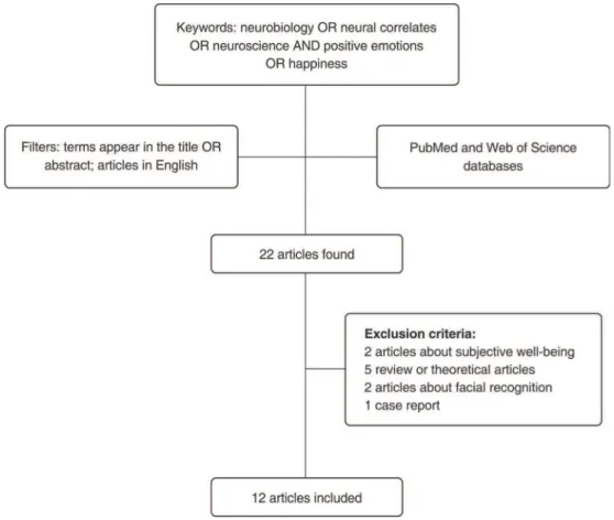
The search for papers was conducted in January 2016 in two different databases, MEDLINE and Web of Science. The following keywords were used in both databases: ‘‘neural correlates and positive emotions,’’ ‘‘neurobiology and positive emotions,’’ ‘‘neurobiology and happiness,’’ ‘‘neural correlates and happiness,’’ and ‘‘neuroscience and happiness.’’ Each of these combinations comprised an individual search query. Keywords had to appear in the title or abstract and the search was restricted to publica-tions written in English. No restricpublica-tions were placed on year of publication.
The inclusion criterion for this systematic review was: original articles dealing with use of neuroimaging or EEG
techniques to investigate neural correlates of positive emotions, including happiness. The exclusion criteria were: review articles, theoretical articles, animal studies, studies about neural correlates of subjective well-being, and studies not directly related to neural correlates of positive emotions or happiness (Figure 1). Using these criteria, 10 of the 22 retrieved articles were excluded; the remaining 12 form the basis of this review (Table 1).
Results
In contrast to abundant data on the structural and functional brain changes associated with psychiatric disorders and negative emotions, there is relatively little data on brain changes associated with normal emotional states and positive emotions; however, in recent years, there has been growing interest in the neural correlates of happiness. Of the 12 articles included in this review, five were published during the last 4 years.
The oldest study included in this review was published in 1995 in the American Journal of Psychiatry. Eleven healthy adult women with no history of mental illness were scanned by using H215O positron emission tomography (PET) during happy, sad, and neutral states induced by recalling affect-appropriate life events and looking at happy, sad, or neutral human faces. Emotional changes across tasks were assessed using paired, two-tailed Student’s t tests to compare scores on the Positive
Table 1 Description of results
Author Year Sample Main findings Main limitations
George28 1995 11 healthy and never mentally ill adult women
1) Transient sadness activated bilateral limbic and paralimbic structures (right medial frontal gyrus, left dorsolateral prefrontal cortex, bilateral cingulate gyrus, caudate, putamen, thalamus, fornix, left insula, and left midline cerebellum). 2) Transient happiness was associated with significant and widespread reductions in cortical regional cerebral blood flow, especially in the right prefrontal and bilateral temporoparietal regions.
1) Small sample
2) Only women participated 3) Many memories are
emotionally mixed. Sadness and happiness are not necessarily mutually exclusive
Lane29 1997 12 healthy women 1) Happiness, sadness, and disgust were each
associated with increases in activity in the thalamus and medial prefrontal cortex (BA9). 2) Happiness, sadness, and disgust were also associated with activation of anterior and posterior temporal structures.
3) Recalled sadness was associated with increased activation in the anterior insula. 4) Happiness was distinguished from sadness by greater activity in the vicinity of the ventral medial frontal cortex.
1) Small sample
2) Only women participated
Pelletier30 2003 Nine professional actors Relative to an emotionally neutral state, both sad
and happy states were associated with significant loci of activation, bilaterally, in the orbitofrontal cortex and in the left medial prefrontal cortex, left ventrolateral prefrontal cortex, left anterior temporal pole, and right pons.
1) Small sample
2) Sample limited to professionally trained (i.e., expert) individuals, which may present certain problems, given that distinctions between experts and typical untrained individuals can be quite marked
Habel31 2005 26 healthy subjects 1) Sad and happy mood, in contrast to the control task, produced similarly significant activations in the amygdala-hippocampal area extending into the parahippocampal gyrus, as well as in the prefrontal and temporal cortex, the anterior cingulate, and the precuneus. 2) During sadness, more activation was demonstrated in the ventrolateral prefrontal cortex, anterior cingulate cortex, transverse temporal gyrus, and superior temporal gyrus. 3) Happiness produced stronger activation in the dorsolateral prefrontal cortex, cingulate gyrus, inferior temporal gyrus, and cerebellum.
Blocked designs are prone to habituation effects.
Kim32 2007 10 healthy, right-handed female volunteers (ages 18-29)
1) Increasing negative and positive emotion engaged primarily left-lateralized prefrontal regions, whereas decreasing emotion activated bilateral prefrontal regions.
2) Regulation effects on amygdala activity were larger for positive than for negative stimuli, potentially reflecting a greater malleability of positive emotional reactions.
1) Small sample
2) Only women were included 3) Psychiatric disorders were not measured
Cerqueira33 2008 11 healthy subjects
(six men, average age 32.4 years) selected according to their ability to vividly recall personal experiences
1) In the happiness relative to the neutral condition, there was increased signal in the left dorsal prefrontal cortex, left insula, left anterior cingulate gyrus, mid-portions of the left middle temporal gyrus, left hypothalamus, and thalamus bilaterally.
2) In happiness relative to irritability, there was increased activity in the left anterior insula, left hypothalamus, thalamus bilaterally, inferior and middle temporal gyri bilaterally, and right posterior insula.
1) Small sample
2) Remote period (6-12 months) used for selection of situations
Mak34 2009 12 healthy women 1) Left superior and lateral frontal regions
(BA8/9) are common neural correlates of regulation of positive and negative emotions. 2) Positive emotions: increased activation in the prefrontal regions (left superior medial, BA8, and dorsolateral, BA9, gyri) and decreased activation in the left insula, the right rolandic operculum (BA6), and the lingual (BA18) gyri.
1) Small sample
Table 1.(continued)
Author Year Sample Main findings Main limitations
3) Negative emotions: increased activation in the left orbitofrontal gyrus (BA11), left superior medial frontal gyrus (BA8), anterior cingulate gyrus (BA19), and right precuneus (BA30); decreased activation in the right precentral gyrus (BA6) and bilateral parietal gyrus (R:BA2; L: BA40). 4) Participants appeared to be more effective in regulating positive than negative emotions. Yu35 2012 Two groups of
18 participants each: very happy group and not very happy group (nine males and nine females each); mean age 21.63 years. All had no history of psychiatric illness
The negative emotional priming effect was greater in participants in the not very happy group. Thus, participants in the very happy group were more sensitive and subject to the influence of external stimuli, particularly negative emotional stimuli.
1) Relatively small sample 2) The method used to rule out the presence of neuropsychiatric disorders in participants was not specified
3) The authors did not compare the effect of priming at different levels
Yuan36 2012 16 extraverts and 16 ambiverts
1) Significant emotion effects for highly positive and moderately positive stimuli at the P2 and P3 components in extraverts, but not in ambiverts. 2) Ambiverts displayed a significant emotion effect for moderately positive stimuli at the N2 and P3 components that was absent in extraverts. 3) The posterior cingulate cortices may mediate the extravert-specific emotion effect for pleasant stimuli.
4) Extraverts are less susceptible to unpleasant stimuli of mild intensity than are ambiverts, while extraverts have additional enhanced sensitivity to pleasant stimuli, regardless of emotion intensity.
Relatively small sample
Luo37 2014 50 young adults 1) Decreased regional homogeneity (ReHo) in unhappy relative to happy individuals was observed within the prefrontal cortex, medial temporal lobe, superior temporal lobe, and retrosplenial cortex.
2) Increased ReHo in unhappy relative to happy individuals was observed within the dorsolateral prefrontal cortex, middle cingulate gyrus, putamen, and thalamus.
3) ReHo within the left thalamus was negatively correlated with Chinese Happiness Inventory scores in the happy group.
1) Relatively small sample 2) Only examined traits of general happiness
Greening38 2014 19 non-medicated patients
with major depressive disorder and 19 controls
1) Controls were significantly better at modulating both negative and positive emotions.
2) Both groups recruited regions of the dorsolateral prefrontal cortex and ventrolateral prefrontal cortex when regulating negative emotions, but only in controls was this accompanied by reduced activity in the sensory cortices and amygdala.
3) Both groups showed enhanced activity in the ventrolateral prefrontal cortex and ventral striatum when enhancing positive affect; however, only in controls was ventral striatum activity correlated with regulation efficacy.
Small sample
Luo39 2016 148 healthy participants 1) Increased functional connectivity in the
Default Mode Network (DMN) was associated with lower levels of happiness.
2) Relative to happy people, unhappy people exhibited greater functional connectivity in the anterior medial cortex (bilateral), posterior medial cortex regions (bilateral), and posterior parietal cortex (left).
3) Increased functional connectivity of the medial prefrontal cortex, posterior cingulate cortex, and inferior parietal lobule correlated positively with inclination to ruminate.
and Negative Affect Schedule and a 25-point visual analogue scale before and after the emotion induction tasks. Transient happiness was associated with wide-spread reductions in cortical regional cerebral blood flow, especially in the right prefrontal and bilateral temporopar-ietal regions. In contrast, transient sadness activated bilateral limbic and paralimbic structures (right medial frontal gyrus, left dorsolateral prefrontal cortex, bilateral cingulate gyrus, caudate, putamen, thalamus, fornix, left insula, and left midline cerebellum). This was one of the first studies to show that happiness and sadness have divergent effects on different brain regions and not just opposite effects on activity in the same brain regions.28 Years later, Lane et al.29used the same technique and a similar method to evaluate the neural correlates of happiness, sadness, and disgust, and found that although these emotions are associated with a common group of regions, each has distinctive associations with specific brain regions. For example, happiness was distinguished from sadness because it was associated with greater activity in the vicinity of the ventral medial frontal cortex.29 Both studies used all-female samples. Another group tried to establish whether the primary emotions share similar neural substrates by investigating nine profes-sional actors of both genders using functional magnetic resonance imaging (fMRI). The actors were scanned during self-induced states of sadness and happiness and blood oxygen level-dependent (BOLD) signal changes were measured in three conditions: sad, happy, and emo-tionally neutral. During the preceding week, subjects were asked to recall powerful, personal emotional episodes involving sadness or happiness (the saddest episode of their life and the happiest episode of their life), as well as an emotionally neutral episode. During the experiment, subjects were asked to invoke the target emotional state (sad, happy, or neutral) by recalling and re-experiencing the appropriate personal episode. Unlike in the first two experiments, the sad and the happy states were associa-ted with specific bilateral activation loci in the orbitofrontal cortex, left medial prefrontal cortex, left ventrolateral prefrontal cortex, left anterior temporal pole, and right pons. Taking into account the small sample size and that the subjects were professional actors, the authors concluded that happiness and sadness may be asso-ciated with similar brain regions, but distinct subregions and neural circuits.30
In the years that followed, other researchers continued to study the basic emotional states, especially happiness and sadness. Negative and positive emotions were found to be associated with different cortical activation foci within a common neural network, probably discriminating diffe-rent emotional feelings.32-37Compared with a control task, tasks designed to induce sad or happy mood produced similar activation of the amygdala-hippocampal area, with activation extending into the parahippocampal gyrus, as well as activation of the prefrontal, temporal, and anterior cingulate cortices and the precuneus. However, happiness produced stronger activation than sadness in the dorso-lateral prefrontal cortex, the cingulate gyrus, the inferior temporal gyrus, and the cerebellum.31Emotions that elicit withdrawal behaviors (negative emotions), such as fear
and disgust, are thought to be primarily processed in right anterior brain regions, whereas emotions driving approach behaviors (positive emotions), such as happiness, are processed predominantly in the left hemisphere.33
Now, increasing negative and positive emotions are understood to primarily engage left prefrontal regions, whereas decreasing emotion activates prefrontal regions bilaterally. On the other hand, regulation effects on amyg-dala activity are larger for positive than for negative stimuli, suggesting that positive emotional reactions may be more malleable. In summary, there is overlap in the regions involved in regulation of positive and negative emotions, but substantial differences have also been found at the cortical and subcortical level, both between negative and positive emotions and between upregulation and downregulation.32
Understanding how different emotions are regulated is important to understanding illness and well-being. For example, happiness has a profound influence on human emotion processing35and extraverts are less susceptible to unpleasant stimuli of mild intensity than are ambiverts, but more sensitive to pleasant stimuli, regardless of intensity.36 Furthermore, mood disorders are associated with an inability to modulate intense emotions. A com-parison of 19 non-medicated patients with major depres-sive disorder and 19 controls demonstrated that controls were better at modulating both negative and positive emotions. During regulation of negative emotions, both groups recruited regions of the dorsolateral prefrontal cortex and ventrolateral prefrontal cortex, but only in controls was this accompanied by reduced activity in the sensory cortices and amygdala. Both groups showed increased activity in the ventrolateral prefrontal cortex and ventral striatum when enhancing positive affect; however, only in controls was ventral striatal activity correlated with regulation efficacy.38
Although the regulation of both positive and negative emotions is important, there have been few studies of the regulation of positive emotions, and their results have been inconsistent, perhaps because of the complexity and heterogeneity of the phenomenon.34,38Left superior and lateral frontal regions are implicated in regulation of both positive and negative emotions. Regulation of positive emotions is associated with increased activation in prefrontal regions (left superior medial and dorsolateral gyri) and decreased activation in the left insula, the right Rolandic operculum, and the lingual gyri. Furthermore, humans seem to be better at regulating positive emotions than negative emotions.34
study, Luo et al.39showed that functional connectivity in the DMN was negatively associated with happiness and positively associated with the inclination to ruminate. These results highlight the importance of the DMN to work on the neural correlates of happiness, and suggest that rumination may play an important role in perception of happiness.39
Discussion
The studies included in this review converge on the conclusion that there is overlap in the brain regions involved in the formation and regulation of positive and negative emotions. However, positive emotions, such as happiness, also activate specific brain regions. Perhaps, this pattern of activity could be used to distinguish them from negative emotions, such as sadness and disgust.
The brain areas specifically associated with happiness and positive emotions were not the same across the studies reviewed herein; differences in methodology and emotional stimuli may account for this heterogeneity. It is also possible that differences are due to the complex nature of emotions, particularly happiness and positive emotions. One must also consider that the stimuli used may be involved in a mix of emotions. Therefore, it is worth focusing our discussion on the more commonly identified neural correlates of happiness.
Formation and regulation of happiness and positive emotions are associated with significant reductions in activity in the right prefrontal cortex and in the tempor-oparietal cortex bilaterally, as well as with increased activity in the left prefrontal regions (especially the dorso-lateral and medial prefrontal cortices), the cingulate gyrus, the inferior and middle temporal gyri, the amygdala, and the ventral striatum.
Additionally, increased functional connectivity between the medial prefrontal cortex, posterior cingulate cortex, and inferior parietal lobule (areas of the DMN) were found to correlate negatively with happiness. Previous theore-tical studies have suggested associations between areas of the DMN and happiness.40
The frontal lobe is functionally and architecturally hete-rogeneous.25,41 Since David Ferrier’s pioneering studies of brain stimulation in the frontal lobe in the 1870s, it has been known that ablation of the frontal lobe causes dra-matic changes in animal behavior.18Based on analysis of the famous case of Phineas Gage, Damasio et al.42showed that lesions in the ventromedial prefrontal cortex produce severe impairments in rational decision-making and emotion processing.42 The prefrontal cortex can be viewed as the site of the confluence of two functions: working memory/ executive function/attention and behavior.41,43
Although cerebral lateralization of emotions is not a consensus, most studies show that the right prefrontal cortex is associated with negative emotions, while the left prefrontal cortex is associated with positive emotions.16,25 Patients with panic disorder show asymmetric frontal activation; right frontal activation is associated with acute activation of avoidance-withdrawal responses and with negative emotions.44 Left prefrontal damage has been associated with secondary depression, and prefrontal
lobe hypoactivity has been found in patients with primary depression.28 The association between left hemisphere damage and depression is more consistent when damage extends beyond the prefrontal cortex.25,44Research on non-depressed people has also shown that films which induce negative emotions elicit increased activity in the right prefrontal cortex.25,45 Damage to right side of the brain, particularly the temporal lobe, has been implicated in cases of secondary mania. Furthermore, during morphine- and cocaine-induced euphoria, there are profound reductions in regional activity in the prefrontal and parietotemporal cortices. Reductions in activity in secondary association cortices may be common to both transient happiness and profound euphoria28; however, a recent study of 96 patients with frontotemporal dementia and 34 healthy controls showed otherwise. Participants watched film clips designed to elicit happiness or sadness while their facial behavior, physiological reactivity, and self-reported emotional state were monitored. Whole-brain voxel-based morphometric analyses revealed that atrophy predominantly confined to the left frontostriatal areas was associated with greater facial responses to happiness. There were no associations between atrophy in specific regions and reactivity to sadness, which suggests that left frontostriatal atrophy is selectively associated with dysregulation of happiness. The authors argued that, although it has been proposed that left frontal injury decreases positive emotional res-ponses, selective disruption of left-hemisphere emotion regulation systems can impair the ability to suppress positive emotions such as happiness.46
Less evidence is available on positive emotions. There are several possible reasons for this. Positive emotions are much harder to elicit in laboratory settings than negative emotions. The body tends to react more strongly to negative stimuli than to positive stimuli, perhaps as a consequence of evolutionary pressures; this is sometimes referred to as the negativity bias.25It has been hypothe-sized that the right hemisphere is specialized for emo-tional and visuospatial functions that are important to the survival of the species.44
Another model of how the brain mediates emotions in humans takes into account concomitant motor responses. It posits that ‘‘approach emotions’’ (e.g., happiness) are lateralized to the left side and ‘‘withdrawal emotions’’ (e.g., disgust) to the right, particularly in the frontal and prefrontal areas.29,44 The studies included in this review seem to support this view of hemispheric specialization, especially in relation to the prefrontal areas. Negative and positive emotions also appear to be associated with various behavioral and developmental functions. Negative emo-tions such as fear and anger clearly have adaptive value, as they help to ensure survival and safety. Nevertheless, these benefits are considered in the short term.47 The ‘‘broaden and build’’ theory states that positive emotions amplify cognition and behavior to optimize performance. According to this model, positive emotions have long-term developmental benefits.48
temperament factors have largely been related to limbic and subcortical structures, whereas character has an extensive relationship with cerebral cortex activation.50 That character and happiness are related to wide cortical activation helps explain why a study of 1,102 volunteers found positive associations between the three character factors and perceived happiness.6 These data are also consistent with the proposal that personality disorders arise as a result of flaws in character structure.51
Together, the cingulate cortex and parahippocampal gyrus constitute what Broca called the great limbic lobe. The posterior cingulate cortex is important for the successful retrieval of autobiographical memories, and cognitive impairment is correlated with alterations in posterior cingu-late activity. The connections of the anterior cingucingu-late cortex mean it is well placed to filter and control the flow of information between the emotional limbic system and autonomic nervous system, and it plays an important role in error detection and the appreciation and expression of emotions.44 The studies included in this review suggest there is an association between happiness and cingulate cortex activity, and this is corroborated by other studies.17,52 However, as many of the studies we reviewed highlight its role in negative emotions, it seems likely that the cingulate cortex plays a key role both in positive and in negative emotions.25 Actually, much of the circuitry associated with emotions, including the limbic system, is involved in emo-tional responses generally, regardless of the valence of the emotion or the nature of the eliciting stimulus. The amygdala, for instance, is involved not only in negative emotions such as fear, but also in positive emotions.17 Indeed, this structure is considered the ‘‘heart and soul’’ of the brain’s emotional network.44 Findings are inconsistent regarding the activity of the amygdala in positive emotions, with some studies showing reduced17,27and others showing increased44activity.
The ventral striatum, also referred to as the limbic striatum, includes a number of basal forebrain structures, such as the nucleus accumbens and the olfactory tubercle.44 This area is widely known to be involved in pleasure,24,40,53 which has a major influence on life satisfaction and psychological well-being.5,54Many theo-retical papers have postulated that the ventral striatum is involved in subjective happiness,17,24,40,52,54 and the studies reviewed here support that view. Brain mechan-isms involved in ‘‘fundamental’’ pleasures (e.g., food and sexual pleasures) overlap with those for ‘‘higher-order’’ pleasures (e.g., artistic, musical, altruistic, and transcen-dent pleasures).54Interestingly, the ventral striatum can also be activated by listening to sad music.55
The limitations of the studies reviewed herein must be taken into consideration. Most used small samples and were cross-sectional. The use of different imaging modalities and the variety of stimuli may have led to heterogeneous results. Furthermore, stimuli may result in a mixture of positive and negative emotions. There is also a lack of consensus on the nature of emotions them-selves. For example, some proposals place anger, rather than sadness, as the opposite of happiness.16,17 In addition, although positive emotions describe a wider range of events, most of the articles reviewed assessed
happiness as the key representative of positive emotions, sometimes almost interchangeably with the concept of positive emotion. Finally, some conclusions of the studies included in this review require additional replication. For example, only a few of the studies included neutral stimuli to differentiate positive emotions from negative emotions. Future research should include similar measures to address whether happiness truly differs from the absence of sadness, as demonstrated in some studies included in this review.
It is too early to claim that there is an established understanding of the neuroscience of positive emotions and happiness. However, although there is overlap in the brain regions involved in the formation and regulation of positive and negative emotions, we conclude that positive emotions, such as happiness, activate specific brain regions. Thus, positive emotions and happiness do not appear to be elicited solely by disabling areas which are activated by negative emotions and sadness. This modern perspective challenges the existentialist view that life is dominated by anguish generated by the fear of death, which is interrupted only briefly by moments of joy. Perhaps, life involves moments of both sadness and happiness, with some people experiencing a higher propensity for positive emotions and happiness, and others, a higher propensity for negative emotions and sadness. Positive emotions must receive greater recognition by clinicians and researchers in the field of psychiatry.
Acknowledgements
The corresponding author thanks Dr. Silvia Laurentino for her neuroscience teachings, which contributed to the writing of this article.
Disclosure
The authors report no conflicts of interest.
References
1 Seˆneca. Da vida retirada, da tranquilidade da alma e da felicidade-Porto Alegre: L&PM Pocket, 2009.
2 Tobgay T, Dophu U, Torres CE, Na-Bangchang K. Health and Gross National Happiness: review of current status in Bhutan. J Multidiscip Healthc. 2011;4:293-8.
3 Tobgay T, Dorji T, Pelzom D, Gibbons RV. Progress and delivery of health care in Bhutan, the Land of the Thunder Dragon and Gross National Happiness. Trop Med Int Health. 2011;16:731-6.
4 Seligman MEP. Felicidade auteˆntica: usando a nova psicologia positiva para a realizac¸a˜o permanente. Rio de Janeiro: Objetiva;
2010.
5 Seligman MEP. Florescer: Uma nova compreensa˜o sobre a natureza da felicidade e do bem-estar. Rio de Janeiro: Objetiva; 2012. 6 Cloninger CR, Zohar AH. Personality and the perception of health
and happiness. J Affect Disord. 2011;128:24-32.
7 Machado L, Tavares H, Petribu´ K, Pinto T, Cantilino A. Happiness and defense styles in psychiatrists. J Nerv Ment Dis. 2016;204:181-7. 8 Baruch Y, Swartz M, Sirkis S, Mirecki I, Barak Y. Staff happiness and work satisfaction in a tertiary psychiatric centre. Occup Med (Lond). 2013;63:442-4.
9 Cloninger CR. Feeling good: the science of well-being, New York: Oxford University; 2004.
11 Machado L, Tavares H, Petribu´ K, Zilberman M, Torres RF, Cantilino A. Happiness and health in psychiatry: what are their implications? Arch Clin Psychiatry (Sa˜o Paulo). 2015;42:100-10.
12 Ferraz RB, Tavares H, Zilberman ML. Felicidade: uma revisa˜o. Rev Psiquiatr Clin. 2007;34:234-42.
13 Naber D, Karow A, Lambert M. Subjective well-being under the neuroleptic treatment and its relevance for compliance. Acta Psy-chiatr Scand Suppl. 2005;111:29-34.
14 Jeste DV, Palmer BW, Rettew DC, Boardman S. Positive psychiatry: its time has come. J Clin Psychiatry. 2015;76:675-83.
15 Seligman ME, Csikszentmihalyi M. Positive psychology. An intro-duction. Am Psychol. 2000;55:5-14.
16 Arciniegas DB. Emotion. In: Arciniegas DB, Anderson CA, Filley CM, eds. Behavioral neurology and neuropsychiatry.Cambridge: Cambridge University; 2013.
17 Chemali ZN, Chahine LM, Naassan G. On happiness: a minimalist perspective on a complex neural circuitry and its psychosocial con-structs. J Happiness Stud. 2008;9:489-501.
18 Wickens AP. A history of the brain: from stone age surgery to modern neuroscience. New York: Psychology; 2015.
19 Matsunaga M, Isowa T, Yamakawa K, Fukuyama S, Shinoda J, Yamada J, et al. Genetic variations in the human cannabinoid receptor gene are associated with happiness. PLoS One. 2014;9: e93771.
20 De Neve JE. Functional polymorphism (5-HTTLPR) in the serotonin transporter gene is associated with subjective well-being: evidence from a US nationally representative sample. J Hum Genet. 2011;56: 456-9.
21 Rietveld CA, Cesarini D, Benjamin DJ, Koellinger PD, De Neve JE, Tiemeier H, et al. Molecular genetics and subjective well-being. Proc Natl Acad Sci U S A. 2013;110:9692-7.
22 Tay L, Kuykendall L. Promoting happiness: the malleability of indivi-dual and societal subjective wellbeing. Int J Psychol. 2013;48:159-76. 23 Chen H, Pine DS, Ernst M, Gorodetsky E, Kasen S, Gordon K, et al. The MAOA gene predicts happiness in women. Prog Neuropsycho-pharmacol Biol Psychiatry. 2013;40:122-5.
24 Berridge KC, Kringelbach ML. Building a neuroscience of pleasure and well-being. Psychol Well Being. 2011;1:1-3.
25 Davidson RJ. Well-being and affective style: neural substrates and biobehavioral correlates. Philos Trans R Soc Lond B Biol Sci. 2004;359:1395-411.
26 Helliwell J, Layard R, Sachs J. World happiness report [Internet]. 2012 Nov 23 [cited 2016 Jul 22]. eprints.lse.ac.uk/47487/.
27 Burgdorf J, Panksepp J. The neurobiology of positive emotions. Neurosci Biobehav Rev. 2006;30:173-87.
28 George MS, Ketter TA, Parekh PI, Horwitz B, Herscovitch P, Post RM. Brain activity during transient sadness and happiness in healthy women. Am J Psychiatry. 1995;152:341-51.
29 Lane RD, Reiman EM, Ahern GL, Schwartz GE, Davidson RJ. Neuroanatomical correlates of happiness, sadness, and disgust. Am J Psychiatry. 1997;154:926-33.
30 Pelletier M, Bouthillier A, Le´vesque J, Carrier S, Breault C, Paquette V, et al. Separate neural circuits for primary emotions? Brain activity during self-induced sadness and happiness in professional actors. Neuroreport. 2003;14:1111-6.
31 Habel U, Klein M, Kellermann T, Shah NJ, Schneider F. Same or different? Neural correlates of happy and sad mood in healthy males. Neuroimage. 2005;26:206-14.
32 Kim SH, Hamann S. Neural correlates of positive and negative emotion regulate. J Cogn Neurosci. 2007;19:776-98.
33 Cerqueira CT, Almeida JR, Gorenstein C, Gentil V, Leite CC, Sato JR, et al. Engagement of multifocal neural circuits during recall of autobiographical happy events. Braz J Med Biol Res. 2008;41: 1076-85.
34 Mak AK, Hu ZG, Zhang JX, Xiao ZW, Lee TM. Neural correlates of regulation of positive and negative emotions: An fmri study. Neurosci. Lett. 2009;457:101-6.
35 Yu GM, Li B. How subjective well-being affects emotional processing: the role of event-related potentials. Soc Behav Pers. 2012;40:1285-92. 36 Yuan J, Zhang J, Zhou X, Yang J, Meng X, Zhang Q, et al. Neural mechanisms underlying the higher levels of subjective well-being in extraverts: pleasant bias and unpleasant resistance. Cogn Affect Behav Neurosci. 2012;12:175-92.
37 Luo Y, Huang X, Yang Z, Li B, Liu J, Wei D. Regional homogeneity of intrinsic brain activity in happy and unhappy individuals. PLoS One. 2014;9:e85181.
38 Greening SG, Osuch EA, Williamson PC, Mitchell DG. The neural correlates of regulating positive and negative emotions in medication-free major depression. Soc Cogn Affect Neurosci. 2014;9:628-37. 39 Luo Y, Kong F, Qi S, You X, Huang X. Resting-state functional
connectivity of the default mode network associated with happiness. Soc Cogn Affect Neurosci. 2016;11:516-24.
40 Kringelbach ML, Berridge KC. The neuroscience of happiness and pleasure. Soc Res (New York). 2010;77:659-78.
41 Mesulam M. Behavioral neuroanatomy: large-scale networks, asso-ciation cortex, frontal syndromes, the limbic system, and hemispheric specializations. In: Mesulam M editors. Principles of behavioral and cognitive neurology.2nd ed.Oxford: Oxford University; 2000. p. 41-9. 42 Damasio H, Grabowski T, Frank R, Galaburda AM, Damasio AR. The return of Phineas Gage: clues about the brain from the skull of a famous patient. Science. 1994;264:1102-5.
43 Arciniegas DB. Executive function In:Arciniegas DB, Anderson CA, Filley CM, editors. Behavioral neurology and neuropsychiatry. Cam-bridge: Cambridge University; 2013. p. 225-49.
44 Clark DL, Boutros NN, Mendez MF. The brain and behavior. 3rd ed. New York: Cambridge University; 2010.
45 Davidson RJ, Chapman JP, Chapman LJ, Henriques JB. Asymme-trical brain elecAsymme-trical activity discriminates between psychomeAsymme-trically- psychometrically-matched verbal and spatial cognitive tasks. Psychophysiology. 1990;27:528-43.
46 Sturm VE, Yokoyama JS, Eckart JA, Zakrzewski J, Rosen HJ, Miller BL, et al. Damage to left frontal regulatory circuits produces greater positive emotional reactivity in frontotemporal dementia. Cortex. 2015;64:55-67.
47 Kobau R, Seligman ME, Peterson C, Diener E, Zack MM, Chapman D, et al. Mental health promotion in public health: perspectives and strategies from positive psychology. Am J Public Health. 2011;101: e1-9.
48 Fredrickson BL. The broaden-and-build theory of positive emotions. Philos Trans R Soc Lond B Biol Sci. 2004;359:1367-78.
49 Cloninger CR, Svrakic DM, Przybeck TR. A psychobiological model of temperament and character. Arch Gen Psychiatry. 1993;50: 975-90.
50 Kedia S, Cloninger CR. Personality. In: Arciniegas DB, Anderson CA, Filley CM, editors. Behavioral Neurology and Neuropsychiatry. New York: Cambridge University; 2013. p. 299-309.
51 Tavares H. Perspectivas futuras dos transtornos da personalida-deInLouza˜ Neto MR, Corda´s TA, editores. Transtornos da persona-lidade. Porto Alegre: Artmed; 2011. p. 337-52.
52 Funahashi S. Brain mechanisms of happiness. Psychologia. 2011;54: 222-33.
53 Esperidia˜o-Antonio V, Majeski-Colombo M, Toledo-Monteverde D, Moraes-Martins G, Fernandes JJ, Assis MB, et al. Neurobiologia das emoc¸o˜es. Rev Psiquiatr Clin. 2007;35:55-65.
54 Kringelbach ML, Berridge KC. The functional neuroanatomy of pleasure and happiness. Discov Med. 2010;9:579-87.