660
Rev Soc Bras Med Trop 49(5):660-662, September-October, 2016 doi:10.1590/0037-8682-0437-2015
Case Report
Corresponding author: Dra. Marcela Sena Teixeira Mendes. e-mail: [email protected]
Received 28 December 2015 Accepted 20 April 2016
Human
T-cell lymphotropic virus-1 infection: three
infected generations in the same family
Marcela Sena Teixeira Mendes
[1], Mariana Carvalho Costa
[2]and Izelda Maria Carvalho Costa
[1][1]. Programa de Pós Graduação Senso Stricto em Ciências da Saúde, Universidade de Brasília, Brasília, Disrito Federal, Brasil. [2]. Departamento de Dermatologia, Hospital Universitário de Brasília, Brasília, Distrito Federal, Brasil.
Abstract
The human T-cell lymphotropic virus-1 (HTLV-1) affects worldwide population; the estimated number of currently infected individuals is 10-20 million. In this report, we describe the clinical indings of three family members with vertical transmission of HTLV-1. This case report highlights the importance of healthcare providers who have optimal knowledge about HTLV-1 including its transmission and pertinent attributes, and who are able to provide affected individuals with adequate information regarding their condition.
Keywords: HTLV-1 infection. Infective dermatitis. HTLV-1 associated myelopathy.
INTRODUCTION
The human T-cell lymphotropic virus (HTLV) is a retrovirus that affects 10-20 million people worldwide. In Brazil, about 2.5 million people are currently infected(1). Despite high prevalence, this disease is neglected with a consequent lack of effective preventive measures aimed at its reduction. In this paper, we report the clinical indings of the vertical transmission of HTLV-1 in three members of the same family, each with different clinical presentations of this infection.
CASE REPORT
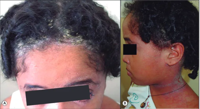
An 11-year-old female patient presented to our department with a 7-year history of erythematous desquamative plaques in the axillae, neck, and retro-auricular areas. She also presented with rhinorrhea, bilateral conjunctival erythema, and crusts on the nostrils (Figure 1A and Figure 1B).
There were no signiicant indings in her prenatal history. The patient was born via cesarean section due to cephalopelvic disproportion and was breastfed until the age of 3 years. The patient’s mother was 32 years and had a known HTLV-1 infection, which was diagnosed at 11 years. The infection manifested as HTLV-1-associated myelopathy (HAM) with a progressive course. The patient’s mother was born vaginally and was breastfed until the age of 4 years. The mother also stated that the patient’s grandmother was an asymptomatic carrier
of the virus, diagnosed at the same time as she was, 21 years previously. The grandmother could not provide any information on how long she breastfed the patient’s mother.
The child was tested for HTLV-1 based on the clinical presentations and on her family’s positive HTLV-1 status. Enzyme-linked immunosorbent assay (ELISA) and western blot analysis were performed, conirming the infection. A viral load of 10,700 copies was detected.
DISCUSSION
HTLV-1 affects worldwide population(1) (2). The majority of carriers are asymptomatic, with only 3-7% of affected people presenting with clinical manifestations(2). The clinical spectrum of the disease encompasses infective dermatitis (IDH), HTLV-1-associated myelopathy/tropical spastic paraparesis (HAM/ TSP), and adult T-cell leukemia lymphoma (ATLL)(3).
Concerning the present case, we suggest that the patient’s grandmother was only a carrier of the virus, similar to most affected individuals. However, the patient’s mother presented with HAM at a young age, unlike the typical HAM presentation, which generally occurs during the fourth or ifth decades of life ; the patient presented with HTLV-1-associated IDH at the age of two years.
Infective dermatitis was irst described by Sweet in 1966 as a chronic form of eczema among Jamaican children, years before the discovery of HTLV, which occurred in 1980(4) (5). In 1998, the criteria to aid the diagnosis of this disease were established by La Granade et al.(6) (7), and later modiied by de Oliveira et al. (Figure 2).
661
Mendes MST et al. - HTLV-1 infection
A B
FIGURE 1. (A): Crusts on the scalp and conjunctival erythema, at the age of 7 years. (B): Crusts, erythema, and maceration on the child’s neck.
La Grenade et al. 1997 De Oliveira et al. 2012
Major:
1. Eczema of scalp, axillae and groin, external ear and
retroauricular areas, eyelid margins, paranasal skin, and/or neck
2. Chronic watery nasal discharge without other signs of
rhinitis and/or crusting of the anterior nares
3. Chronic relapsing dermatitis with prompt response to
appropriate therapy but prompt recurrence on withdrawal of the use of antibiotics
4. Usual onset in early childhood
5. Human T-lymphotropic virus type I antibody
seropositivity
Minor or less specific
- Positive cultures for Staphylococcus aureus and/or
β-hemolytic streptococci from the skin or anterior nares
- Generalized fine papular rash (in most severe cases)
- Generalized lymphadenopathy with dermopathic
lymphadenitis
- Anemia
- Elevated erythrocyte sedimentation rate
- Hyperimmunoglobulinemia (IgD and IgE)
- Elevated CD4 count, CD8 count and CD4/CD8 ratio
Major:
1. Presence of erythematous-scaly, exudative, and
crusted lesions of the scalp, retroauricular areas, neck, axillae, groin, paranasal and perioral skin, ears, thorax, abdomen and other sites
2. Crusting of nostrils
3. Chronic relapsing dermatitis with prompt
response to appropriate therapy but prompt recurrence on discontinuation of antibiotics
4. Diagnosis of HTLV-1 infection (via serological or
molecular biological testing)
Of the five major criteria, four are required for diagnosis with the mandatory inclusion of 1, 2, and 5. To fulfill criteria 1, the involvement of at least 2 of the sites is required.
Of the four major criteria, three are required for diagnosis, with the mandatory inclusion of 1, 3, and 4. To fulfill criteria 1, the involvement of ≥ 3 of the sites is required, including the involvement of the scalp and retroauricular areas.
FIGURE 2. Diagnostic Criteria for Infective Dermatitis(6) (7). HTLV-1: human T-cell lymphotropic virus; IgD: immunoglobulin D; IgE: immunoglobulin E;
662
Rev Soc Bras Med Trop 49(5):660-662, September-October, 2016
crusted plaques on the scalp, postauricular region, popliteal, and antecubital fossa; blepharoconjunctivitis; rhinorrhea; and crusts on the nostrils(3) (4) (6). Another feature of IDH is its association with an increased risk of ATLL and HAM(8). In families infected with HTLV, 93% of the cases of IDH and HAM/TSP occur in 2 generations of the same family(9). Hence, close follow-up of children presenting with IDH is important, even after remission of this condition.
In the present case, the prolonged breastfeeding period was identiied as the probable source of infection. The most prevalent form of transmission among affected children is vertical (transplacental, birth canal, and breastfeeding), but transmission can also occur through sexual intercourse, use of intravenous drugs, or blood transfusions(2) (3). Intrauterine and peripartal virus transmissions occur in less than 5% of cases but rise to 10-25% when breastfeeding is involved(2). Prolonged breastfeeding (>6 months) has been associated with a greater risk of infection compared to other forms of transmission(2) (4) (8). This was a key aspect of our case, since both the patient and
her mother were breastfed for long periods. However, we could not establish if the lack of symptoms in the grandmother was related to a shorter period of breastfeeding.
In conclusion, we suggest the importance of adequate prenatal care, during which HTLV serology tests may be performed. The care should also include providing patients and caregivers information concerning the risks of transmission through breastfeeding and consequences of infection. We believe that this case illustrates the importance of healthcare providers who are knowledgeable about HTLV-1, which consequently can lead to improvement in eficiency and accuracy of disease diagnosis and prevention.
Conlict of Interest
The authors declare that there is no conlict of interest.
REFERENCES
1. Carneiro-Proietti AB, Ribas JG, Catalan-Soares BC, Martins ML, Brito-Melo GE, Martins-Filho OA, et al. Infection and disease caused by the human T cell lymphotropic viruses type I and II in Brazil. Rev Soc Bras Med Trop 2002; 35:499-508.
2. Amano M, Setoyama M, Grant A, Kerdel FA. Human T-lymphotropic virus 1 (HTLV-1) infection – dermatological implications. Int J
Dermatol 2011; 50:915-920.
3. Gessain A. Le rétrovirus humain oncogène HTLV-1: épidémiologie descriptive et moléculaire, origine, évolution et aspects diagnostiques et maladies associées. Bull Soc Pathol Exot 2011; 104:167-180.
4. Bittencourt AL, Oliveira MF. Cutaneous manifestations associated
with HTLV infection. Intern J Dermatol 2010; 49:1099-1110.
5. de Oliveira MF, Brites C, Ferraz N, Magalhaes P, Almeida F,
Bittencourt AL. Infective dermatitis associated with the human T cell lymphotropic virus type I in Salvador, Bahia, Brazil. Clin Infect
Dis 2005; 40:e90-96.
6. de Oliveira MF, Fatal PL, Primo JR, da Silva JL, Batista ES,
Farré L, et al. Infective dermatitis associated with human T-cell lymphotropic virus type 1: evaluation of 42 cases observed in Bahia, Brazil. Clin Infect Dis 2012; 54:1714-1714.
7. La Grenade L, Manns A, Fletcher V, Derm D, Carberry C, Hanchard B, et al. Clinical, pathologic, and immunologic features of human T-lymphotrophic virus type I-associated infective dermatitis in
Children. Arch Dermatol 1998; 134:439-444.
8. Hlela C, Bittencourt A. Infective dermatitis associated with HTLV-1 mimics common eczemas in children and may be a prelude to severe systemic diseases. Dermatol Clin 2014; 32:237-248.
9. da Silva JLS, Primo JRL, de Oliveira MF, Batista ES, Moreno-Carvalho O, Farré L, et al. Clustering of HTLV-1 associated