ISSN 0100-879X
BIOMEDICAL SCIENCES
AND
CLINICAL INVESTIGATION
www.bjournal.com.br
www.bjournal.com.br
Volume 43 (9) 812-913 September 2010
Institutional Sponsors
The Brazilian Journal of Medical and Biological Research is partially financed by
Hotsite of proteomics metabolomics developped by:
Braz J Med Biol Res, September 2010, Volume 43(9) 821-827
doi: 10.1590/S0100-879X2010007500084
Caspase-8 and p38MAPK in DATS-induced apoptosis of human CNE2
cells
Caspase-8 and p38MAPK in DATS-induced
apoptosis of human CNE2 cells
C. Ji
1, F. Ren
1and M. Xu
21College of Chemistry and Chemical Engineering, 2Research Institute for Molecular Pharmacology and Therapeutics,
Central South University, Changsha, Hunan, China
Abstract
Nasopharyngeal carcinoma is a common malignancy in Southern China of uncertain etiologic origin. Diallyl trisulfide (DATS), one of the major components of garlic (Allium sativum), is highly bactericidal and fungicidal. In this study, we investigated the function of p38 mitogen-activated protein kinase (MAPK) and caspase-8 in DATS-induced apoptosis of human CNE2 cells us-ing MTT [3-(4,5-dimethylthiazol-2-yl)-2,5-diphenyltetrazolium bromide], flow cytometry assay, and Western blottus-ing. After CNE2 cells were treated with DATS (50, 100, or 150 μM) for 24 h, cell viability rates were 75.9, 63.4 and 39.6%, and apoptosis rates were 24.5, 36.9, and 62.4%, respectively. The data showed that DATS induced CNE2 cell death in a dose-dependent manner. After human CNE2 cells were treated with 100 μM DATS and inhibitors (10 μM SB203580 and Z-LETD-FMK for p38MAPK and caspase-8, respectively), changes in cell viability and apoptosis and in p38MAPK and caspase-8 activity were detected.Cell viability rates were 66.5 and 68.1% and decreased 9.9 and 11.5% compared with inhibitor treatment alone. Apoptosis rates were 31.53 and 29.98% and increased 9.1 and 10% compared with inhibitor treatment alone.The results indicated that DATS activates p38MAPK and caspase-8, but both inhibitors have an effect on P38MAPK and caspase-8 activity. In conclusion, our data indicate that p38MAPK and caspase-8 are involved in the process of DATS-induced apoptosis in human CNE2 cells and interact with each other.
Key words: DATS; p38 MAPK; Caspase-8; Inhibitor; Apoptosis
Introduction
Correspondence: F. Ren, College of Chemistry and Chemical Engineering, Central South University, Changsha, Hunan 410083, China. Fax: +86-731-8883-6993. E-mail: [email protected]
Received April 15, 2010. Accepted August 5, 2010. Available online August 27, 2010. Published September 13, 2010.
Mitogen-activated protein kinase (MAPK), an important intracellular signal transduction system, has a marked ef-fect on the regulation of gene expression and cytoplasmic functional activities (1-3). The p38 signaling pathway is an important branch of the MAPK pathway, playing a significant role in a variety of physiological and pathological processes, such as inflammation, cell stress, apoptosis, cell cycle and growth, and so on (4,5). Caspase is an inactive enzyme zymogen under normal circumstances, but once activated it will trigger the caspase cascade, eventually leading to apoptosis. In the central control and effective stage of apoptosis, activated caspase-8 can lead directly to the appearance of apoptotic structural characteristics in cells, and play a key role in the process of apoptosis (6,7).
Nasopharyngeal carcinoma (NPC) is a malignant tu-mor of high incidence in the Southeastern region of Asia. In clinical practice, NPC is treated by radiation, but the therapeutic effect is not satisfactory (8-10). In recent years,
822 C. Ji et al.
that p38MAPK and caspase-8 are involved in the process of DATS-induced apoptosis in human CNE2 cells and interact with each other.
Material and Methods
Material
DATS (99% purity) was purchased from Chia-tai Tianq-ing Pharmaceutical Co., Ltd. (China). RPMI1640, BSA and SB203580 were purchased from Sigma (USA). Z-LETD-FMK was purchased from Biovision (USA), goat horserad-ish peroxidase (HRP)-conjugated anti-rabbit secondary antibody was purchased from Santa Cruz Biotechnology (USA). Antibodies to p38, phospho-p38 (p-p38), and cas-pase-8 were purchased from Cell Signaling (USA).
Cell culture
CNE2, a human NPC cell line, was provided by the Xiangya School of Medicine and cultured in RPMI1640 containing 10% heat-inactivated fetal bovine serum (FBS), benzylpenicillin (100 kU/L) and streptomycin (100 mg/L) at 37°C in an incubator containing humidified air with 5% CO2.
Cell viability assay
Cells were seeded onto 96-well plates at 1 x 10⁴ cells per
well 24 h before treatment. The cultures were then rinsed in phenol-free RPMI1640 medium and incubated with the respective test substances in phenol-free and serum-free RPMI1640 for 24 h. In the inhibition test, cells were treated with DATS after being treated with inhibitors for 30 min. At the end of this time, 20 µL (5 mg/mL) MTT [3-(4,5-dimeth -ylthiazol-2-yl)-2,5-diphenyltetrazolium bromide] was added to each well, and after incubation at 37°C for 4 h the MTT solution was removed and 200 µL dimethylsulfoxide (DMSO) was added to dissolve the crystals. The absorbance of each well at 570 nm was measured.
Flow cytometry analysis
Cells (12 x 106) were seeded into 100-mL cell culture bottles 24 h before treatment and then treated as described above and incubated for 24 h. Cells were then collected and
single cell suspensions were prepared and centrifuged at 800 g for 5 min. The supernatant was discarded and cells were washed three times with cool PBS and fixed for 24 h with cool alcohol at 4°C. One-milliliter cell suspension (106/mL) was washed three times with cool PBS, treated with RNase for 30 min at 37°C, stained with propidium iodide (PI) for 30 min at 37°C in the dark, and used for flow cytometry analysis.
Western blotting
Cells in the logarithmic growth phase were treated as described above and incubated for 24 h. After fragmentation on ice for 20 min, the lysates were centrifuged at 15,000 g
for 10 min at 4°C, the protein was collected, quantitated by the bicinchoninic acid (BCA) method, electrophoresed and isolated by 10% SDS-PAGE using the electrotransfer method, blocked, and hybridized on cellulose nitrate film. The protein expression of the cells was detected using the ECL Western blotting method. The densities of protein bands were calculated using the Quantyone software.
Statistical analysis
Data are reported as means ± SD of three independent experiments and were evaluated by one-way analysis of variance (ANOVA). Differences were considered to be significant at P < 0.05.
Results
Changes in cell activity
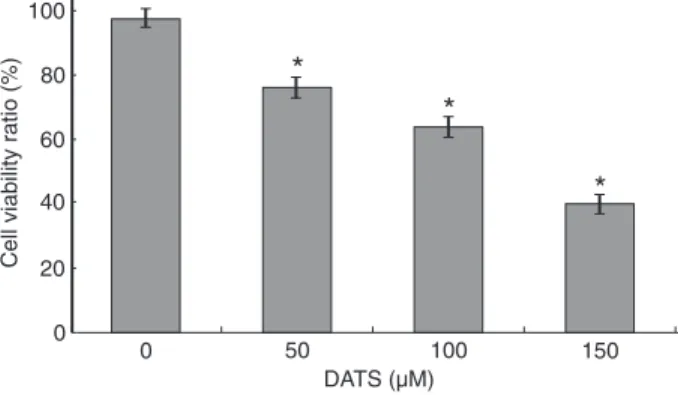
We tested the effect of DATS on human CNE2 cells. The cells were treated with various concentrations of DATS for 24 h and cell viability was determined by the MTT assay. As shown in Figure 1, 50 μM DATS induced a 75.9% decrease in cell viability. When the cells were incubated with 100 and 150 μM DATS, cell viability decreased by about 63.4 and 39.6%, respectively, at 24 h. DATS had a dose-dependent effect.
The MTT conversion assay was used to test the effects of inhibitors on the viability of CNE2 cells. After treatment with 10 μM SB203580 or Z-LETD-FMK for 30 min, 100 μM DATS induced a 66.5 or 68.1% decrease in cell viability at 24 h. When the cells were incubated with 10 μM SB203580
Figure 1. Effect of diallyl trisulfide (DATS) on cell viability. CNE2
or Z-LETD-FMK, cell viability decreased by about 76.4 or 79.6% at 24 h (Figure 2).
Flow-cytometry analysis of apoptosis
To further examine the effects of DATS on apoptosis, flow
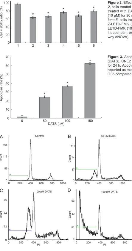
cytometry was used to quantify the apoptotic state. We found that 24.5, 36.9, and 62.4% of CNE2 cells became apoptotic when they were exposed to 50, 100, 150 μM DATS for 24 h (Figures 3 and 4). These results supported the view that DATS induces apoptosis of CNE2 cells in a
concentration-Figure 3. Apoptosis of CNE2 cells treated with diallyl trisulfide
(DATS). CNE2 cells were treated with 0, 50, 100, 150 μM DATS for 24 h. Apoptosis was determined by flow cytometry. Data are reported as means ± SD of three independent experiments. *P < 0.05 compared to control (one-way ANOVA).
Figure 2. Effect of inhibitors on cell viability. Lane 1, Control; lane 2, cells treated with diallyl trisulfide (DATS; 100 μM); lane 3, cells treated with DATS (100 μM) after being treated with SB203580 (10 μM) for 30 min; lane 4, cells treated with SB203580 (10 μM); lane 5, cells treated with DATS (100 μM) after being treated with
Z-LETD-FMK (10 μM) for 30 min; lane 6, cells treated with
Z-LETD-FMK (10 μM). Data are reported as means ± SD of three independent experiments. *P < 0.05 compared to control (one-way ANOVA).
Figure 4. Effect of diallyl trisulfide (DATS)
824 C. Ji et al.
dependent manner.
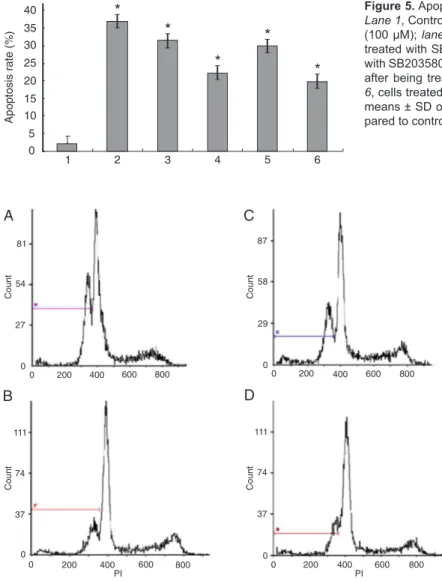
We also determined the effects of both inhibitors on apop -tosis by flow cytometry. Combined treatment with SB203580 plus DATS (100 μM) increased the proportion of apoptotic CNE2 cells by an absolute value of 9.1% when compared with inhibitor treatment alone. CNE2 cell apoptosis was increased by 10% with Z-LETD-FMK plus DATS (100 μM) compared with inhibitor treatment alone (Figures 5 and 6).
Protein expression
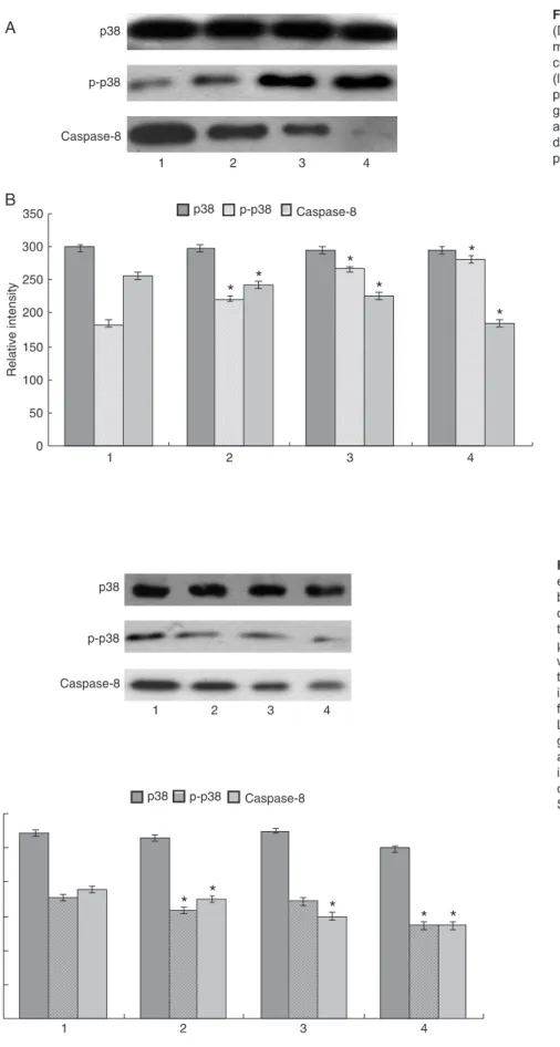
We tested the effects of DATS on p-p38 and caspase-8 activity as a function of dose by Western blotting. As shown in Figure 7, p-p38 and caspase-8 were markedly activated in a dose-dependent manner, with an accumulation of both after 24 h of DATS treatment. These data show that p-p38 and caspase-8 may play important roles in DATS-induced CNE2 cell apoptosis.
To test whether inhibitors block DATS-induced activation of p38 and caspase-8 in CNE2 cells, we also examined
pro-tein expression by Western blot analysis. When SB203580 or Z-LETD-FMK was added to CNE2 cells 30 min prior to DATS treatment, the activation of p-p38 and caspase-8 was markedly decreased. Similar results were observed with inhibitor treatment alone (Figure 8).
Discussion
Apoptosis is a type of physiological cell death that maintains body stability. The apoptosis process is strictly controlled by multiple genes, and malignant cell transfor-mation is due, to a large extent, to the dysfunction of cell proliferation and death (23). The MAPK signal transduc-tion pathway is an important signal transductransduc-tion system for mediating extracellular stimulation of an intracellular response. A variety of extracellular stimuli can cause the phosphorylation chain reaction of the MAPK system, and this reaction can regulate cell proliferation, differentiation, apoptosis, and interactions (24,25). In recent years,
stud-Figure 5. Apoptosis of CNE2 cells according to each treatment.
Lane 1, Control; lane 2, cells treated with diallyl trisulfide (DATS)
(100 μM); lane 3, cells treated with DATS (100 μM) after being
treated with SB203580 (10 μM) for 30 min; lane 4, cells treated with SB203580 (10 μM); lane 5, cells treated with DATS (100 μM)
after being treated with Z-LETD-FMK (10 μM) for 30 min; lane 6, cells treated with Z-LETD-FMK (10 μM). Data are reported as
means ± SD of three independent experiments. *P < 0.05 com -pared to control (one-way ANOVA).
Figure 6. Effect of inhibitors on diallyl trisul-fide (DATS)-induced apoptosis of CNE2 cells.
A, Cells treated with DATS (100 μM) after
being treated with SB203580 (10 μM) for 30 min; B, cells treated with SB203580 (10 μM); C, cells treated with DATS (100 μM) after be -ing treated with Z-LETD-FMK (10 μM) for 30 min; D, cells treated with Z-LETD-FMK (10
Figure 7. Effect of diallyl trisulfide
(DATS) on protein expression deter-mined by Western blotting. A, CNE2 cells were treated with 0 (lane 1), 50 (lane 2), 100 (lane 3), and 150 μM p-p38 DATS for 24 h. B, Representative graphs of Western blot densities. Data are reported as means ± SD of three in-dependent experiments. *P < 0.05 com -pared to control (one-way ANOVA).
Figure 8. Effect of inhibitionon protein expression determined by Western blotting. A, Lane 1, cells treated with diallyl trisulfide (DATS) (100 μM) af -ter being treated with SB203580 (10 μM) for 30 min; lane 2, cells treated with SB203580 (10 μM); lane 3, cells treated with DATS (100 μM) after be -ing treated with Z-LETD-FMK (10 μM) for 30 min; lane 4, cells treated with
826 C. Ji et al.
References
1. Donepudi M, MacSweeney A, Briand C, Grutter MG. Insights into the regulatory mechanism for caspase-8 activation. Mol Cell 2003; 11: 543-549.
2. Choi BM, Yoo KH, Bae IS, Oh MH, Hong YS, Lee JW, et al. Angiotensin-converting enzyme inhibition modulates mitogen-activated protein kinase family expressions in the neonatal rat kidney. Pediatr Res 2005; 57: 115-123. 3. Tian W, Zhang Z, Cohen DM. MAPK signaling and the kid
-ney. Am J Physiol Renal Physiol 2000; 279: F593-F604.
4. Davis RJ. Signal transduction by the JNK group of MAP kinases. Cell 2000; 103: 239-252.
5. Johnson GL, Lapadat R. Mitogen-activated protein kinase pathways mediated by ERK, JNK, and p38 protein kinases.
Science 2002; 298: 1911-1912.
6. Li SY, Yu B, An P, Wei JC, Zuo FY, Cai HY. Influence of FasL gene expression on hepatic metastasis of colorectal carci-noma. Hepatobiliary Pancreat Dis Int 2004; 3: 226-229.
7. Nagaraj NS, Anilakumar KR, Singh OV. Diallyl disulfide causes caspase-dependent apoptosis in human cancer cells through a Bax-triggered mitochondrial pathway. J Nutr Biochem 2010; 21: 405-412.
8. Qin L, Zhang X, Zhang L, Feng Y, Weng GX, Li MZ, et al. Downregulation of BMI-1 enhances 5-fluorouracil-induced apoptosis in nasopharyngeal carcinoma cells. Biochem Biophys Res Commun 2008; 371: 531-535.
9. Xie SM, Fang WY, Liu Z, Wang SX, Li X, Liu TF, et al. Lenti -virus-mediated RNAi silencing targeting ABCC2 increasing the sensitivity of a human nasopharyngeal carcinoma cell line against cisplatin. J Transl Med 2008; 6: 55.
10. Zhang YW, Wen J, Xiao JB, Talbot SG, Li GC, Xu M. Induc -tion of apoptosis and transient increase of phosphorylated MAPKs by diallyl disulfide treatment in human nasopha -ryngeal carcinoma CNE2 cells. Arch Pharm Res 2006; 29:
1125-1131.
11. Kern M, Pahlke G, Balavenkatraman KK, Bohmer FD, Marko D. Apple polyphenols affect protein kinase C activity and the onset of apoptosis in human colon carcinoma cells. J Agric Food Chem 2007; 55: 4999-5006.
12. Yoon H, Liu RH. Effect of selected phytochemicals and apple extracts on NF-kappaB activation in human breast cancer MCF-7 cells. J Agric Food Chem 2007; 55: 3167-3173.
13. Gayathri R, Gunadharini DN, Arunkumar A, Senthilkumar K, Krishnamoorthy G, Banudevi S, et al. Effects of diallyl disulfide (DADS) on expression of apoptosis associated proteins in androgen independent human prostate cancer cells (PC-3). Mol Cell Biochem 2009; 320: 197-203. 14. Sundaram SG, Milner JA. Diallyl disulfide induces apoptosis
of human colon tumor cells. Carcinogenesis 1996; 17:
669-673.
15. Kwon KB, Yoo SJ, Ryu DG, Yang JY, Rho HW, Kim JS, et al. Induction of apoptosis by diallyl disulfide through activa -tion of caspase-3 in human leukemia HL-60 cells. Biochem Pharmacol 2002; 63: 41-47.
16. Druesne-Pecollo N, Pagniez A, Thomas M, Cherbuy C, Duee PH, Martel P, et al. Diallyl disulfide increases CDKN1A promoter-associated histone acetylation in human colon tumor cell lines. J Agric Food Chem 2006; 54: 7503-7507.
17. Bottone FG Jr, Baek SJ, Nixon JB, Eling TE. Diallyl disulfide (DADS) induces the antitumorigenic NSAID-activated gene (NAG-1) by a p53-dependent mechanism in human colorec-tal HCT 116 cells. J Nutr 2002; 132: 773-778.
18. Wen J, Zhang Y, Chen X, Shen L, Li GC, Xu M. Enhance -ment of diallyl disulfide-induced apoptosis by inhibitors of MAPKs in human HepG2 hepatoma cells. Biochem Phar-macol 2004; 68: 323-331.
19. Park EK, Kwon KB, Park KI, Park BH, Jhee EC. Role of
ies have shown that the apoptosis signal transduction is closely related to the activation of caspase, whose family exist in mammalian cells, with 16 members of the family having been identified thus far. The caspase enzyme family is a central effector of apoptosis. The undynamic caspase will trigger apoptosis when it is activated, and play a very important role as the central effector of apoptosis when the cells start the apoptotic process (26-30).
To probe the relationship between p38MAPK and caspase-8 in the apoptosis process of human CNE2 cells induced by DATS, we pretreated CNE2 cells with SB203580 and Z-LETD-FMK, phosphospecific inhibitors of p38 and caspase-8, and then added DATS. The results presented in the present study established a potential role for inhibitors of p38MAPK and caspase-8 in DATS-induced apoptosis. First, the inhibitors (SB203580 or Z-LETD-FMK) showed inhibitory activity on p38MAPK and caspase-8. Second, a combined treatment with DATS and inhibitors (SB203580 or Z-LETD-FMK) reduced the inhibition and the apop-totic activity of CNE2 cells increased compared to cells treated with DATS alone (Figures 1, 2, 3, 5, 7, and 8). The
combined effect suggests a co-chemocytotoxic activity against human NPC. In conclusion, our results show that p38MAPK and caspase-8 are involved in the process of DATS-induced apoptosis in human CNE2 cells and interact with each other.
Ca(2+) in diallyl disulfide-induced apoptotic cell death of HCT-15 cells. Exp Mol Med 2002; 34: 250-257.
20. Hong YS, Ham YA, Choi JH, Kim J. Effects of allyl sulfur compounds and garlic extract on the expression of Bcl-2, Bax, and p53 in non small cell lung cancer cell lines. Exp Mol Med 2000; 32: 127-134.
21. Nakagawa H, Tsuta K, Kiuchi K, Senzaki H, Tanaka K, Hioki K, et al. Growth inhibitory effects of diallyl disulfide on human breast cancer cell lines. Carcinogenesis 2001; 22: 891-897.
22. Lu HF, Sue CC, Yu CS, Chen SC, Chen GW, Chung JG. Dial -lyl disulfide (DADS) induced apoptosis undergo caspase-3 activity in human bladder cancer T24 cells. Food Chem Toxicol 2004; 42: 1543-1552.
23. Widmann C, Gibson S, Jarpe MB, Johnson GL. Mitogen-activated protein kinase: conservation of a three-kinase module from yeast to human. Physiol Rev 1999; 79: 143-180.
24. Tortora G, Bianco R, Daniele G, Ciardiello F, McCubrey JA, Ricciardi MR, et al. Overcoming resistance to molecularly targeted anticancer therapies: Rational drug combinations based on EGFR and MAPK inhibition for solid tumours and haematologic malignancies. Drug Resist Updat 2007; 10: 81-100.
25. Marks N, Berg MJ. Recent advances on neuronal caspases in development and neurodegeneration. Neurochem Int
1999; 35: 195-220.
26. Henkels KM, Turchi JJ. Cisplatin-induced apoptosis pro
-ceeds by caspase-3-dependent and -independent pathways in cisplatin-resistant and -sensitive human ovarian cancer cell lines. Cancer Res 1999; 59: 3077-3083.
27. Chen YC, Shen SC, Lee WR, Hsu FL, Lin HY, Ko CH, et al. Emodin induces apoptosis in human promyeloleukemic HL-60 cells accompanied by activation of caspase 3 cascade but independent of reactive oxygen species production.
Biochem Pharmacol 2002; 64: 1713-1724.
28. Hofmanova J, Vaculova A, Kozubik A. Polyunsaturated fatty acids sensitize human colon adenocarcinoma HT-29 cells to death receptor-mediated apoptosis. Cancer Lett 2005; 218: 33-41.
29. Wen J, Wang XC, Zhang YW, Nie YL, Talbot SG, Li GC, et al. Mitogen-activated protein kinase inhibitors induce apoptosis and enhance the diallyl disulfide-induced apoptotic effect in human CNE2 cells. J Health Sci 2008; 54: 129-136.
30. Xiao D, Choi S, Johnson DE, Vogel VG, Johnson CS, Trump DL, et al. Diallyl trisulfide-induced apoptosis in human prostate cancer cells involves c-Jun N-terminal kinase and extracellular-signal regulated kinase-mediated phosphoryla-tion of Bcl-2. Oncogene 2004; 23: 5594-5606.
31. Fan Y, Chen H, Qiao B, Luo L, Ma H, Li H, et al. Opposing effects of ERK and p38 MAP kinases on HeLa cell apoptosis induced by dipyrithione. Mol Cells 2007; 23: 30-38. 32. Wu JH, Hong LC, Tsai YY, Chen HW, Chen WX, Wu TS. Mi